Introduction
Bamboo has been the focus of research and development in recent years. They are considered to be among the fastest growing plant on earth and the best possible alternative to replace timber in the future. Research and development which covers all aspects in silviculture, propagation, processing, properties and utilization of bamboo found naturally growing wild in the forest and cultivated has been intensified. However, study on cultivated bamboo stands has so far mostly confined to selected species in silviculture and fertilizers application to enhance growing (Azmy et al. 2007). Information on the properties such as anatomical and structural properties is rather limited.
The anatomy and physical properties of bamboo culms have been known to have significant effects
on their durability and strength (Latif & Tamizi, 1993; Liese, 1985; Razak, 1998). Studies on the anatomical and physical properties of cultivated Bambusa vulgaris conducted by Razak et al. (2010) support this statement. Information generated on the anatomical properties pf bamboo can be used to determine their possible proper utilization. Currently, bamboo used for making traditional products such as handicraft, basketry, and high-value added products of panels, parquets, furniture and construction materials. Gigantochloa species of bamboo are among the most popular tropical bamboo species for plantation. These bamboo are easily cultivated and possess thick culms wall, and having uniform sizes between the nodes and internodes. This makes them perfect as materials for industrial usage.
Anatomical Properties and Microstructures Features Of Four Cultivated Bamboo Gigantochloa Species
Abstract
Author
Mohd Tamizi Mustafa
Senior Research Officer
Forest Research Institute Malaysia, Kepong 52109 Kuala Lumpur Malaysia. E-mail: [email protected]
Razak Wahab
Profesor at Faculty Agro Industry & Natural Resources Universiti Malaysia
Kelantan, 16100 Pengkalan Chepa,
Kelantan, Malaysia. E-mail: [email protected]
Mahmud Sudin
Assoc. Prof. at School of International Tropical Forestry Universiti Malaysia Sabah, 88999 Kota Kinabalu, Sabah Malaysia. E-mail: [email protected]
Izyan Khalid
Lecturer at Faculty Agro Industry &
Natural ResourcesUniversiti Malaysia
Kelantan, 16100 Pengkalan Chepa,
Kelantan Malaysia.
E-mail: [email protected]
Nurul Ain’ Mohd Kamal
Lecturer at Faculty Agro Industry &
Natural ResourcesUniversiti Malaysia
Kelantan, 16100 Pengkalan Chepa,
KelantanMalaysia.
E-mail: nurul’[email protected]
Key words: Cultivated Gigantochloa
species, anatomy, vascular bundles, fibers, microstructure features.
The objectives of the study were to determine the differences in the anatomical and structural properties between four (4) G. brang, G. levis, G. scotechinii and G. wrayi. The anatomical structures were investigated due to their relationship with strength, preservative absorption, distribution and likely pathways for colonisation by micro-organisms (Razak et al. 2005, 2002).
Material and Methodology
Materials Samples of the four bamboo species from genera
Gigantachloa namely G.brang, G.levis,
G.scortechinii and G.wrayi were harvested from The Bambusetum Plot, Forest Research Institute Malaysia (FRIM), Kepong, Selangor, Malaysia. Culms of 3-year-old were selected for the study as the culms of this age was found to be most suitable as material for industrial uses. The bamboos had their age verified from the tags and had been monitored since the sprouting stage. The plants were harvested in January 2010.The bamboo culms were cut at about 30 cm above ground level. These culms were taken from randomly selected clumps with diameter range from 8-17 cm diameter, depend on species. Each stem was marked and cut at nodes and internodes 8. An end-coating paint was applied to the cut surfaces before the samples were transported to the laboratory. This was done to minimize evaporation and prevent fungal and insect attacks on the bamboo. The number of specimens taken were 10 culms per species.
Sample Preparation
The bamboo culms were divided according to species, and further sub-divided into node and internodes, position in the bamboo culms wall (outer layer, middle layer and inner layer). Specimen blocks intended for anatomical investigations and were fixed in formalin-acetic acid (FAA) immediately after felling and kept in closed bottles. The mixture of FAA consists of 90% ethanol (conc. 70%), 4% glacial acetic acid and 6% formaldehyde (conc. 37-48%) (Razak, 1998). Each culm was consistently cross cut into position with and without nodes with the sampling preparation protocol for each respective investigation.
Anatomy Assessment The technique used by Latif and Tamizi (1993) was used with some modification in measuring and counting in the distribution of the vascular bundles on the bamboo surface at the cross section. The anatomical characteristic of the four (4) bamboo species with two locations (node and internode) and three (3) positions (outer, middle and inner layers) of the bamboo culm were studied.
Vascular Bundles
Method of measuring the vascular bundles distribution and fibre dimensions was adopted from the technique used by Latif and Tamizi (1993).
Vascular Bundle size
The sizes of the vascular bundles were measured by the scanning electron microscope (SEM) images through it measuring tools.
Determination of Fiber Morphology
Bamboo Maceration
The bamboo splits of size 20 mm x 10 mm x thickness were cut tangentially and divided into 3 equal portions (inner, middle and outer layers). Each portion were splits radials into match stick sizes using a sharp knife.
Macerates were prepared from match-stick sizes bamboo by placing them in solution containing glacial acetic acid (M=60.05g/mol) and hydrogen peroxide (30% and M=34.01 g/mol) at ratio 1:1. The bamboo in the solution were heated over a water bath inside a fume chamber for 2-3 hrs until it become soft and white. One or two drops of sodium hydrogen carbonate crystals were added to neutralize the acid before the mixture was decanted and washed with distilled water. A through shaking of the mixture was done to separate the individual fibers. Safranin was used to colour the extracted fiber to red.
One hundred (100) undamaged or unbroken fibers were measured for their length (L), fiber widths (d), lumen diameter (l) and cell wall thickness(w). Quantimeter Image Analyzer equipped with Lecia Microscope and Hipad Digitizer (Quantimet 520, Cambridge Instruments) was used to observed and measured at computer images at 10 x (length), 100 x (diameter) and 100 x (lumen) magnifications. The calculations of Runkel’s ratio (2w/l), coefficient of suppleness or flexibility ratio (l/d) and felting factor (L/d) were carried out using the equations (1) and (2) below:
Cell wall thickness = (Fiber
Diameter-Lumen diameter) / 2 ………(1)
Runkle’s ratio = (2 x fiber wall thickness (µm)) / (lumen diameter (µm))
..……. (2)
walls. For SEM analysis, the samples were then selected and cut into a smaller size for shorter duration of pre vacuum process. The surface portion of samples was cut using high speed microtome blade to ensure the smooth surface. The samples went through pre vacuum process on thin plate before the Aurum coating process took place (about 20 nm) to ensure the efficient conductivity for the analysis process. The apparatus for the coating process is called ‘sputters coater’ Fison SC 515. Scanning analysis was performed using ‘Leica Cambridge S - 360’, with magnification up to 4000 times.
The samples for TEM analysis were dehydrated in an ethanol series and embedded in Spurr resin. For cell wall structure of bamboo fiber, they were chosen according to species and position in bamboo culm and cut into pieces of 2 x 3 blocks. Samples were then dehydrated in an ethanol series and embedded in Spurr resin (Epon), which polymerized for 24 hours at 60°C. Transverse sections (1µm) were cut from the embedded material, using the Sorvall ultra microtome (MT 5000) and stained with 1% Toluidine Blue for lignin distribution determination. This gives a high contrast to lignin rich structure such as middle lamellas and cell corners. The section was viewed under polarized microscope (Nikon YS2-H). Ultra-thin section (0.1µm) were obtained from embedded samples, stained with 2% uranyl acetate and lead citrate and finally viewed under TEM (energy filter - Zeiss Libra®120).
Result and Discussion
Vascular bundle distribution
The result for the vascular bundles distribution on the four (4) selected Gigantochloa species are shown in Table 1. The mean number of vascular bundle for G. scortechinii was 6.38 bundle/4 mm2 follow by G.wrayi at 6.84 bundle/4 mm2 and G. brang at 6.38 bundle/4 mm2 under one group and the lowest was G. levis at 4.33 bundle/4 mm2. These were in agreementwith Latif (1991) finding of the number of vascular bundles in G. scortechinii. The anatomical features within and between culm of different or even the same bamboo species may vary as the individual characteristic of the bamboo itself (Pattanath, 1972; Soeprayitno et al., 1990).
Different number of vascular bundle in the node and the internode sections were observed in the bamboo culm. The distributions of vascular bundles in the internodes were higher than the nodes. The vascular bundles were also observed to be higher in
number and more compacted at the outer layers of the bamboo culm than those at the inner layers. This is acknowledged by other researchers (Liese, 1992, Latif & Tamizi 1993, Hisham et al, 2006). Li (2004) in his studies on a monopodial bamboo P.
pubescens found that the number of vascular
bundles were higher compared to the simpodial bamboo species. Hisham et al (2006) studied on the anatomical, physical and chemical properties the characterization of bamboo G. scortechinii at different ages found that the number of vascular bundles increases from the inner zone towards the outer zone. Similar trend was reported in sympodial bamboo Phyllostachys pubescens (Wenyue et al. 1981). This indicates that bamboo possesses long and small vascular bundle at the outer zone, but short and big towards inner the inner zone (Liese, 1985).
Vascular Bundle Length
The results on the the measurement of the vascular bundles length is shown in Table 2. The higher mean of vascular bundle length at internodes were
G. levis (1171.14 µm) followed by G. brang
(788.82 µm), G. scortechinii (787.19 µm), and G. wrayi (754.06 µm).
The mean average for vascular bundle length at the nodes were G. levis (1193.89 µm) followed by G. scortechinii (1078.20 µm), G. brang (1000.27 µm), and G. wrayi (963.41 µm). The vascular bundles length were longer at the node than the internodes. The mean average of vascular bundle length for outer layer position was 748.54 µm, middle layer 1013.25 µm and for inner layer was 1131.42 µm. The vascular bundles length were longer at the middle than at the outer and inner periphery.
Vascular Bundle Width
The results on the measurement of the vascular bundles width is showed in Table 3. The higher mean of vascular bundle width was G. levis (798.26 µm), G.wrayi (532.88 µm), G. brang (509.47 µm) and G. scortechinii ( 501.38 µm).
Table 1: Mean number of vascular bundle (per 4 mm2 ) of various Gigantochloa species
Position Position G. brang G.levis G.scortechinii G.wrayi
Outer 9.65 (±1.54) 7.46 (±1.72) 13.24 (±1.75) 8.91(± 1.73) Internode Middle 6.04 (±1.15) 3.00 (±0.54) 6.44 (±1.12) 6.27 (±1.01) Inner 3.45 (±0.69) 2.54 (±0.40) 3.50 (±0.64) 5.35 (±1.35) Mean 6.38 (±1.13) 4.33 (±0.87) 7.73 (±1.17) 6.84 (±1.36) Outer 6.06 (±1.27) 5.94 (±3.40) 10.55(±1.77) 6.69 (±3.08) Node Middle 4.18 (±1.11) 3.56 (±1.03) 5.80 (±1.40) 3.81 (±1.20) Inner 3.46 (±1.00) 2.87(± 0.88) 2.75 (±1.13) 3.44 (±1.23) Mean 4.57 (±1.13) 4.12 (±1.77) 6.37 (±1.43) 4.65 (±1.84)
Values in bracket represent the standard deviation,
Table 2: Mean vascular bundle length (µm) of various Gigantochloa species
Position Position G. brang G.levis G.scortechinii G.wrayi
Outer 706.34 (±141.07) 928.73 (±303.07) 625.77 (±232.89) 685.45 (± 56.25) Internode Middle 828.67 (±71.07) 1176.49 (±144.23) 882.32 (±74.07) 692.74 (± 49.88) Inner 831.46 (±121.93) 1408.20(± 210.11) 853.60 (±110.02) 818.66 (±52.57) Mean 788.82 (±113.67) 1171.14 (±219.13) 787.19 (±138.99) 754.06 (±52.90) Outer 752.50 (±123.57) 769.09 (± 129.79) 785.40 (±193.88) 735.06 (±125.33) Node Middle 1102.92(±117.22) 1387.66 (±65.41) 999.55 (±157.05) 1035.68(±142.97) Inner 1145.39(±180.68) 1424.92 (±99.85) 1449.64(±172.69) 1119.49(±114.70) Mean 1000.27 (±140.49) 1193.89 (±98.35) 1078.20 (±174.54) 963.41 (±127.67)
Values in bracket represent the standard deviation,
Table 3: Mean vascular bundle width (µm) of various Gigantochloa species
Position Position G. brang G.levis G.scortechinii G.wrayi
Outer 357.15 (±69.33) 610.61 (±129.41) 382.41 (±141.22) 383.27 (±43.91) Internode Middle 438.81 (±94.14) 723.01 (±93.36) 494.11 (±73.87) 582.74 (±75.64) Inner 732.46 (±43.89) 1061.18 (±103.56) 627.62 (±101.20) 632.62 (±43.07) Mean 509.47 (±69.12) 798.26 (±108.78) 501.38 (±105.43) 532.88 (±54.21) Outer 430.60 (±48.21) 570.89 (± 89.88) 478.04 (±46.60) 524.83 (±94.20) Node Middle 594.08 (±60.97) 752.93 (±76.08) 593.74 (±72.24) 619.67 (±90.79) Inner 628.23 (±98.00) 837.48 (±73.18) 691.88 (±92.30) 846.01 (±68.39) Mean 551.14(±69.06) 720.43 (±79.71) 587.89 (±70.38) 685.82 (±84.46)
Values in bracket represent the standard deviation
Fiber morphology
Fiber Length
The results for the fiber lengths study of the various Gigantochloa species are showed in Table 5. The longest fiber length were obtained from the G. levis (2039.98 µm) follow by G. brang (1909.68 µm), G. wrayi (1798.79 µm) and G. scortechinii (1745.27 µm).
for fiber length in genera Gigantochloa from this study was 1600 - 2000 µm. The result obtained by Hisham (2006) studies on G. scotechinii was between 2350-2630 µm. While Ireana 2009, study on B. blumeana found the length of fiber was around 2900 µm. B.vulgaris fiber length is 3600-4700 µm (Razak, 2010), 1940 - 2430 µm (Latif, 1995).
The results from this study showed that bamboo fiber length from Gigantochloa genera was longer than the fiber from P. Pubescens which growth in large areas of China, Japan, Taiwan and Indochina, The fiber length for this species was about 1300 µm length (Liese, 1992) compared with the genera
Gigantochloa (1750-2040 µm). Walter Liese (1992)
studied the structure of bamboo in relation to its properties and utilization. They reported that the fibers contribute 60-70% by weight of the total culm tissue. Certain species generally have shorter fibers, such as Phyllostachys edulis (1.5 mm), Ph.
pubescens (1300 µm), other longer ones like
Dendrocalamus giganteus (3200 µm),
Oxytenanthera nigrocilliata (3600 µm), D.
membranaceus (4300 µm).
The fiber length of Gigantochloa (1600-2000 µm) genera was clearly shorter than the Softwood fiber length (3600 µm), but still longer than hardwood (1200 µm). The bamboo fiber length were in fact it is longer than Eucalytus spp (960-1.0400 µm) a popular as a source of fiber pulp for paper industry (Horn & Setterholm, 1990; Ververis et al., 2004). The fiber pulp using by paper mill in Malaysia are mostly imported from Brazil (Eucalytus spp.) and Canada (Softwood). This shows that the Gigantochloa fiber has a potential to be use as a pulp for in the future.
Fiber diameter
The results on the fiber diameter study in the Gigantochloa genera are showed in Table 5. The larger mean average of fiber diameter were G. brang (22.75 µm), followed by G. levis (22.67 µm), G. wrayi (17.86 µm) and G. scortechinii (17.26 µm). The fiber diameter at the node at greater than at the internodes. Significant different existed between the fiber diameter in position at the internodes and nodes. The fiber diameter at different position showed that the middle layer has greater in sizes followed by the inner and outer layer. The fiber diameter of the Gigantochloa genera in this study ranged between 17-22.8 µm. The previous studies on the fiber diameter for G.
scortechinii were 26 µm (Hisham, 2006), 23-37 µm
(Abd. Latif, 1995), while studies on the species of bambusa genera found that; fiber diameter for B. blumeana were 12.0 µm (Ireana, 2009), B.vulgaris was 16.9-18.0 µm (Razak, 2010), 20-42 µm (Latif, 1995). This study found that the fiber diameter
were smaller than the previous studies. The diameter of the fiber of this study was 17-22.8 µm and is smaller than the Softwood (35 µm) and hardwood (25 µm). The comparison between the fiber diameter on this study showed that the fiber diameter Gigantochloa genera (17-22.8µm) was bigger than Eucalytus spp (15.5 - 16.3µm).
Lumen diameter
The lumen diameter for G. scortechinii, G. brang,
G. levis and G. wrayi were8.60 µm, 4.75 µm, 4.75
µm and 4.75 µm respectively (see Table 5). The results obtained in G. scortechinii were almost the same as obtained by Hisham (2006), but smaller than those obtained by Latif (1995). The lumen diameter for Bambusa were 1.6 µm for B. blumeana (Ireana, 2009), 2.3-2.6 µm for B. vulgaris (Razak, 2010). The lumen diameter for Eucalytus spp was 8.5-9.5 µm. The lumen diameter at the nodes were langer than those at the internodes. The lumen diameter at the inner layer were larger than those at the middle and outer layers.
Wall thickness
The results on the measurement of the wall thickness of various Gigantochloa species are shown in Table 5. G. levis (9.34 µm) possess thicker wall compare to the G. brang (9.02 µm), G. wrayi (7.02 µm) and G. scortechinii (4.30 µm). The fiber wall thickness were thicker at the nodes than at the internodes. The wall thickness at the middle layer were thicker at the middle followed by the outer and inner layers respectively. From this study the fiber wall thickness for Gigantochloa genera ranged 4.3-9.34 µm. The fiber wall thickness for G. scortechinii obtained by Hisham et al (2006) was 8-10 µm and Latif (1995) was 12.5-30.1 µm. The fiber wall thickness of G.wrayi (9.02 µm), G.brang (9.34 µm) was thicker than B. blumeana which was 5.01 µm (Ireana, 2009), B. vulgaris which was 7.1-7.6 µm (Razak, 2010), 2.5-13.3 µm (Latif, 1995). As a comparison, the fiber wall thickness of G.scortechinii almost similar with fiber wall thickness of Eucalytus spp which was 4.3 µm and 3.29-3.86 µm (Viane at el., 2009), respectively.
Analysis of Varianace
Fiber Runkle’s ratio
The results on the fiber Runkle’s ratio of various Gigantochloa species in Table 6. The higher mean of fiber Runkle’s ratio was G. levis (5.32) followed by G. brang (4.90), G. wrayi (4.13) and the lowest was G. scortechinii (1.35). The mean average of fiber Runkle’s ratio for internode was 4.17 and for node was 3.68. It shows there was significant different of the Fiber Runkle’s ratio between position at node and internode of the bamboo.
The result showed the fiber Runkle’s ratio was bigger at the node as compare to the internodes and it was a significantly difference between this two position. Table 6 showed the value of fiber Runkle’s ratio at node and internode. The mean average for fiber Runkle’s ratio at difference position showed that at the outer layer was 7.03, middle 8.43 and at the inner layer was 6.80. The result showed that the fiber Runkle’s ratio is bigger at the middle and thinner toward the inner and outer layer. It was a significantly difference between this three position. Table 6 showed the fiber Runkle’s ratio value for every species, position and position. Runkle's ratio of fiber in this study was higher than
1.0 for G.brang, G.wrayi and G.levis.
G.scortechinii was the only species has the value of Runkle’s ratio less than 1.0. The Runkle's ratio value more than one, this main the fiber properties was hard and difficult to felting during the paper production. The quality of the paper will be gross and poor bonding if Runkle’s ratio value more than one. If the Runkel’s ratio less than one, it indicates the fiber has a thin fiber wall and easily to felting. The quality of the paper will be better and bonding will be good. This indicates that G.scortechinii could be a source to replace short-fiber pulp that was imported from abroad. Eucalytus spp, the Runkle's ratio is less than 1.0, namely 0.7 and 0.8 (Viena et. al., 2009) was even shorter fiber than G.scortechinii. The Runkle's for hardwood and Softwood was 0.4-0.7 and 0.35 respectively. Kenaf has Runkle's ratio of 0.5-0.7 to prove they are good fiber felting power. Runkle's ratio for G.scortechinii was lowest than value one, which was 0.97 to prove it can still be used.
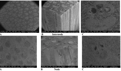
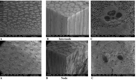
Ultrastructure Study
The ultrastructure studies on the four (4) selected bamboo from Gigantochloa genera are shown in Figures 1 to 8. Figures 1 to 4 shows the ultrastructures of the internodes and nodes focusing at the vascular bundles of the G. brang, G. levis, G. scortechinii and G. wrayi taken using the SEM.
Figures 5 to 8 shows the images of the bomboo species taken using TEM focusing at the fibers cells. The cells wall of the fibers clearly shows that they possess more than two layers, which were S1, S2, S3 and Sn.. All the four bamboo species shows clear that they belong to bamboo of are bamboo in class type. They possesses of a vascular sheath fiber and one fiber strand. Eventhough all the cells are similar in shape but they are however different in sizes in position at internodes and nodes, and position in the bamboo at either the outer, middle and the inner layers.The distribution of the vascular bundles per mm2, vascular length, mvascular bundle width are shown in Tables 1, 2, 3 and 4. The fibers length, diameter, lumen diameters, wall thickness and the Rumkle’s ratio are given in Tables 5 and 6.
Conclusion
The anatomical structure varies significantly with the species. The distribution and the size of vascular bundle are differences between species and even in the same genera. The vascular bundle of four species almost similar and were classified under namely Type III consisting of single vascular sheath fiber and one fiber strand. But, the vascular bundle size was significantly different between position (node and internode) and position (outer, middle and inner layers).
The fiber morphologi for each species has a different measure of size in terms of length, diameter, lumen diameter and wall thickness. The study identified that there was differences in fiber dimensions on the position (node and internode) and position (outer, middle and inner layer) in the same species. Fiber length was longer at the internode node. While the middle layer has the longest fiber length compare to the outer and inner layer.
Table 4: Analysis of variance for anatomical properties between bamboo species, position & position. Anatomical Properties
No. Vascular bundle Vascular bundle length Vascular bundle Width
Species G. brang 5.47b 894.55c 530.22c
G. levis 4.23c 1182.51a 759.35a
G. scortechinii 7.05a 932.71b 544.63c
G. wrayi 5.75b 847.84d 598.02b
Position Internode 6.32a 869.87b 585.42b
Node 4.93b 1058.94a 630.70a
Position Outer layer 8.56a 748.54c 467.23c
Middle layer 4.89b 1013.25b 599.76b
Inner layer 3.42c 1131.42a 757.19a
Table 5: Analysis of variance for fibre morphology between species, position & position. Fibre Morphology
Fibre Length
Fiber Diameter
Lumen Diameter
Wall Thickness
Runkle’s Ration
Species G. brang 1909.68b 22.75a 4.75b 9.02a 4.90b
G. levis 2039.98a 22.67a 4.00c 9.34a 5.32a
G. scortechinii 1745.27c 17.26b 8.66a 4.30c 1.35d
G. wrayi 1798.79c 17.86b 3.83c 7.02b 4.13c
Position Internode 2074.24a 18.23b 4.43b 6.90b 4.17a
Node 1672.62b 22.04a 6.18a 7.02a 3.68b
Position Outer layer 1698.52c 18.49c 5.44c 7.03b 4.04b
Middle layer 2060.41a 22.36a 5.51b 8.43a 4.29a
Inner layer 1861.35b 19.56b 5.96a 6.80c 3.45c
Values followed by the same letter in a column is not significant different at 95% probability level.
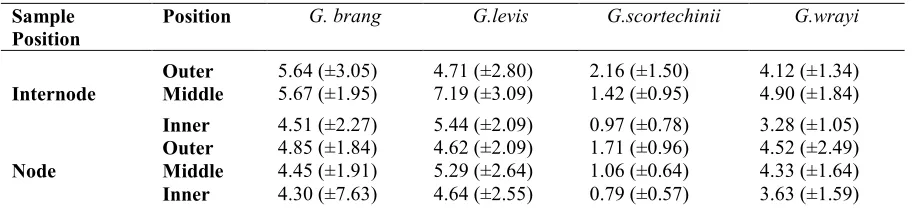
Table 6: Runkle’s ratio of various Gigantochloa species Sample
Position
Position G. brang G.levis G.scortechinii G.wrayi
Outer 5.64 (±3.05) 4.71 (±2.80) 2.16 (±1.50) 4.12 (±1.34) Internode Middle 5.67 (±1.95) 7.19 (±3.09) 1.42 (±0.95) 4.90 (±1.84) Inner 4.51 (±2.27) 5.44 (±2.09) 0.97 (±0.78) 3.28 (±1.05) Outer 4.85 (±1.84) 4.62 (±2.09) 1.71 (±0.96) 4.52 (±2.49) Node Middle 4.45 (±1.91) 5.29 (±2.64) 1.06 (±0.64) 4.33 (±1.64) Inner 4.30 (±7.63) 4.64 (±2.55) 0.79 (±0.57) 3.63 (±1.59)
A B Internode C
A B Node C
Figure 1: SEM images of the vascular bundles at internodes and nodes of the G. brang
A B Internode C
A B Node C
A B Internode C
A B Node C
Figure 3: SEM images of the vascular bundles at internodes and nodes of the G. scortechinii
A B Internode C
A B Node C
Figure 4: SEM images of the vascular bundles at internodes and nodes of the G. wrayi
A B C Figure 5: Fibre cells (A, B) and middle lamella (C) at internodes of the G. brang
A B C
Figure 6: Fibre cells (A, B) and middle lamella (C) at internodes of the G. levis
A B C
Figure 7: Fibre cells (A, B) and middle lamella (C) at internodes of the G. scortechinii
A B C
References
Abd. Latif, M. (1995) Some selected properties of two Malaysia bamboo species in relation to age, height, site and seasonal variation. PhD. Thesis, Universiti Putra Malaysia. 282p.
Abd. Latif M., & Tamizi, M., (1993) “Variation in anatomical properties of three Malaysian bamboos from natural stands” Journal Tropical Forest Science Vol.5, No.1, pp. 90-96.
Azmy, M., Hall, J.B., Othman, S., Razak, W., & Rashidah, A.B.K. (2007) “Quality management of the bamboo resource and its contribution to environmental conservation in Malaysia. Management of Environmental Quality” An International Journal. Vol. 18, No. 6, pp. 643-656.
Horn R.A., & Setterholm V.C. (1990) Fiber Morphology and New Crops. In: Janick J. and Simon J.E. (eds), Advances in new crops. Timber Press, Portland, Origen. P 270-275.
Ireana, Y., (2009) Cell Wall Architecture, Properties and Characteristics of Bamboo, Kenaf and Rice Straw Fibers. M.Sc Thesis, USM.
Liese., W. (1985). Anatomy and properties of bamboo. Recent research on bamboos. Proceedings of the International Bamboo Workshop. October 6-14, 1985, Hangzhou, China.
Liese, W., (1992). The structure of bamboo in relation to its properties and utilization. In Zhu, S., Li, W., Zhang, X. Wang, Z. ed., Bamboo and its use. Proceedings of the International symposium on Industrial Use of Bamboo, Beijing, China, 7-11 December 1992. International Tropical Timber Organization: Chinese Academy of Forestry, Beijing, China. Pp 96-100.
Liese, W., & Grosser, D. (1972) “Untersuchungen zur Variabilitat der Faserlange bei Bambus (Variation of fibre length and fibre width within one internodes in bamboo species)” Holzforsch. Vol.26, No.6, pp.202-211.
Norul Hisham, H., Othman, S., Rokiah, H., Latif, M., Ani, S., & Tamizi, M. (2006) “Characterization of Bamboo Gigantochloa Scortechinii at different ages” Journal of Tropical Forest Science, Vol.18, No.4,pp.236-242.
Pattanath, P.G. (1972) “Trend of variation in fibre length in bamboos” Indian Forester. Vol.98, No.4, pp.241-243.
Razak, W., Tamizi, M., Othman, S., Aminuddin, M., Affendy, H., & Izyan, K. (2010) “Anatomical and Physical Properties of Cultivated Two- and Four-year-old Bambusa vulgaris” Sains Malaysiana Vol.39, No.4, pp.571–579.
Razak, W., Mahmud, S., & Hashim, W.S. (2005) “Fungal colonization and decay in tropical bamboo species” Journal of Applied Science, Vol.5, No.5, pp.897-902.
Razak, W., Hashim, W.S., & Azmy, M. (2002). “Properties of boards from 3-layers laminated and composite-ply from tropical bamboo Gigantochloa scortechinii” Journal of Borneo Science, Vol.12, No. 2, pp.43-50.
Razak, W. (1998). Effect of selected preservatives on the durability of Gigantochloa scortechinii. A PhD thesis, University of London.
Soeprayitno, T., Tobing L., & Widjaja E. A. (1990) “Why the sundanese of West Java Prefer Slope-inhabiting Gigantochloa pseudoarundinacea to those growing in the valley”. In International Workshop on Bamboo held in Cochin, 14-15 Nov. 1988: Proceeding edited by I.R.R. Rao, R. Gnanaharam and C.B. Sastry. Peechi : Kerala Forest Research Institute and International Development Research Centre, Pp. 215-217.
Ververis C., Georghiou K., Christodoulakis N., Santas P., & Santas R. (2004) “Fiber dimensions, lignin and cellulose content of various plant materials and their suitability for paper production” Industrial Crops and Products an International Journal Vol.19, No. 3, pp. 245-254.
Viena L.S, Trugilho P.F, Gherardi Hein P.R., Lima J.T., & Mareiro da Silva J.R. (2009) “Predicting the morphology characteristics and basic density of Eucalytus Wood using the NIRS technique” Cerne, Vol.15, No.4, pp 421-429. Wenyue, H., Shiyi Q., & Youfen L. (1981) “The anatomical structure of culms of Phyllostachys pubescens Mazel exh. de Lehaie” Bamboo Research Vol.1, pp.58–65