Archives • 2017 • vol.1 • 19-27
CHEMICAL ANALYSIS AND NORMO-GLYCEMIC EFFECT OF OPUNTIA FICUS INDICA
Patricia I. Manzano
1,2, Oswaldo G. Pesantes
1, Glenda M. Sarmiento
1, Ivan Chóez-Guaranda
3,
Zoraida C. Burbano
1, Galo M. Duran
4, María C. Villacres
1*
1 Universidad de Guayaquil. Facultad de Ciencias Químicas. Cdla. Universitaria “Salvador Allende”. Malecón del Salado entre Av. Delta y Av. Kennedy. Guayaquil, Ecuador
2 Escuela Superior Politécnica del Litoral, ESPOL, Facultad de Ciencias de la Vida, Campus Gustavo Galindo Km 30.5 Vía Perimetral, P.O. Box 09-01-5863, Guayaquil, Ecuador
3 Escuela Superior Politécnica del Litoral, ESPOL, Centro de Investigaciones Biotecnológicas del Ecuador, Campus Gustavo Galindo Km 30.5 Vía Perimetral, P.O. Box 09-01-5863, Guayaquil, Ecuador
4 Universidad de Guayaquil, Facultad de Ciencias Administrativas, Cdla. Universitaria “Salvador Allende”. Malecón del Salado entre Av. Delta y Av. Kennedy. Guayaquil, Ecuador
*mcvillacres@gmail.com
Abstract
Products from Opuntia ficus indica have been used in ancestral medicine for treatment of hyperglycemia, lipidaemia, inflammation, infection, among others. This study aimed to validate the effect of Opuntia ficus indica
on acute hyperglycemia and identify the component/s mediating this effect. Crude extracts were analyzed by gas chromatography mass spectrometry (GC-MS) and phytochemical screening. Several saturated and two mono-unsaturated fatty acids were identified, as well as one diterpene and one carbohydrate. Water soluble and organic-solvent soluble compounds were obtained from the stem pulp of Opuntia ficus indica. Phytochemical screening indicated the presence of glycosides and coumarins, with weak reactions for tannins and flavonoids. The most abundant water soluble components were identified as polysaccharides. Bioactivity on blood glucose levels by several preparations of Opuntia ficus indica was readily detected using an acute model of induced hyperglycemia in Wistar rats. Dose-response experiments demonstrated normo-glycemic activity in the range 50-300 mg/kg. In conclusion products from Opuntia ficus indica stems display potent capacity to reduce postprandial levels of glycemia in an animal model of acute hyperglycemia.
Introduction
Diabetes type 2 (DT2), also called insulin-independent or non-genetic, constitutes a major health problem worldwide, reaching epidemic proportions in some countries (1). Predictions from the International Diabetes Federation estimates the frequency of diabetes at 382 million worldwide in 2013, with a projected frequency of 4.4% by year 2035 (2). Such a high level of disease requires ever growing medical resources, increasing overall the costs of healthcare.
Unhealthy dietary habits and a sedentary life style are considered mayor contributors to the escalating frequency of diabetes (3). Genetic factors appear to increase the risk of DT2 in specific human groups, including Hispanics, and about 36 genes –each one with rather low risk, but accumulative- have been identified as risk factors (4). Mortality is increased among affected individuals, and the associated morbidities include obesity, dyslipidemias, arterial hypertension, cardiovascular disease and disability (5, 6). Diabetes has been detected in humans since antiquity, and different ethnic groups have used a wide variety of products, mainly of vegetal origin, to treat and alleviate this disease. In addition, complementary and alternative medicine based on some of these natural products has been increasingly consumed, reaching high percentages of users in some communities (7,8,9).
Both the fruits and pulp of the prickly pear cactus (family Cactaceae) are popularly consumed as food (10,11). Also, preparations from several varieties of these cacti (genus Opuntia) have been attributed a number of biological actions, including anti-diabetic, anti-hyperlipidemic, antioxidant and antiviral activities (12). The dried pulp of the cladodes from Opuntia ficus indica (OFI) has been widely used by native people in Mexico and Central America for control of DT2 (13). Moreover, some early but limited clinical studies supported the notion of an anti-diabetic effect in OFI (13,14) as well as in Opuntia streptacantha (14,15,16)
Several compounds have been identified as biologically active in members of the Opuntia genus. For example, flavonoids, sterols and alpha-pyrones were reported in Opuntia dillenii (17,18,19). Polyphenols (20,21), flavonoids (20), proanthocyanidins (22), beta-sterols (23), and a glycoprotein with potential normo-lipemic effect (24) were found in OFI. Polysaccharides from the same species had an anti-diabetic effect in diabetic subjects (6), dermal healing in rats (25), and an anti-inflammatory effect evaluated in chondrocyte cultures (26). More recently, an interesting study demonstrated modulatory activity of Opuntia polyacantha on macrophages in vitro, providing a mechanism for its anti-inflammatory effects (27).
However, the biological effects of polysaccharides and other components of OFI are still in need of more extensive characterization. We investigated in this study the chemical components of Opuntia ficus indica by gas chromatography and mass spectra analysis, and evaluated its potential in glycemia control compared to a standard treatment drug, in a rat model of acute hyperglycemia.
Methods
Extract Preparation.
Stems of OFI (0.5 kg) were washed with H2O, peeled and the pulp cut in squares about 1.5 cm long. This material was processed in a food blender until the mass was homogeneous in texture. The resulting mix was spun at 2000 rpm for 10 min at 4oC. The crude extract was used for the lyophilization -silanization procedure described below. An acetone-treated extract for biological assays was generated by treating 1 ml crude extract with 2 ml acetone. The white precipitate was further rinsed with fresh acetone and diluted in H2O. Ethanol extracts were also prepared for the phytochemical screening.
Lyophilization and silanization.
300 mg of crude OFI extract were freeze dried in a LABCONCO freeze-drier system of 4.5 L capacity (Houston, Texas). Then, dried OFI extracts were mixed with 200 ul of BSTFA (-N,O-Bis (trimethylsilyl) trifluoroacetamide), Sigma-Aldrich and maintained at 80oC for 2 h to reaction as described before (28).
Analysis by gas chromatography mass spectrometry (GC-MS)
identified by comparison of their mass spectra and mass reference of Wiley 9th with NIST 2011 MS Library.
Phytochemical screening
Samples were tested for glycosides, coumarins, tannins, flavonoides, and alkaloids. For glycosides, 1 g of crude extract was dissolved in 5 ml H2O and filtered; 5 ml Benedict’s reagent was added to 1 ml clear filtrate; an orange read precipitate was read as a positive reaction (Benedict’s test). A confirmatory test for carbohydrates was performed with the water-soluble phase of a crude extract, adding 1 ml α-naphtol and 4 ml concentrated sulfuric acid to a 1 ml saturated OFI water solution (Molisch’s test). For coumarins, about 1 g of the extract was covered with filter paper adsorbed with diluted NaOH and heated in a water bath for 4-5 min; a fluorescent yellow reaction under UV light observation was considered positive. The ferric chloride test was used for determination of tannins: ferric chloride (3-4 drops of 5% sol) was added to 1 ml extract; a green color indicated a positive reaction. For flavonoids, a small piece of magnesium foil was added to a vial containing 1 g of crude extract dissolved in 5 ml ethanol. HCl acid was added drop wise until red color development (Shinoda’s test). For alkaloids, 5 g of crude extract were suspended in 5 ml HCl 1%, stirred on a water bath and filtered; for the Dragendroff’s test, 1 ml of clear filtrate was mixed with 1 ml Dragendroff’s reagent, and an orange-red precipitate was indicative of a positive reaction. A Wagner’s test was performed by mixing 1 ml clear filtrate with 2 ml of Wagner’s reagent, and a red-brown precipitate was read as a positive reaction.
Biological assay for glycemia
Wistar rats about 200 g ± 20 g were housed in regular laboratory conditions (22 ± 3oC, relative humidity 50-55%, 12 h light/dark cycles) and had access to water and standard animal food ad libidum. Animals were restricted on food for the last 12 h before assays. OFI extracts for biological assays were prepared as indicated above. Wistar rats about 200 g were randomly assigned to groups, with 4-5 per group and orally administered saline solution (No treatment control group), and Metformin (Sanofi-Aventis, Cali, Colombia) as positive control of treatment or OFI extracts. Metformin was given at 10 mg/kg and OFI extracts were given at the doses indicated in the graphs. Treatments were orally administered at a volume of 400 µl/rat. A glucose (D-glucose, Mallinckrodt Baker, Phillipsburg, NJ) solution
containing 400 mg/rat was given orally 30 minutes later to all groups. A negative control group received glucose only. Blood samples from control and test groups were taken at baseline, 15 min and 60 after bulk glucose ingestion using a glucometer Accu-Chek® (Roche Diagnostics GmbH, Mannheim, Germany). Animal management was performed according to the NIH Guide for the Care and Use of Laboratory Animals (NIH publications No. 80-23), revision of 1996.
Statistical Analysis
Data from the GC-MS chromatograms was analyzed using the INFOSTAT software. Data from bioassays was analyzed applying the SPSS statistical software package, version 22.0 (SPSS, Chicago, IL). Values at p<0.05 in two-tailed pair-wise comparisons were considered statistically significant.
Results
Chromatographic analysis
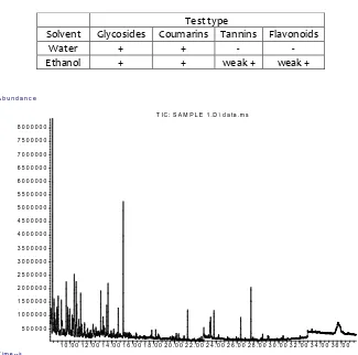
The analysis of the chemical structures in the samples was established by comparison of the mass spectra focusing on those with certainty >90%. Several of the compounds identified were saturated fatty acids: butanedioic acid, nonanedioic acid, hexadecanoic acid, octadecanoic acid, and eicosanoic acid (Table 1 and Figure 1). There were two mono-unsaturated fatty acids: palmitelaidic acid and trans-13-octadecenoic acid, a diterpenoid (5,8-epoxy-15-nor-labdane) and one carbohydrate (D-mannitol).
Phytochemical screening
Testing for phytochemical screening was performed in parallel, in water-based extracts and in ethanol-extracts. Reactions for glycosides and coumarins were positive in both the water-base extracts and ethanol-based extracts (Table 2). A confirmatory carbohydrate determination (Molisch’s test) indicated a strong positive reaction. The ethanol extract gave also a weak reaction for tannins and flavonoids. Reaction for alkaloids was negative in both types of extracts.
Effect of OFI extracts on glycemia
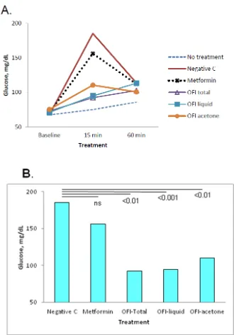
was used as positive control of treatment, and a group receiving glucose only as a negative control of treatment. Compared with the negative control of treatment (glucose only), all groups receiving Metformin or OFI extracts (50 mg/rat) had reduced levels of glycemia at 15 min post administration of the glucose challenge, whereas the values declined at 60 min after glucose challenge and the differences tended to disappear (Figure 2a). Pair-wise comparisons of values determined at 15 min using the negative control as a reference indicated that the differences were statistically significant for the Opuntia total extract (p < 0.01, a stem crude extract), Opuntia liquid phase (p < 0.001) and Opuntia acetone precipitate (p < 0.01, Figure 2b).
Dose response to OFI extracts in glucose challenge
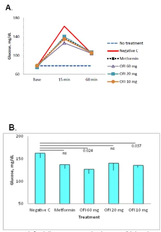
Dose response experiments were performed to investigate the potency of treatment with the OFI acetone precipitate. Six groups with five rats each were randomly distributed in control groups as described above and OFI acetone precipitate was tested at 60, 20 and 10 mg /rat. Overall, all treated groups presented reduced glycemia at min 15 post challenge, and all values tended to normalize by min 60 (Figure 3a). Pair-wise comparison of the values determined at 15 min post challenge indicated statistically significant differences for the 60 mg/rat (p = 0.024) and 10 mg/rat doses (p = 0.037) using the glucose-only group (negative control of treatment) as a reference (Figure 3b). Pair-wise comparison of values of the three doses from OFI-treated groups with the Metformin-treated control indicated no statistically significant differences.
Discussion
Several of the compounds identified in the chromatographic analysis were saturated fatty acids. Of these, nonanedioic acid, also known as azelaic acid, has reportedly antibacterial activity (29), and is being actively investigated for its anti-inflammatory properties, that appear to include increased expression and transcriptional activity of PPARγ (30).
Of the two mono-unsaturated fatty acids detected in the chromatographic analysis, palmitelaidic acid may have some application on weight management and health by reducing adiposity, modulating fatty acid synthesis in adipocytes (31)
The analysis also detected a labdane-type diterpene. This is an interesting finding, as the diterpenes are a large family commonly found in the plant kingdom, with some of them having shown broad biological effects (32). At least some of those promising effects
appear to be anti-inflammatory and involve regulation of components of the NFkB complex, a key step in immune activation (33).
Polysaccharides are abundant metabolites in members of the Opuntia genus. In agreement with previous studies, we detected a substantial concentration of polysaccharides in OFI samples in the phytochemical screening, and confirmed by specific chemical reaction. Of note, detection of compounds other than glycosides was highly efficient by gas chromatography, but less so for glycosides. As reported earlier, crude extracts of OFI present significant capacity to control the postprandial spike on blood glucose (2), as well as in
Opuntia streptacantha (7). A recent work on O.
streptacantha indicated immunomodulatory action by
its polysaccharides, supporting the notion that this family of compounds is important for the biological effects observed (27).
In our study, the effect of OFI in control of postprandial glycemia was readily observed in biological assays with several OFI extracts. The dose response experiments showed a high efficiency of the OFI acetone extract to control glucose levels in blood, indicated by levels similar to those obtained by the anti-diabetes drug Metformin. This observation is important, since it suggests that even low doses of OFI may help to control postprandial glycemia in humans.
The need of new approaches to help control glycemia levels increases as the frequency of diabetes type 2 continues to rise worldwide (14,16). This is particularly pressing in countries with developing economies, where the frequency of diabetes escalates at a faster rate, but with limited resources. Moreover, large segments of populations in these highly affected countries are inclined to attend their medical needs using traditional medicine. Therefore, studies like ours are justified to develop and evaluate procedures that target communities in need. It seems also feasible to explore the use of natural products as OFI as a dietetic help, reducing the dose of pharmaceutical chemicals needed to achieve healthy blood glucose levels. We conclude that further studies are justified to investigate the cellular events mediating glycemia control.
Acknowledgments
References
1 Kahn SE, Cooper ME, Del Prato S. Pathophysiology and treatment of type 2 diabetes: perspectives on the past, present, and future. Lancet. 2014; 383(9922): 1068-1083 2 Lee CMY, Colagiuri S. Risk scores for diabetes
prediction: the International Diabetes Federation PREDICT-2 project. Diabetes Res Clin Pract. 2013; 100(2): 285-286.
3 Schwarz PE, Greaves CJ, Lindström J, Yates T, Davies MJ. Nonpharmacological interventions for the prevention of type 2 diabetes mellitus.
Nat Rev Endocrinol. 2012; 8(6): 363-373.
4 Grarup N, Sandholt CH, Hansen T, Pedersen O. Genetic susceptibility to type 2 diabetes and obesity: from genome-wide association studies to rare variants and beyond. Diabetologia.
2014; 57(8): 1528-1541.
5 Gallagher EJ, LeRoith D. Epidemiology and molecular mechanisms tying obesity, diabetes, and the metabolic syndrome with cancer.
Diabetes Care. 2013; 36 Suppl 2: S233-239. 6 Shi Y, Hu FB. The global implications of
diabetes and cancer. Lancet. 2014; 383(9933): 1947-1948.
7 Kim C, Kwok YS. Navajo use of native healers.
Arch Intern Med. 1998; 158(20): 2245-2249. 8 Mull DS, Nguyen N, Mull JD. Vietnamese
diabetic patients and their physicians: what ethnography can teach us. West J Med. 2001; 175(5): 307-311.
9 Noel PH, Pugh JA, Larme AC, Marsh G. The use of tradicional plant medicines for non-insulin depednent diabetes mellitus in south Texas.
Phytother Res. 1997; 11(7): 512-517
10 Feugang JM, Konarski P, Konarski P, et al. Nutritional and medicinal use of Cactus pear (Opuntia spp.) cladodes and fruits. Front Biosci.
2006; 11: 2574-2589.
11 Stintzing FC, Carle R. Cactus stems (Opuntia spp.): a review on their chemistry, technology, and uses. Mol Nutr Food Res. 2005; 49(2): 175-194.
12 El-Mostafa K, El Kharrassi Y, Badreddine A, et al. Nopal cactus (Opuntia ficus-indica) as a source of bioactive compounds for nutrition, health and disease. Molecules. 2014; 19(9): 14879-14901.
13 Alarcon-Aguilar FJ, Valdes-Arzate A, Xolapa-Molina S, et al. Hypoglycemic activity of two polysaccharides isolated from Opuntia ficus-indica and O. streptacantha. Proc West Pharmacol Soc. 2003; 46: 139-142.
14 Frati AC, Gordillo BE, Altamirano P, et al. Acute hypoglycemic effect of Opuntia streptacantha Lemaire in NIDDM. Diabetes Care. 1990; 13(4): 455-456.
15 Frati-Munari AC, Gordillo BE, Altamirano P, Ariza CR. Hypoglycemic effect of Opuntia streptacantha Lemaire in NIDDM. Diabetes Care. 1988; 11(1): 63-66.
16 Ibanez-Camacho R, Roman-Ramos R. Hypoglycemic effect of Opuntia cactus. Arch Invest Med (Mex). 1979; 10(4): 223-230.
17 Ahmed MS, El Tanbouly ND, Islam WT, Sleem AA, El Senousy AS. Antiinflammatory flavonoids from Opuntia dillenii (Ker-Gawl) Haw. flowers growing in Egypt. Phytother Res.
2005; 19 (9): 807-809.
18 Jiang J, Li Y, Chen Z, Min Z, Lou F. Two novel C29-5beta-sterols from the stems of Opuntia dillenii. Steroids. 2006; 71(13-14): 1073-1077. 19 Qiu YK, Zhao YY, Dou de Q, Xu BX, Liu K. Two
new alpha-pyrones and other components from the cladodes of Opuntia dillenii. Arch Pharm Res. 2007; 30(6): 665-669.
20 Galati EM, Mondello MR, Giuffrida D, et al. Chemical characterization and biological effects of Sicilian Opuntia ficus indica (L.) mill. Fruit juice: antioxidant and antiulcerogenic activity. J Agric Food Chem. 2003; 51(17): 4903-4908.
21 Lee JC, Kim HR, Kim J, Jang YS. Antioxidant property of an ethanol extract of the stem of Opuntia ficus-indica var. saboten. J Agric Food
Chem. 2002; 50(23): 6490-6496.
22 Sreekanth D, Arunasree MK, Roy KR, et al. Betanin a betacyanin pigment purified from fruits of Opuntia ficus-indica induces apoptosis in human chronic myeloid leukemia Cell line-K562. Phytomedicine. 2007; 14(11): 739-746. 23 Park EH, Kahng JH, Lee SH, Shin KH. An
anti-inflammatory principle from cactus.
Fitoterapia. 2001; 72(3): 288-290.
24 Oh PS, Lim KT. Glycoprotein (90 kDa) isolated from Opuntia ficus-indica var. saboten MAKINO lowers plasma lipid level through scavenging of intracellular radicals in Triton WR-1339-induced mice. Biol Pharm Bull. 2006; 29(7): 1391-1396.
26 Panico AM, Cardile V, Garufi F, et al. Effect of hyaluronic acid and polysaccharides from Opuntia ficus indica (L.) cladodes on the metabolism of human chondrocyte cultures. J Ethnopharmacol. 2007; 111(2): 315-321.
27 Schepetkin IA, Xie G, Kirpotina LN, et al. Macrophage immunomodulatory activity of polysaccharides isolated from Opuntia polyacantha. Int Immunopharmacol. 2008; 8(10): 1455-1466.
28 Merchant A, Peuke AD, Keitel C, et al. Phloem sap and leaf delta13C, carbohydrates, and amino acid concentrations in Eucalyptus globulus change systematically according to flooding and water deficit treatment. J Exp Bot. 2010; 61(6): 1785-1793.
29 Sieber MA, Hegel JK. Azelaic acid: Properties and mode of action. Skin Pharmacol Physiol.
2014; 27 Suppl 1: 9-17.
30 Mastrofrancesco A, Ottaviani M, Aspite N, et al. Azelaic acid modulates the inflammatory response in normal human keratinocytes through PPARgamma activation. Exp Dermatol.
2010; 19(9): 813-820.
31 Kadegowda AK, Burns TA, Miller MC, Duckett SK. Cis-9, trans-11 conjugated linoleic acid is endogenously synthesized from palmitelaidic (C16:1 trans-9) acid in bovine adipocytes. J Anim Sci. 2013; 91(4): 1614-1623.
32 Peters RJ. Two rings in them all: the labdane-related diterpenoids. Nat Prod Rep. 2010; 27(11): 1521-1530.
33 de las Heras B, Hortelano S. Molecular basis of the anti-inflammatory effects of terpenoids.
Table 1. Compounds identified by gas chromatography/mass spectroscopy in crude extracts of Opuntia ficus indica
Tr (min) Compounds %
abundance
1 9.76 Butanedioic acid 1.54
2 17.83 Nonanedioic acid 0.41
3 20.18 D-Mannitol 0.32
4 20.92 Palmitelaidic acid 0.17
5 21.26 Hexadecanoic acid 1.26
6 23.47 Trans-13-Octadecenoic acid 1.44
7 23.79 Octadecanoic acid 1.66
8 26.12 Eicosanoic acid 0.09
9 35.90 5,8-Epoxy-15-nor-labdane 0.58 34
Table 2. Phytochemical screening of crude stem extracts from Opuntia ficus indica
Test type
Solvent Glycosides Coumarins Tannins Flavonoids
Water + + - -
Ethanol + + weak + weak +
1 0 . 0 0 1 2 . 0 0 1 4 . 0 0 1 6 . 0 0 1 8 . 0 0 2 0 . 0 0 2 2 . 0 0 2 4 . 0 0 2 6 . 0 0 2 8 . 0 0 3 0 . 0 0 3 2 . 0 0 3 4 . 0 0 3 6 . 0 0 5 0 0 0 0 0
1 0 0 0 0 0 0 1 5 0 0 0 0 0 2 0 0 0 0 0 0 2 5 0 0 0 0 0 3 0 0 0 0 0 0 3 5 0 0 0 0 0 4 0 0 0 0 0 0 4 5 0 0 0 0 0 5 0 0 0 0 0 0 5 5 0 0 0 0 0 6 0 0 0 0 0 0 6 5 0 0 0 0 0 7 0 0 0 0 0 0 7 5 0 0 0 0 0 8 0 0 0 0 0 0
T im e --> A b u n d a n c e
T I C : S A M P L E 1 . D \ d a t a . m s
Figure 2. Effect of Opuntia ficus indica preparations on postprandial glycemia in Wistar rats. Groups of 4 rats were given glucose only (Negative control), or treatment with Metformin or OFI total extract, OFI liquid preparation, or OFI acetone-precipitate, all extracts at 50 mg/rat. A) Overall glycemia values were elevated at min 15 and declined by min 60 after glucose
Figure 3. Dose response to Opuntia ficus indica acetone preparations in postprandial glycemia. Groups of five rats were randomly assigned to control groups (as described in Figure 2) or OFI acetone-extract at doses of 60, 20 or 10 mg/rat. A) The
3 doses of OFI tested mediated regulation of glycemia at min 15, and values declined by min 60. B) Compared with the glucose-only group (Negative control), differences in glycemia levels were statistically significant for the groups receiving the