Research article Available online www.ijsrr.org
ISSN: 2279–0543
International Journal of Scientific Research and Reviews
Navicular Drop Gender Differences Among College Students: A
Cross Sectional Study
Malik Manoj
1, Kaur Jaspreet
1, Punia Sonu
1*and Bhagesh
2Assistant Professor, Deptt. of Physiotherapy, GJUST, Hisar-125001
ABSTRACT
Knee injury associated with abnormal foot biomechanics can lead to foot, knee injury because knee and foot were working simultaneously so it was clinically important to know about abnormal foot biomechanics in the prevention and treatment of injury.1 During standing position or full weight bearing, all the joint of lower limb working as interactive segments means with foot pronation, internal rotation of the tibia occurring simultaneously. Aim of present study was to investigate the influence of foot length, age, gender, and BMI on the navicular drop in weight bearing relaxed erect position. 20 college going students were recruited from hisar City College; Present study was cross sectional study; 18-25 years normal young adults of both sex and they were cooperative and obey command. Demographic details of student were taken. 30 students (15 M; 15 F) with mean age 20.97±1.85 years participated in the study. The data was not normal so we have done non parametric correlation (spearman’s) of navicular drop with weight, height, BMI, age, gender and foot length was examined. The correlation of Right ND with left ND and age was significant, but no significant correlation was found between RND and height, weight, foot length and BMI. The correlation of Left ND with height, weight, foot length, age and BMI of individuals was not found significant. The present study revealed the incidence of navicular drop more than normal value (6mm-9mm) in college aged students.
KEYWORDS:
Gender; Adults; Navicular drop*Corresponding author
Sonu Punia
Assistant Professor,
Department of Physiotherapy, GJUST, Hisar-125001.
INTRODUCTION
Knee injury associated with abnormal foot biomechanics can lead to foot, knee injury
because knee and foot were working simultaneously so it was clinically important to know about
abnormal foot biomechanics in the prevention and treatment of injury.1-3 During standing position or
full weight bearing, all the joint of lower limb working as interactive segments means with foot
pronation, internal rotation of the tibia occurring simultaneously.2
Normally navicular bone in humans is situated medially, and articulates talus proximally, cuneiform
distally and cuboid laterally and only one muscle (tibialis posterior) attached with this bone. 2%-14%
general population may have an accessory navicular bone.4-5
Foot pronation was measured by Navicular drop clinically which is the change in height of
the navicular bone when the subject’s foot transferred from neutral position of foot to weight bearing
in erect position3. Normal navicular drop was calculated as 6-8 mm and it may expresses as
excessive or abnormal if it was greater than 10-12 mm.6-7
Abnormality of navicular drop has been associated with anterior cruciate ligament patients,
medial tibial stress syndrome and patellofemoral pain syndrome. Other researcher also revealed
excessive navicular drop with plantar intrinsic muscle fatigue.
Position of measurement of navicular drop also affected foot biomechanics; therefore it may helpful
for patients with overuse symptoms of the lower extremity.8-13
Navicular drop occurs due to structural abnormality and atrophy of the muscles that supports
the arch. Navicular drop characterized by visible swelled reddened bony prominence medially and
severe pain in mid of the foot. Navicular drop may be influenced length of foot, age, gender, and
Body Mass Index (BMI). 14-16
So our aim of present study was to investigate the influence of foot length, age, gender, and
BMI on the navicular drop in weight bearing relaxed erect position.
METHODOLOGY
Study Participants
20-collage going students were recruited from hisar City College. Present study was cross
osteoarthritis of knee, past history of lower limb injury, Congenital abnormalities of foot, pregnant
females, abnormality in limb length and amputation of Lower limb amputation. Demographic details
(age, height, weight, BMI) of each subjects was taken. Then Subjects were selected for study by
giving consent to study and they have explained whole procedure of study.
Navicular Drop Test
It was a clinical instrument to measure the navicular height in sagittal plane first given by
Brody 1982 which described pronation of foot. Reliability and validity of test was already calculated
and it was valid test for calculation. Measurement in sitting and standing position and the difference
between two test position measurements is called navicular drop. Instrument required for test
measurement was a pen, card, measure tape and markers and all the measurement were recorded on
data collection form.13, 16-17
Measurement procedure
Subjects were sitting in a comfortable position with flat feet on normal surface. The subject
was asked to flex knee to 900 and placed ankle joints in neutral position and we palpated most
prominence of the navicular bone tubercle in neutral sitting position and marked with a pen on card
that besides the foot. Then the same procedure was applied in standing weight bearing position
without change in distribution of equal weight on feet again same reading was taken in the standing
position and relative position of prominence of navicular tubercle bone was marked on the card. At
last, the difference between the height of prominence of navicular tubercle bone in relaxed sitting
position and weight bearing erect position was noted with a measuring tape revealing the navicular
drop. Displacement of more than 10 mm in erect position weight bearing is considered as significant
overpronation of the foot. Repeat the procedure with other leg and compared the difference.
Data analysis
Data analysis was done with help of SPSS 16.0 version. The Kolmogorov’s Smirnov test
was used for assessing normality of data. Mean and IQR were assessed for the demographic
characteristics and ND since the data was not normally distributed. Comparison of male and female
ND was done with Mann-Whitney U test because data was not normal. Correlation of navicular
drop with age, height, weight, foot length and BMI was calculated with spearman’s test. A p-value
RESULTS
30 students (15 M; 15 F) with mean age 20.97±1.85 years (18-25 years) participated in the
study. Descriptive statistics of demographic variables were shown in Table 1.
Table 1: Descriptive statistics
Range Mean Std. Deviation
Age 18-25 20.97 1.847
Weight(kg) 42-80 60.87 12.235
Height(cm) 152-185 172.70 8.133
BMI 15-29 21.39 3.375
Foot length(cm) 14-27 22.63 3.538
Right ND(mm) 5-14 9.21 2.094
Left ND(mm) 5-16 9.27 2.651
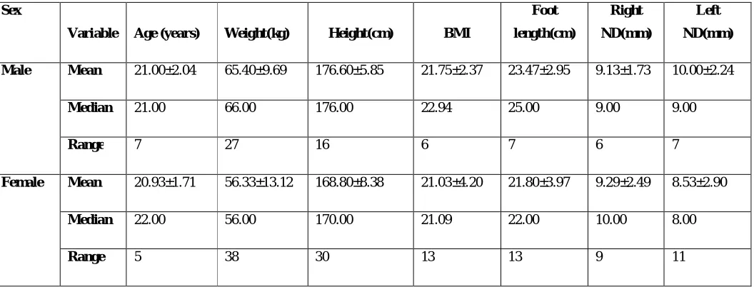
Table 2: Outcome variables Sex
Variable Age (years) Weight(kg) Height(cm) BMI
Foot length(cm)
Right ND(mm)
Left ND(mm)
Male Mean 21.00±2.04 65.40±9.69 176.60±5.85 21.75±2.37 23.47±2.95 9.13±1.73 10.00±2.24
Median 21.00 66.00 176.00 22.94 25.00 9.00 9.00
Range 7 27 16 6 7 6 7
Female Mean 20.93±1.71 56.33±13.12 168.80±8.38 21.03±4.20 21.80±3.97 9.29±2.49 8.53±2.90
Median 22.00 56.00 170.00 21.09 22.00 10.00 8.00
Range 5 38 30 13 13 9 11
Study variables (height, weight, BMI, foot length, Right ND, Left ND) among different sex
were calculated as shown in table 2. The Kolmogorov’s Smirnov test was used for assessing
normality of data. Mean and IQR were assessed for the demographic characteristics and ND since
the data was not normally distributed.
Comparison of male and female ND was done with Mann Whitney U test because data was
not normal. The result was not statistically significant on right side but significant on left side as
Comparison of male and female ND was done with Mann Whitney U test because data was
not normal. The result was not statistically significant on right side but significant on left side as
shown in table 3
Table 3: Comparison of right leg with left leg navicular drop
Right ND Left ND
Z statstics -.291 -1.97
P-value .771 .049
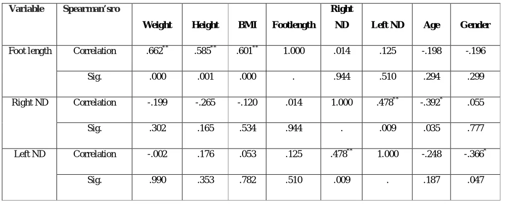
The data was not normal so we have done non parametric correlation (spearman’s) of
navicular drop with weight, height, BMI, age, gender and foot length was examined. The correlation
of Right ND with left ND and age was significant, but no significant correlation was found between
RND and height, weight, foot length and BMI. The correlation of Left ND with height, weight, foot
length, age and BMI of individuals was not found significant as shown in table 4.
Table 4: correlation between different outcome variables
Variable Spearman’sro
Weight Height BMI Footlength
Right
ND Left ND Age Gender
Foot length Correlation .662** .585** .601** 1.000 .014 .125 -.198 -.196
Sig. .000 .001 .000 . .944 .510 .294 .299
Right ND Correlation -.199 -.265 -.120 .014 1.000 .478** -.392* .055
Sig. .302 .165 .534 .944 . .009 .035 .777
Left ND Correlation -.002 .176 .053 .125 .478** 1.000 -.248 -.366*
Sig. .990 .353 .782 .510 .009 . .187 .047
DISCUSSION
Our study findings suggest that hypothesis of study was not accepted as output of study
outcome variables was found insignificant. Present study results were in agreement with Ashok
Aenumulapalli et al., 2017 study results that ND difference between gender were statistically
In addition, some other researchers have used different types of measurement methods to
calculate Navicular Difference and they found navicular difference of 15 mm13, 13 mm17 and 10
mm19. Ashok Aenumulapalli et al., 2017 found the median values with inter quartile range for
navicular drop for male students was [right leg 6 mm (4-8); left leg 6 mm (4-9)] and for female
students was [right leg 6mm (4-10) ; left leg 7mm (3-8)].18
Fukano M et al have also claimed that navicular difference was affected by various different
variables like height, weight, foot length, age, gender and BMI of subjects.20-23 But this study did
not found any correlation with weight, height, foot length and BMI but significant correlation with
gender (left ND) and age (right ND).
Based on research evidences we stated that excessive navicular difference results in
abnormal overpronation of foot which was associated with abnormal biomechanics of lower limb at
pelvis, hip and knee joint. So we planned some rehabilitation protocol to correct out faulty
biomechanics at subtalar joint.
CONCLUSION
The present study revealed that the prevalence of navicular drop was high in age group of
18 to 25 years showed gender differences for navicular drop and influence of demographic
characteristic (age, height, weight and BMI) on navicular drop showed no significant findings.
REFERENCES
1. Mary K. Allen, Ward M. Glasoe. Metrecom Measurement of Navicular Drop in Subjects
with Anterior Cruciate LigamentInjury .Journal of Athletic Training 2000; 35(4):403-406.
2. Donatelli RA. Normal anatomy and biomechanics. In: Donatelli RA, ed.The Biomechanics
of the Foot and Ankle. 2nd ed. Philadelphia, PA: FA Davis; 1996:3-31.
3. Nawoczenski DA, Cook TM, Saltzman CL. The effect of foot orthotics on
three-dimensional kinematics of the leg and rearfoot during running.J Orthop Sports Phys Ther.
1995;21:317-327.
4. McPoil TG, Cornwall MW, Medoff L, Vicenzino B, Forsberg K, Hilz D. Arch height
change during sit-to-stand: an alternative for the navicular drop test. Journal of foot and
ankle research. 2008 Jul 28;1(1):1.
6. Coughlan GF, Fullam K, Delahunt E, Gissane C, Caulfield BM. A comparison between
performance on selected directions of the star excursion balance test and the Y balance test.
Journal of athletic training. 2012 Aug; 47(4):366.
7. Cornwall MW, Lane C, Norwood J, Patterson S, Strauss D. Reliability and validity of the
Sit-To-Stand Test to assess Global Foot Mobility. J Sports Med Ther. 2017; 2: 066-073.
8. Sahin N, Ozturk A, Atıcı T. Foot mobility and plantar fascia elasticity in patients with plantar
fasciitis. Acta Orthop Traumatol Turc. 2010; 44: 385-391.
9. Reilly KA, Reilly K, Barker KL, Barker K, Shamley D, et al. Influence of foot characteristics
on the site of lower limb osteoarthritis. Foot Ankle Int. 2006; 27: 206-211.
10. Bandholm T, Boysen L, Haugaard S, Zebis MK, Bencke J. Foot medial longitudinal-arch
deformation during quiet standing and gait in subjects with medial tibial stress syndrome. J
Foot and Ankle Surg. 2008; 47: 89-95.
11. Bennett JE, Reinking MF, Pluemer B, Pentel A, Seaton M, et al. Factors contributing to the
development of medical tibial stress syndrome in high school runners. J Orthop Sports Phys
Ther. 2001; 31: 504-510.
12. Loudon JK, Jenkins W, Loudon KL. The relationship between static posture and ACL injury
in female athletes. J Orthop Sports Phys Ther. 1996; 24: 91-97.
13. Brody DM. Techniques in the evaluation and treatment of the injured runner. Orthop Clin
North Am. 1982; 13: 541-558.
14. Evans AM, Copper AW, Scharfbillig RW, Scutter SD, Williams MT. Reliability of the foot
posture index and traditional measures of foot position. J Am Podiatr Med Assoc. 2003; 93:
203-213.
15. Picciano AM, Rowlands MS, Worrell T. Reliability of open and closed kinetic chain subtalar
joint neutral positions and navicular drop test. J Orthop Sports Phys Ther. 1993; 18: 553-558.
16. Schultz S, Nguyen DM, Windley T, Kulas AS, Botic T, et al. Intratester and intertester
reliability of clinical measures of lower extremity anatomic characteristics: Implications for
muticenter studies.Clin J Sports Med. 2006; 16: 155-161.
17. Mueller MJ, Host JV, Norton BJ. Navicular drop as a composite measure of excessive
pronation. Journal of the American Podiatric Medical Association. 1993;83(4):198-202.
18. Aenumulapalli A, Kulkarni MM, Gandotra AR.Prevalence of Flexible Flat Foot in Adults: A
Cross-sectional Study. Journal of Clinical and Diagnostic Research. 2017 Jun, Vol-11(6):
AC17-AC21.
19. Beckett ME, Massie DL, Bowers KD, Stoll DA. Incidence of Hyperpronation in the ACL
20. Fiolkowski P, Brunt D, Bishop M, Horodyski M. Intrinsic pedal musculature support of the
medial longitudinal arch: an electromyography study. The Journal of Foot and Ankle
Surgery. 2003;42(6):327-33.
21. Nakhaee Z, Rahimi A, Abaee M. The relationship between the height of the medial
longitudinal arch (MLA) and the ankle and knee injuries in professional runners. The Foot.
2008;18(2):84-90.
22. Adhikari U, Arulsingh W, Pai G. Normative values of Navicular drop test and the effect of
demographic parameters – A cross sectional study. Annals of Biological Research.
2014;5(7):40-48.
23. Fukano M, Fukubayashi T. Motion characteristics of the medial and lateral longitudinal arch
during landing. European Journal of Applied Physiology. 2009;105(3):387-92.