Open Access Rheumatology: Research and Reviews
Dove
press
R e v i e w
open access to scientific and medical research
Open Access Full Text Article
Animal models of systemic sclerosis: their utility
and limitations
Carol M Artlett
Department of Microbiology and immunology, Drexel University College of Medicine, Philadelphia, PA, USA
Correspondence: Carol M Artlett Department of Microbiology and immunology, Drexel University College of Medicine, 2900 Queen Lane, Philadelphia, PA 19129, USA
Tel +1 215 991 8585 Fax +1 215 848 2271
email [email protected]
Abstract: Without doubt, animal models have provided significant insights into our
understanding of the rheumatological diseases; however, no model has accurately replicated all aspects of any autoimmune disease. Recent years have seen a plethora of knockouts and trans-genics that have contributed to our knowledge of the initiating events of systemic sclerosis, an autoimmune disease. In this review, the focus is on models of systemic sclerosis and how they have progressed our understanding of fibrosis and vasculopathy, and whether they are relevant to the pathogenesis of systemic sclerosis.
Keywords: systemic sclerosis, animal models, vasculopathy, fibrosis, chicken, mouse
Introduction
Systemic sclerosis is an autoimmune disease characterized by fibrosis of the skin and internal organs. It is a rare disease with orphan status in the USA. It has a prevalence of 240 cases per million adults, with an annual incidence of 20 cases per million adults.1 The initiating events leading to systemic sclerosis are currently unknown,
so understanding the factors driving this disease is difficult and animal models have therefore provided significant insights into its pathology. The hallmarks of systemic sclerosis are inflammation and autoimmunity, endothelial cell dysfunction leading to widespread vasculopathy, and progressive fibrosis.2 Some of the earliest events
in the pathology of systemic sclerosis result in microvascular abnormalities, such as endothelial injury leading to a perivascular infiltrate and neointimal thickening of the vessels. In addition, autoantibodies develop. Taken together, these factors culminate in progressive fibrosis that can affect the skin and internal organs, including the kidney, heart, lung, and gastrointestinal tract.
Systemic sclerosis is further divided into two subsets of disease that describe the pattern of skin fibrosis. Limited cutaneous systemic sclerosis includes a subset of patients with fibrosis limited to the distal extremities and with facial involvement, whereas diffuse cutaneous systemic sclerosis is characterized by fibrosis that is proxi-mal to the knees and elbows. Internal organ involvement can occur in both subsets, and patients with diffuse cutaneous disease more frequently have organ involvement early on in their disease course and these patients have sooner morbidity and mortality than those with limited disease.
The autoantibody profile found in patients with systemic sclerosis defines the sub-set of disease (diffuse cutaneous or limited cutaneous) and can often predict internal organ involvement. Three dominant mutually exclusive autoantibody profiles are found to be associated with systemic sclerosis.3 These are the topoisomerase-1/Scl70
Open Access Rheumatology: Research and Reviews downloaded from https://www.dovepress.com/ by 118.70.13.36 on 26-Aug-2020
For personal use only.
Number of times this article has been viewed
Dovepress
Artlett
autoantibody associated with diffuse disease, the centromeric autoantibody associated with limited disease, and the autoan-tibodies against RNA polymerase I and III that are strongly associated with patients who have the diffuse subset and develop hypertensive renal disease.4
Animal models are important and have been used to study numerous aspects of disease that otherwise cannot be studied at length within the human population. They provide the opportunity to investigate genes or therapeutics without initial risk to humans. Rodents have a physiology and anatomy similar to that of man, and most models employ mice, given that over 95% of the mouse genome is similar to that in humans. Rodents have an accelerated lifespan, are small, require less space to house, and are cost-effective; as a result, a multitude of reagents have been developed for use. Rodent genes can be easily manipulated and genetically engineered for selective tissue expression to replicate human disease more accurately. However, animal models do not always replicate disease faithfully, and in many instances they are predictive such that they exhibit one or two features of a disease, rather than being homologous to the human illness by demonstrating all features of the disease.
Investigators researching systemic sclerosis have often borrowed models from other pathological diseases that exhibit features similar to those of fibrosis; for example, the models that were developed for the study of idiopathic pulmonary fibrosis. However, while idiopathic pulmonary fibrosis may be more faithfully replicated in those models, it often falls short as a model for systemic sclerosis due to the systemic nature of the disease. The systemic nature of systemic sclerosis is probably the most profound difference between this and other fibrosing disorders where usually a specific organ is affected.
Not encompassing of all the models used to study the pathogenesis of systemic sclerosis, this review critically focuses on the benefits and disadvantages of some of the more commonly used models and a number of the recently developed animal models (Table 1). We also discuss the advantages and disadvantages of the avian model of systemic sclerosis.
Genetic models of systemic
sclerosis
There are three models of systemic sclerosis that have arisen by spontaneous mutations to the genome, resulting in increased extracellular matrix deposition. Two spontaneous mutations arose in mice and one in the chicken. All these models have proven to be useful in studying fibrosis and vasculopathy; however, the mutations present in these animals
do not appear to be relevant to human disease from a genetic standpoint given that systemic sclerosis is not hereditary.
Tight skin 1 mouse model
This mouse was first reported in 1976 by Green et al,5 occurred
as a result of spontaneous mutation in the B10.-D2(58N)/Sn strain, and was detected by a feeling of tethering in the skin in the interscapular region. The mutation is homozygous lethal and mice are bred as heterozygotes. These mice have thickened skin that is firmly bound to the subcutaneous tissue. There is loss of elasticity in the skin, although the mice have increased deposition of elastin.6 Due to the increased elastin,
the mouse also displays an emphysema-like pathology in the lung tissue.7 The tight skin 1 (Tsk1/+) mouse has an enlarged
heart and has been studied in the context of cardiomyopa-thy.8 It has also been reported to have an enlarged skeleton,
with increased bone and cartilage growth.5 The mutation
was found to be a partial duplication of the fibrillin-1 gene on chromosome 2.9 This mutation is thought to sequester
increased levels of transforming growth factor (TGF)-β in the extracellular matrix, driving collagen synthesis.10 With
identification of this mutation in the Tsk1/+ mouse and the presence of autoantibodies to fibrillin-1,11 clinicians and
researchers have tried to correlate this mouse better with sys-temic sclerosis. Autoantibodies found in patient serum were reported to be directed against fibrillin-1 in a study cohort of Choctaw Native American Indians.12 A follow-up study failed
to demonstrate fibrillin-1 autoantibodies in patients of other ethnicities with systemic sclerosis.13 Another study reported
an association with a microsatellite marker near the fibrillin-1 gene in Choctaw Indians and Japanese patients with systemic sclerosis,14,15 but this was not reproduced in other cohorts.16,17
Further studies in systemic sclerosis have shown that the fibroblasts are able to assemble fibrillin-1 microfibrils but these fibrils are unstable, suggesting that there is an inherent defect in fibrillin-1 in systemic sclerosis.18
More recent analyses of fibrillin-1 have suggested that the mutations associated with this protein are correlated with stiff skin syndrome, which is an autosomal dominant congenital form of systemic sclerosis. Stiff skin syndrome is characterized by increased synthesis of fibrillin, elastin, and collagen. Microfibrillar assembly is increased but abnor-mal, and the microfibrils appear to be shorter. There is also increased activation of the TGF-β pathway, as indicated by the nuclear localization of Smad2 and expression of con-nective tissue growth factor.19 A mouse model employing
the fibrillin-1 missense mutation identified in stiff skin syndrome was found to recapitulate fibrosis in the dermis,
Open Access Rheumatology: Research and Reviews downloaded from https://www.dovepress.com/ by 118.70.13.36 on 26-Aug-2020
Dovepress Animal models of scleroderma
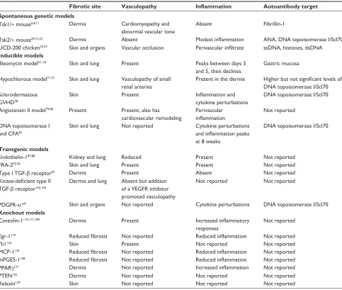
Table 1 Prominent features of animal models of systemic sclerosis
Fibrotic site Vasculopathy Inflammation Autoantibody target Spontaneous genetic models
Tsk1/+ mouse5,8,11 Dermis Cardiomyopathy and
abnormal vascular tone
Absent Fibrillin-1
Tsk2/+ mouse29,31,32 Dermis Absent Modest inflammation ANA, DNA topoisomerase i/Scl70
UCD-200 chicken35,37 Skin and organs vascular occlusion Perivascular infiltrate ssDNA, histones, dsDNA
Inducible models
Bleomycin model51–53 Skin and lung Present Peaks between days 3
and 5, then declines
Gastric mucosa
Hypochlorous model71,73 Skin and lung vasculopathy of small renal arteries
Present in the dermis Higher but not significant levels of
DNA topoisomerase i/Scl70 Sclerodermatous
GvHD78
Skin Present Inflammation and
cytokine perturbations
DNA topoisomerase i/Scl70
Angiotensin ii model79,80 Present Present, also has cardiovascular remodeling
Perivascular
inflammation
Not reported
DNA topoisomerase i and CFA83
Skin and lung Not reported Cytokine perturbations
and inflammation peaks
at 8 weeks
DNA topoisomerase i/Scl70
Transgenic models
endothelin-187,88 Kidney and lung Reduced Present Not reported
FRA-295,96 Skin and lung Present Present Not reported
Type i TGF-β receptor97 Dermis Present Absent Not reported
Kinase-deficient type II
TGF-β receptor102,104
Dermis and lung Absent but addition of a veGFR inhibitor promoted vasculopathy
Not reported Not reported
PDGFR-α107 Skin and organs Not reported Cytokine perturbations DNA topoisomerase i/Scl70
Knockout models
Caveolin-1110,111,180 Dermis Present Increased inflammatory
responses
Not reported
egr-1119 Reduced fibrosis Not reported Reduced inflammation Not reported
Fli1123 Skin Present Not reported Not reported
MCP-1124 Reduced fibrosis Not reported Reduced inflammation Not reported
mPGeS-1128 Reduced fibrosis Not reported Reduced inflammation Not reported
PPARγ131 Dermis Not reported Increased inflammation Not reported
PTeN136 Dermis Not reported Not reported Not reported
Relaxin139 Skin Not reported Not reported Not reported
Abbreviations: ANA, antinuclear antibody; CFA, Freund’s complete adjuvant; MCP-1, macrophage chemoattractant protein-1; PPARγ, peroxisome proliferator-activated receptor-gamma; PTeN, phosphatase and tensin homolog; FRA-2, Fos-related antigen-2; egr-1, early growth response gene; Fli1, Friend leukemia integration factor-1; TGF-β, transforming growth factor-beta; PDGFR, platelet-derived growth factor receptor; ssDNA, single-stranded DNA; dsDNA, double-stranded DNA; veGFR, vascular endothelial growth factor receptor; GvHD, graft versus host disease; Tsk1/+, tight skin 1; Tsk2/+, tight skin 2; UCD-200, University of California at Davis line 200; mPGeS-1, microsomal prostaglandin e2 synthase-1.
and this signaling was via α5β1 and αvβ3 integrins, which was driven by TGF-β.20 Intriguingly, the stiff skin model
had autoantibodies toward topoisomerase I.20 Inflammation
in the skin was also prominent and there was enrichment of plasmacytoid dendritic cells and Th2, Th17, and Th9 CD4 subpopulations.20 The authors argued that the abnormal
fibrillin-1 results in microfibrillar aggregates that fail to make adequate contact with cells but are able to retain the ability to sequester latent TGF-β, which becomes activated due to stiffness in the extracellular matrix resulting in a feed-forward signaling mechanism to drive fibrosis.20 Integrins are known
to sense the extracellular environment, and by that sensing are able to dictate the response of the fibroblast.21–24 The stiff skin
syndrome model hints at dysregulated signaling mechanisms driven by integrins that may be relevant for the pathology of systemic sclerosis; however, it does not address the etiology of systemic sclerosis because these fibrillin-1 mutations are not identified in the disease.
The Tsk1/+ mouse has been used to study the efficacy of various drugs for the treatment of fibrosis, and the model has proved valuable in this regard. Imatinib is a small-molecule tyrosine kinase inhibitor that is able to inhibit both TGF-β
and platelet-derived growth factor (PDGF) pathways. Imatinib prevents the development of inflammation-driven experimental fibrosis mediated by the drug bleomycin. Furthermore, imatinib reduced dermal thickening in the
Open Access Rheumatology: Research and Reviews downloaded from https://www.dovepress.com/ by 118.70.13.36 on 26-Aug-2020
Dovepress
Artlett
Tsk1/+ model,25 suggesting the involvement of TGF-β
and PDGF in the development of fibrosis in this model. Interferon-gamma therapy administered by nanoparticles also successfully reduced skin thickness in the Tsk1/+ mouse,26
and edaravone, a free radical scavenger, decreased collagen synthesis,27 as did rapamycin.28
Tight skin 2 mouse model
The tight skin 2 (Tsk2/+) mouse was first described in 1986, and arose in the offspring of a 101/H mouse exposed to the mutagenic agent ethylnitrosourea. As with Tsk1/+, tight skin is found in the interscapular region.29 The gene mutation
is located on chromosome 1 between 42.5 and 52.5 Mb,30
and to date the genetic defect has not been published. Like Tsk1/+, Tsk2/+ traits are highly penetrant and are homozy-gous embryonic lethal. Tsk2/+ mice replicate many features of systemic sclerosis, including tight skin, increased deposi-tion of dermal extracellular matrix, and autoantibodies.31,32
Tsk2/+ shows increased transcription rates of type I and III collagen in dermal fibroblasts,33 suggesting that the increased
collagen synthesis is cell autonomous rather than dependent on the integrity of the skin as a whole. Tsk2/+ mice were also reported to have increased autoimmunity, and a majority of the mice developed multiple autoantibodies specific for systemic sclerosis, including Scl70, centromere, and DNA.32
A restricted T-cell receptor bias was noted in skin infiltrates that encompasses specific T-cell receptor subtypes.34 Until
the genetic mutation has been identified, the value of this model cannot be fully realized. Another caveat to this model is that few laboratories choose to study it, possibly because the gene mutation is yet to be reported.
UCD-200 chicken
University of California at Davis line 200 (UCD-200) chickens develop a hereditary connective tissue disease that is similar in many ways to systemic sclerosis. It is character-ized by a perivascular lymphocytic infiltration,35 endothelial
injury resulting in vascular occlusion,36 speckled antinuclear
antibodies,37,38 and fibrosis of the skin and visceral organs.37
During experiments to map the genetic mutation in UCD-200 chickens crossed with red jungle fowl chickens, it was noted that the F1 generation were disease-free, whereas 36% of the back-crossed population (F2 generation) had early onset avian systemic sclerosis and 61% had late onset avian systemic sclerosis. The incidence of avian systemic sclerosis was sig-nificantly higher in male chickens than in female chickens.39
This suggests that the genetic defect in the UCD-200 line has an autosomal recessive mode of inheritance, or is
com-prised of multiple genetic loci or multiple loci interactions that are required to drive disease.39 Five genes have been
identified that confer a high association with the disease, and include TGFBR1, IGFBP3, EXOC2/IRF4, CCR8, and SOCS1. All of the genes are located on chromosome 2, with the exception of SOCS1, which is located on chromosome 14. All of these genes have been reported to have significance in the pathology of systemic sclerosis or to play a role in autoimmunity.40–44 In addition, COL1A2 was found to be
located in close proximity to the quantitative trait loci on chromosome 2, suggesting that this gene may play a role in avian systemic sclerosis. A shorter mRNA transcript has been identified in the early inflammatory stage in the comb and esophagus of diseased chickens.39
Of the three known spontaneous genetic animal mod-els, this model probably represents systemic sclerosis the closest in terms of its pathological features. Especially important to systemic sclerosis and the UCD-200 model is the perivascular infiltrate that precedes vasculopathy. Inflam-mation of the skin occurs early on in the disease, and this is predominantly found in the comb, which becomes swollen and erythematous. The vasculopathy appears to be driven by endothelial apoptosis.36,38 Histology of the comb demonstrates
that there is vessel occlusion with neointimal formation and a perivascular inflammatory infiltrate.36 The model also has
internal organ involvement. Autoantigens present in the sera include single-stranded DNA, double-stranded DNA, histones, poly(G), and cardiolipin; however, they did not reflect the antinuclear antibody spectrum that is observed in diffuse systemic sclerosis.45 The caveats to this model are that
few animal facilities are equipped to house these chickens, which require significantly more room than mice and have to be housed separately. Few laboratories have studied this model because most researchers prefer smaller animals with greater reagent resources.
Inducible models of fibrosis
Animal models predicated on inducing fibrosis in the mouse are of great value because they allow for study of the initiat-ing events in fibrosis. These models have proven to be very useful. However, induction of fibrosis in animals is limited to a small number of chemicals, and sclerodermatous graft versus host disease (GVHD) is difficult to work with.
Bleomycin model of fibrosis
The bleomycin mouse model of fibrosis is commonly used to replicate systemic sclerosis, or dermal or pulmonary fibrosis. Bleomycin is a glycopeptide antibiotic produced by
Open Access Rheumatology: Research and Reviews downloaded from https://www.dovepress.com/ by 118.70.13.36 on 26-Aug-2020
Dovepress Animal models of scleroderma
Streptomyces verticillus and has been used to treat Hodgkin’s lymphoma, squamous cell carcinoma, and testicular cancer, and is usually used in combination with other antineoplastic drugs.46 It has also been used to treat plantar warts.47 However,
use of bleomycin is not without significant risks, and a systemic sclerosis-like syndrome can ensue in susceptible individuals.48–50 This observation paved the way for
develop-ment of the bleomycin mouse model of fibrosis by Yamamoto et al.51 Local dermal injections of bleomycin in mice induced
collagen synthesis at the injection site over 4 weeks. The overall effects were found to be systemic because the lung also showed increased collagen synthesis.51 The sensitivity to bleomycin
was found to be strain-specific, and skin thickening was greater in some strains than in others. Yamamoto et al52 found that
fibrosis was induced by approximately two-fold in C3H/He, DBA/2, B10.D2, and B10.A strains, and that dermal thickness was increased by more than 2.5-fold in A/J, C3H/He, B10.A, and B10.D2 strains. One of the initiating events in bleomycin-induced fibrosis is mediated by inflammation. During the first 3–5 days of injections, there is a peak in the skin inflamma-tory infiltrate which then decreases with extended treatment. Bleomycin has been reported to induce vascular changes in the skin at the injection site.53 Upon withdrawal of bleomycin,
the fibrosis resolves over time. Investigators usually terminate the study prior to resolution, but this model could be utilized in study pathways involved in the resolution of fibrosis. In a similar manner, skin involved in systemic sclerosis appears to soften during the course of the disease.54 The model replicates
some of the most important biomarkers of systemic sclerosis but is by its very nature a model with limited uses.
There are several modes for administration of bleomy-cin that have been used to induce fibrosis depending on the organ to be investigated. As described above, bleomycin can be injected subdermally to induce synthesis of collagen; however, this is labor-intensive and has to be performed daily for 3–4 weeks for a robust fibrotic lesion to develop. In addition to the skin becoming fibrotic, this method also causes lung fibrosis.55 A single administration of bleomycin
via intratracheal instillation in weight-adjusted dosages is frequently used to study pulmonary fibrosis. The fibrotic stage exists until 3–4 weeks post-administration.56 There can
be a poorer survival rate depending on the strain of mouse used.57 Finally, bleomycin can be administered via Alzet®
osmotic pumps (DURECT Corporation, Cupertino, CA, USA) that are embedded under the skin and deliver a constant amount of compound over a certain number of days, making this approach to administration convenient on many levels. When used with bleomycin, the mortality observed in mice is
significantly less than with intratracheal instillation.58 Further,
this method induces a pattern of interstitial lung disease that replicates the lung pathology of systemic sclerosis and produces fibrosis in the skin and internal organs also in a pattern that is similar to systemic sclerosis.58
Bleomycin activates the NLRP3 inflammasome, leading to downstream signaling events in fibroblasts and resulting in synthesis of collagen.55 Many of the signals that result
in activation of the NLRP3 inflammasome are still being elucidated. Reactive oxygen species, oxidative stress, and the mitochondria are integrally involved in activation of the NLRP3 inflammasome.59,60 Bleomycin induces
oxida-tive stress61,62 by activating the mitochondria, and it is this
induction of oxidative stress that activates the inflammasome. Further evidence in support of this finding is that cells depleted of mitochondria are resistant to bleomycin-induced activation.63 To further underscore the importance of reactive
oxygen species and oxidative stress in this model, fibrosis was found to be abrogated when N-acetylcysteine was admin-istered.64 The culmination of inflammasome assembly and
activation results in cleavage of caspase-1, which is then able to cleave interleukin (IL)-1β and IL-18 for secretion, leading to fibrosis.55 IL-1β and IL-18 were found to be elevated in
sys-temic sclerosis serum65,66 and it was not until our laboratory
specifically studied the involvement of the inflammasome in the dermal fibrosis component of systemic sclerosis that the association between an activated inflammasome, fibro-sis, and systemic sclerosis was found.55 Further, we found
that activated caspase-1 was driving the increased collagen synthesis and myofibroblast phenotype.55
Although the bleomycin model replicates some of the earliest signaling patterns observed in systemic sclerosis, the model does not present with the profound autoantibody patterns typical of systemic sclerosis, but the mouse can establish an autoantibody that cross-reacts with the gastric mucosa.67
Hypochlorous model of systemic
sclerosis
Reactive oxygen species have been implicated in the pathogenesis of systemic sclerosis, and systemic sclerosis monocytes68 and fibroblasts69 release increased amounts of
reactive oxygen species, which triggers synthesis of collagen. The endoplasmic reticulum itself can induce reactive oxygen species and can also induce mitochondrial-reactive oxygen species,70 suggesting that increased reactive oxygen
species may play a central role in the fibrosis component of systemic sclerosis.
Open Access Rheumatology: Research and Reviews downloaded from https://www.dovepress.com/ by 118.70.13.36 on 26-Aug-2020
Dovepress
Artlett
The hypochlorous mouse model is a newly developed inducible animal model, and provides further important insights into the initiating signals derived from the reactive oxygen species that promote fibrosis. The hypochlorous mouse model is induced by repeated intradermal injections of hypochlorous acid, which causes generation of hydroxyl radicals leading to increased synthesis of collagen in the skin and lung tissues. In addition, this model mimics the pathological changes observed in the systemic sclerosis kidney and induces anti-topoisomerase antibodies.71 The
downside of this model at the moment is that it is currently only used by two laboratories to study the initiating events of fibrosis,72–74 and it is not fully understood how hypochlorous
acid induces fibrosis. It has been shown that overproduction of reactive oxygen species in this model activates ADAM17, which is involved in NOTCH signaling.73 However, this
model, like the bleomycin model, has proved to be useful in the testing of various candidate molecules for the prevention of fibrosis.73,74
Sclerodermatous GvHD model
This model has been used to understand further some of the initiating factors that lead to systemic sclerosis, and is derived from the initial observations of dermal fibrosis induced in patients with chronic GVHD. Thus, this mouse model was developed and then characterized. The model demonstrates increased collagen synthesis as in systemic sclerosis, and the fibrotic changes were found to be driven by TGF-β because inhibition of TGF-β abrogated the progression of fibrosis.75
Unlike systemic sclerosis, mice with sclerodermatous GVHD have a significant inflammatory infiltrate consisting of T-cells and monocytes/macrophages, and the numbers of inflammatory cells in the lesions are directly dependent on TGF-β.76 Patients with systemic sclerosis generally do not
present with such an infiltrate, and the few T-cells found in the lesions tend to be perivascular.77
A modified model of GVHD was then developed and found to exhibit all the major aspects of systemic sclerosis. GVHD was developed in the RAG-2 knockout mouse by the transfer of B10.D2 spleen cells.78 GVHD develops over
time, and the model shows progressive skin thickening with disease duration. The kidneys and large and small intestines were found to be fibrotic. Vasoconstriction was reported, increased α-smooth muscle actin and endothelin-1 were found in the vessels of the skin and kidney, and anti-Scl70 antibodies were found in the serum. This model characterizes many of the features of systemic sclerosis, and of note, renal crisis was one of the pathologies observed. TGF-β was found
to be elevated, and neutralization of TGF-β with antibodies reduced skin thickness and the ensuing vasculopathy.78
Angiotensin II-inducible model of fibrosis
This is a recently developed model that uses angiotensin II to induce vascular constriction, leading to inflammation and fibrosis.79 Using Alzet osmotic pumps, angiotensin IIwas found to induce dermal fibrosis by activating the TGF-β
pathway, and increases were found in TGF-β2 and TGF-β3 but not in TGF-β1 mRNA. Angiotensin II was also found to increase the numbers of myofibroblasts and fibrocytes in the skin.79 Increased numbers of vascular endothelial
(VE)-cadherin and fibroblast-specific protein-1-positive cells were observed around the small vessels in this mouse model, sug-gesting increased endothelial to mesenchymal transition. The model also includes cardiovascular remodeling.80 However, it
is unknown whether these mice develop autoantibodies specific for systemic sclerosis.79
The hypochlorous mouse model was used to test the hypothesis that angiotensin II receptor blockade could ame-liorate fibrosis. Irbesartan, an angiotensin II receptor inhibitor, was found to reduce dermal thickness and total collagen con-tent, with less inflammation, and to reduce anti-topoisomerase I antibodies.81 This suggests that angiotensin levels can be
regu-lated by oxidative stress and play a critical role in the fibrosis component of systemic sclerosis, and it has been found that a subset of patients have elevated angiotensin II signaling.82
Model induced by DNA topoisomerase i
and Freund’s complete adjuvant
Autoimmunity is a significant feature in systemic sclero-sis, and yet few investigators have developed models that specifically investigate this specific aspect of its pathology. Yoshizaki et al83 demonstrated that subcutaneous
administra-tion of DNA topoisomerase I and Freund’s complete adjuvant for 8 weeks promoted dermal and lung fibrosis. Adjuvant lacking mycobacterial components (Freund’s incomplete adjuvant) did not induce fibrosis. Cytokine perturbations included increased levels of IL-4, interferon-gamma, IL-10, TGF-β, and tumor necrosis factor-alpha.83 Bronchoalveolar
lavage fluid was analyzed and the investigators found that the T-cell profile was skewed toward Th2 and Th17 in mice receiving DNA topoisomerase I and Freund’s complete adju-vant, and in the mice receiving DNA topoisomerase I and Freund’s incomplete adjuvant, and in the IL-6 deficient mice it was skewed toward Th1 and regulatory T-cells. This study raises the provocative possibility that an antibody directed against topoisomerase I could promote fibrosis.
Open Access Rheumatology: Research and Reviews downloaded from https://www.dovepress.com/ by 118.70.13.36 on 26-Aug-2020
Dovepress Animal models of scleroderma
Transgenic models of fibrosis
These models are important for understanding the role of the specific genes that contribute to fibrosis. Many of these models have been utilized from investigators outside the field of systemic sclerosis who study other forms of fibrosis. However, this should not detract from the importance of these models and what they tell us about the role of a particular gene in regulating collagen synthesis.
endothelin-1 mouse model
Damage to endothelial cells causes increased expression of endothelin-1. The effects of overexpression of endothelin-1 were found to be deleterious to the vasculature and to contrib-ute to hypertrophy of the vessels and vascular permeability, leading to vasoconstriction.84 Endothelin-1 also plays a role
in increased cell proliferation, fibrosis, and inflammation.85,86
The transgenic mouse develops glomerulosclerosis and interstitial fibrosis.87,88 Patients with systemic sclerosis have
increased tissue and circulatory endothelin-1 levels as well as increased vasculopathy, and these findings underscore the importance of this protein in the pathology of systemic sclerosis.89,90
Bosentan is an endothelin-1 antagonist and has been used successfully to treat pulmonary hypertension in systemic sclerosis.91,92 Tsk1/+ mice were found to have impaired
aor-tic relaxation, suggesting a role for endothelin-1,93 and this
led to the study of bosentan in these mice.94 With bosentan,
there was partial restoration of the relaxation capability of the vessels in Tsk1/+ mice, suggesting that these mice have endothelial dysfunction.94 More specifically, endothelin-1
promoted increased expression of endothelin receptor B, but not endothelin receptor A, in Tsk1/+ mice.
Fos-related antigen-2 mouse model
The transgenic mouse model overexpressing the Fos-related antigen-2 (FRA-2) gene demonstrates many of the important factors resulting in the vasculopathy of systemic sclerosis. The FRA-2 transgenic animal displays microangiopathy in addition to progressive skin fibrosis. Overall, it was found that this pathology was preceded by apoptosis of endothe-lial cells.95 FRA-2 is a transcription factor activator protein
and belongs to the activator protein-1 family. This family is induced by cellular stress, and as a result, controls many of the downstream responses in the cell, such as cell proliferation, apoptosis, inflammation, tumorigenesis, and wound healing. In light of this, it was found that expression of FRA-2 was independent of the expression of major cytokines such as TGF-β1 and PDGF, and both are important cytokines driving
the pathology of systemic sclerosis. However, overexpression of FRA-2 is apparent in the skin lesions of systemic sclero-sis, suggesting there may be a direct causal involvement in fibrosis95 and that it may also play a role in vasculopathy.96
Type i TGF-
β
receptor transgenic model
A model was developed to specifically upregulate the type I TGF-β receptor in fibroblasts on a Cre-ER transgenic back-ground. In this model, with progressing age, there were increasing levels of collagen deposition in the skin of the mouse. In addition, the ultrastructure of collagen was found to be disorganized. Vascular changes similar to systemic scle-rosis were apparent, with hypertrophy of smooth muscle cells and neointimal thickening of vessel walls. Mice had increased levels of von Willebrand factor, which is similar to what is observed in systemic sclerosis, leading to the hypothesis that sustained activation of TGF-β signaling in fibroblasts can lead to endothelial damage.97 Vascular smooth muscle cells from
this mouse demonstrate enhanced remodeling of collagen gel lattices with altered endothelin-1 and endothelin recep-tor signaling. This model further replicates other features of the pathology of systemic sclerosis in that it has myocardial fibrosis and altered aortic dynamics. As expected, activation of the TGF-β pathway caused increased phosphorylation of Smad2/3 in fibroblasts, leading to increased phosphoryla-tion of ERK1/2 and p38.97 This model suggests that chronic
expression of TGF-β in fibroblasts could cause the pathology observed in systemic sclerosis; however, some investigators believe that damage to endothelial cells is one of the leading culprits in initiating and driving the disease.98–100
Kinase-deficient type II TGF-
β
receptor transgenic model
This model was developed when a fibroblast-specific tran-scriptional enhancer was identified upstream of the COL1A2 collagen gene.101 The enhancer was used to specifically
express a kinase-deficient mutant type II TGF-β receptor that can engage TGF-β but cannot lead to phosphorylation of TGF-β receptor 1 and its downstream signaling pathways.102
The directed expression of the kinase-deficient type II TGF-β
receptor resulted in increased fibrosis in the lung and skin. The transgenic fibroblasts were found to have a TGF-β
activated phenotype with sustained induced expression of connective tissue growth factor and increased phosphoryla-tion of Smad2/3,102 and increased myofibroblast numbers.103
Although this transgenic model allows for the study of TGF-β signaling, caveats are the lack of immune activation and the fact that autoantibodies are not present. In addition,
Open Access Rheumatology: Research and Reviews downloaded from https://www.dovepress.com/ by 118.70.13.36 on 26-Aug-2020
Dovepress
Artlett
the transgenic model does not have vasculopathy; however, when a vascular endothelial growth factor receptor inhibi-tor is administered to these mice, they develop pulmonary arterial hypertension pathology similar to that observed in systemic sclerosis.104
PDGF receptor-
α
transgenic
model of fibrosis
PDGF has been implicated in the development of the fibro-sis and vasculopathy seen in systemic sclerofibro-sis.105,106 Olson
and Soriano investigated the role of the PDGF receptor-α
in fibrosis.107 They designed transgenic mice to carry
con-ditionally expressed PDGF receptor-α activating mutations that promoted fibrosis in the skin and internal organs. The importance of PDGF signaling during fibrosis was further recapitulated utilizing sunitinib and sorafenib,108 two tyrosine
kinase inhibitors that act to block the bioactivity of PDGF receptors and the vascular endothelial growth factor (VEGF)/ VEGF receptor pathway. This study demonstrated that inhibi-tion of phosphorylainhibi-tion of PDGF receptor-β with sunitinib effectively reduced skin and lung fibrosis. Sorafenib was not as effective for reduction of fibrosis; however, it significantly reduced the DNA topoisomerase I autoantibodies, IL-6 and TGF-β.108 As discussed previously, imatinib, a TGF-β and
PDGF pathway inhibitor, effectively reduced Tsk1/+ dermal fibrosis,25 adding further credence to the value of tyrosine
kinase inhibitors.
Knockout models of fibrosis
These models study the specific contribution of a gene in fibrosis. In some instances, the gene is a negative regula-tor of collagen synthesis and its deletion promotes fibrosis, whereas other genes are required for induction of the fibrotic process.
Caveolin-1
Caveolin is found to be expressed within the lipid rafts of the caveolae in the cell membranes and functions to modulate the functioning of various signaling molecules.109 It was
noted that caveolin-1-deficient mice had increased alveolar septa and lung fibrosis. In addition, these mice were found to have altered vasculopathy due to endothelial proliferation.110
On closer examination of the systemic sclerosis phenotype and the phenotypic changes, these mice were observed to have increased collagen and fibronectin accumulation, in addition to increased numbers of myofibroblasts.111 It is now
known that caveolin-1 regulates TGF-β signaling by inter-acting with the type I TGF-β receptor and regulating Smad
signaling pathways by suppression of phosphorylation of Smad2112 and inhibiting ERK signaling.113 However, TGF-β
decreases expression of caveolin-1.114 Further, it has been
found that loss of caveolin-1 can lead to oxidative stress115
and this is a pathology that has been associated with systemic sclerosis.116,117
early growth response protein-1
knockout mouse
Early growth response protein-1 (Egr-1) is a transcription factor that is a zinc finger binding protein and is involved in cell growth, differentiation, and survival. It is induced by stress signals.118 Egr-1-deficient mice were injected
with bleomycin and skin fibrosis was assessed. There were decreased numbers of infiltrating inflammatory cells in the skin in addition to decreased dermal thickness and colla-gen expression.119 The signaling events downstream from
Egr-1 play a role in collagen fibril formation because mice deficient in this protein have disorganized collagen fibers. Furthermore, bleomycin-induced lung fibrosis was attenu-ated in the absence of Egr-1, as was the ability to induce wound healing, and the absence of Egr-1 inhibited wound closure in in vitro scratch wound assays.119 In confirmation
of the importance of Egr-1 in wound healing, an Egr-1 transgenic mouse was tested and found to enhance wound healing. This further demonstrates the importance of this transcription factor in the synthesis of collagen, wound healing, and fibrosis.
Friend leukemia integration factor-1
conditional knockout mouse
This model recapitulates many of the features of systemic sclerosis in that it has abnormalities of the vasculature.120
Friend leukemia integration factor-1 (Fli1) is constitutively expressed in fibroblasts and plays a pivotal role in inhibiting the expression of extracellular matrix proteins, such as con-nective tissue growth factor and type I collagen.121 However,
more recently, Fli1 was demonstrated to modulate endothelial cell function and to be involved during vascular development. The Fli1 conditional knockout mouse has increased vascu-lar permeability, and this closely mimics the permeability observed in patients with systemic sclerosis due to reduced expression of VE-cadherin.122 Collagen fiber formation is
dependent on Fli1123 and this model further mimics the
abnor-malities in collagen fibril formation in systemic sclerosis, further confirming the importance of Fli1 in the pathogen-esis of systemic sclerosis. Phosphorylation of Fli1 and its interaction with p300 transcription factor is also dependent
Open Access Rheumatology: Research and Reviews downloaded from https://www.dovepress.com/ by 118.70.13.36 on 26-Aug-2020
Dovepress Animal models of scleroderma
on TGF-β, thus underscoring further the importance of this cytokine in the pathology of systemic sclerosis.
Macrophage chemoattractant
protein-1 mouse model
The involvement of basic fibroblast growth factor and con-nective tissue growth factor in conjunction with macrophage chemoattractant protein-1 (MCP-1) is required for tissue fibrosis.124 MCP-1-deficient mice show abrogated fibrotic
responses when basic fibroblast growth factor and connective tissue growth factor are injected into the skin. In addition, this model has reduced macrophage recruitment, fewer mast cells, and fewer CD4+ T-cell numbers.124 Connective tissue
growth factor alone does not induce fibrosis, but when admin-istered with basic fibroblast growth factor, there is increased autocrine signaling along with persistent fibrosis. MCP-1, basic fibroblast growth factor, and connective tissue growth factor are proteins that are regulated by TGF-β.125–127 The
correlation and interaction between MCP-1, basic fibroblast growth factor, and connective tissue growth factor indicates the importance of the interplay between these three proteins in fibrosis.
Microsomal prostaglandin
e2 synthase-1 knockout model
Microsomal prostaglandin E2 synthase-1 (mPGES-1) is an inducible enzymatic protein that acts downstream of cyclo oxygenase. It specifically causes the conversion of prosta-glandin H2 to prostaglandin E2. mPGES-1 was found to be overexpressed in systemic sclerosis fibroblasts, suggest-ing that this pathway is dysregulated.128 Mice deficient in
mPGES-1 are resistant to bleomycin-induced fibrosis, have decreased levels of α-smooth muscle actin, and reduced num-bers of macrophages,128 further underscoring the importance
of this gene in fibrosis.
Peroxisome proliferator-activated
receptor-gamma
Peroxisome proliferator-activated receptor-gamma (PPARγ) has been associated with the pathology of numerous dis-eases, including obesity, diabetes, atherosclerosis, and cancer. More recently, decreased PPARγ has been shown to be associated with fibrosis.129 PPARγ is a nuclear receptor
ligand-activated transcription factor that is decreased by TGF-β1 signaling. PPARγ inhibits collagen synthesis,130
whereas TGF-β inhibits PPARγ expression,131 and is thus
involved in the fibrotic response in systemic sclerosis. Kapoor et al further explored the role of PPARγ in fibrosis
and found that conditional PPARγ-deficient mice developed greater skin thickening with bleomycin when compared with wild-type mice.132 Skin thickening was assessed by
analysis of total collagen content, increased α-smooth muscle actin expression, and increased inflammation.132 In
confirmation of this finding, rosiglitazone, a PPARγ agonist, was found to decrease fibrosis in the bleomycin mouse model.129 Further, rosiglitazone was also effective in
reduc-ing already established fibrosis.129 Inhibition of PPARγ with
rosiglitazone and the PPARγ knockout mouse have helped to define the role of this protein in collagen synthesis. It is unknown if this model sees increased vascular remodeling, as it was not reported, but PPARγ is known to play a role in this process.133
PTeN conditional knockout
mouse model
Phosphatase and tensin homolog (PTEN) is a protein lipid phosphatase that activates Akt via phosphorylation, and the phosphoinositide 3-kinase-Akt signaling axis has been implicated in the production of collagen.134 Akt
phospho-rylation is elevated in systemic sclerosis fibroblasts135
and PTEN levels are decreased.136 Regulation of PTEN
is dependent on TGF-β; however, PTEN knockout mice have increased collagen deposition and thickened skin, in addition to increased numbers of proliferating cell nuclear antigen-positive fibroblasts, increased numbers of α-smooth muscle actin-bearing fibroblasts, and increased numbers of connective tissue growth factor-positive fibroblasts.136 The
data from this mouse model suggest that PTEN normally suppresses fibrogenesis and its absence spontaneously pro-motes collagen synthesis.
Relaxin knockout mouse
Relaxin is a small dimeric peptide that is generally asso-ciated with the female reproductive tract, and is known to be a vasodilator but also has antifibrotic effects.137,138
Mice deficient in relaxin were found to have increased collagen deposition in the skin over time. Administration of human recombinant relaxin via osmotic minipumps abrogated fibrosis in this model.139 This knockout mouse
has been used to study other fibrotic diseases, such as liver fibrosis due to administration of carbon tetrachloride,140
and cardiac fibrosis,141 and silica-induced pulmonary
fibrosis.142 In all cases, relaxin was found to ameliorate
collagen synthesis. This suggests that relaxin regulates collagen synthesis and could be used as a therapy for fibrotic diseases.
Open Access Rheumatology: Research and Reviews downloaded from https://www.dovepress.com/ by 118.70.13.36 on 26-Aug-2020
Dovepress
Artlett
Using mouse models for
preclinical testing
There are no formal guidance documents from the US Food and Drug Administration (FDA) requiring the use of animal models of efficacy for preclinical studies of a particular drug candidate prior to human clinical testing. However, the FDA does require various toxicological and pharmacological safety studies to ensure that the drug candidate and the final formulated drug product is reasonably safe before proceeding to human clinical testing. These toxicological and pharma-cological safety studies are usually performed in rodent and canine species, but may include other species (porcine for example) if physiologically relevant to the drug candidate being tested or therapeutic area being targeted. Certainly from this perspective, a model replicating all or some of the aspects of disease is not required. However, in light of this, the efficacy of a potential drug candidate in preclinical stud-ies has usually been required by pharmaceutical companstud-ies prior to investment of significant financial resources in clini-cal trials and in bringing the product to market. In addition, animal models have been used in proof-of-concept studies to investigate the role of a particular protein or pathway, or to test inhibitors or reagents identified more often than not at the coal face by academic researchers. Even so, success in an animal model has not always translated into a successful treatment for the patient (Table 2). Due to the polymorphic nature of the disease, until the cause (or causes) of systemic sclerosis are identified, a drug that treats all symptoms of the disease may not be realistic. Indeed, it may be that multiple treatment regimens are required to manage symptoms.
Anakinra is an IL-1R antagonist and has shown efficacy in treating IL-1-related diseases that are driven by inflam-masome activation. Anakinra was effective at blocking bleomycin-induced pulmonary fibrosis,143 and clinical trials
are currently underway to determine whether it will be effec-tive in systemic sclerosis. Systemic sclerosis was not origi-nally thought of as an IL-1-mediated disease until recently;55
however, IL-1 expression can drive TGF-β signaling, and inhibition of this innate immune signaling pathway may also lead to inhibition of fibrosis in systemic sclerosis. Rilonacept is another drug that interferes with IL-1 signaling. This drug was never studied in preclinical models; however, a clinical trial is currently underway in patients with systemic sclerosis although no results are available.
Several immunosuppressants have been tested in pre-clinical models and in systemic sclerosis pre-clinical trials with mixed success. Cyclosporine demonstrated some efficacy in preventing bleomycin-induced lung fibrosis;144,145 however,
although some patients with systemic sclerosis tolerated the drug,146 there is a significant risk of renal toxicity.147 Likewise,
prednisone can promote renal toxicity in patients and their renal function has to be closely monitored.148
Mycophenolate mofetil is an immunosuppressant that prevents the purine biosynthesis necessary for development of T-cells and B-cells. It is currently used to treat other autoimmune diseases. This drug successfully prevented bleomycin-induced fibrosis149 and was successful in a small
study of patients with systemic sclerosis;150 however, larger
studies are currently underway and it is currently unknown as to whether this drug will hold up to close scrutiny.
Thalidomide has emerged as an immunomodulating therapeutic and is thought to modulate the Th1/Th2 profile by inhibiting tumor necrosis factor-alpha, IL-6, IL-10, and IL-12, and by enhancing the production of IL-2, IL-4, and IL-5 by immune cells.151 It has also been reported to increase
the lymphocyte count, induce costimulation of T-cells, and alter the cytotoxicity of natural killer cells.151 Thalidomide
and its analogs could abrogate fibrosis in mouse models, paving the way for this drug to be trialed in patients. In a small pilot study of patients with systemic sclerosis, thali-domide was found to enhance the healing of digital ulcers.152
However, to date, there has been some difficulty in recruit-ing patients for a long-term clinical trial and the study was terminated.
Colchicine, a drug that inhibits mitosis, was demonstrated to afford some protection against bleomycin-induced pul-monary fibrosis.153 However, when used to treat systemic
sclerosis, there was mixed success in the clinical trials.154,155
Inhibition of tumor necrosis factor-alpha by etanercept was effective in treating bleomycin-induced dermal fibrosis;156
however, when trialed in systemic sclerosis, the drug appeared to effectively reduce the inflammatory joint disease seen in these patients157 but did not appear to be efficacious
against fibrosis,158 and is not without risk for fatal fibrosing
alveolitis.159
Imatinib, a tyrosine kinase inhibitor, was initially promis-ing as a therapeutic and was found to reduce the fibrosis in the Tsk1/+ mouse and the inducible bleomycin model;25
how-ever, when used in patients, adverse events were frequently observed and tolerability was poor.160 Iloprost, a prostacyclin
agonist, also effectively reduced bleomycin-induced fibrosis;161 however, there were heterogeneous responses
in small patient cohorts and this has made the efficacy of iloprost hard to assess.162,163
Halofuginone has been shown to interfere with collagen synthesis. It does so by inhibiting TGF-β signaling through
Open Access Rheumatology: Research and Reviews downloaded from https://www.dovepress.com/ by 118.70.13.36 on 26-Aug-2020
Dovepress Animal models of scleroderma
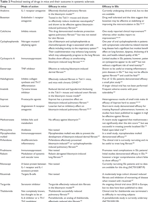
Table 2 Preclinical testing of drugs in mice and their outcome in systemic sclerosis
Drug Mode of action Efficacy in mice Efficacy in SSc
Anakinra iL-1 receptor antagonist Effectively blocked pulmonary fibrosis
in the bleomycin model143
Currently undergoing clinical trial, but no data are available
Bosentan endothelin-1 receptor antagonist
Tested in the Tsk1/+ mouse and shown to effectively reduce moderate vasculopathy94 and shown to be effective against bleomycin-
induced pulmonary fibrosis181
Drug well tolerated and the data suggest that bosentan may be effective at stabilizing or improving the microcirculation in SSc166,167
Colchicine inhibits mitosis This drug demonstrated moderate protection
against pulmonary fibrosis153 but was not tested
for skin fibrosis
One study reported clinical improvement182 whereas other studies report no improvement154,155
Cyclophosphamide Nitrogen mustard alkylating agent
Use of bleomycin and cyclophosphamide as chemotherapeutic drugs is associated with side effects including toxicity to the respiratory system176 Their coadministration may enhance lung toxicity
which may subsequently progress to the lung fibrosis
One year of oral cyclophosphamide in patients with symptomatic scleroderma-related interstitial
lung disease had a significant but modest beneficial
effect on lung function, dyspnea, thickening of the skin, and health-related quality of life177
Cyclosporin A immunosuppressant Studies show efficacy at ameliorating bleomycin induced lung fibrosis144,145
Limited studies in SSc patients; however, patients on cyclosporine appear to do well146 but not
without a significant risk of renal toxicity147 etanercept TNF inhibitor effective at reducing bleomycin-induced
dermal fibrosis156
Shown to be useful at treating inflammatory
joint diseases in SSc157 but may not be effective
against fibrosis158 and could be fatal159 Halofuginone inhibits collagen
synthesis and Th17 differentiation
Effectively reduced fibrosis in Tsk1/+ mice, and mice with chronic GvHD165
Five of 12 SSc patients demonstrated efficacy in
a small pilot study165
A larger clinical trial has not been performed imatinib Tyrosine kinase
inhibitor
Reduced dermal and hypodermal thickening in the Tsk1/+ mouse and reduced overt fibrosis
in the bleomycin mouse model25
Frequent adverse events with poor tolerability160
iloprost Prostacyclin agonist iloprost has a protective effect on
bleomycin-induced pulmonary fibrosis161
Heterogeneous results of small trials make the
efficacy of iloprost hard to assess162,163 Losartan Angiotensin ii receptor
antagonist
Losartan had an inhibitory effect on
bleomycin-induced pulmonary fibrosis168,169
Short-term study demonstrated efficacy for
treating Raynaud’s phenomenon; however, no studies have been published to suggest it might
be effective against fibrosis
Methotrexate inhibits folic acid metabolism
No efficacy against bleomycin175 A recent study suggested that methotrexate did
not significantly alter the skin score173 but seems successful in treating juvenile localized SSc174
Minocycline Antibiotic Not tested Failed open-label trial172
Mycophenolate mofetil
immunosuppressant Mycophenolate mofetil was able to prevent the
development of bleomycin-induced dermal fibrosis149
in a small study, mycophenolate mofetil
demonstrated efficacy150 Pirfenidone
Antifibrotic/anti-inflammatory
Pirfenidone effectively prevented bleomycin-induced183 or cyclophosphamide-
induced pulmonary fibrosis184
The clinical trial is currently recruiting and may
be useful to treat lung fibrosis185
Prednisone immunosuppressant Not tested Promotes renal complications in SSc patients148
Relaxin Modulation of systemic and vascular tone
Relaxin abrogated bleomycin-induced
lung fibrosis138
Initial studies demonstrated efficacy in SSc;186 however a larger comprehensive cohort failed
to show efficacy170 Rilonacept A fusion protein between
iL-1 receptor and iL-1 accessory protein
Not tested Currently recruiting SSc patients and no data
are available for this drug (NCT01538719)
Rituximab Targets B-cells Not tested A moderately large cohort showed reduced
fibrosis and inhibition of worsening of disease
when treated with rituximab187 Terguride Serotonin inhibitor Terguride effectively reduced skin fibrosis
in the bleomycin model188
An ongoing clinical trial since 2010 in europe, but no data have been published to date Thalidomide Not completely known,
but thought to be an iL-6 inhibitor or a Th1/ Th2 modulator
Thalidomide successfully reduced
pulmonary fibrosis189
Pomalidomide, an analog of thalidomide,
effectively reduced skin fibrosis190
Clinical trial for thalidomide was terminated due
to difficulty in recruiting subjects
A pomalidomide study is currently underway (NCT01559129)
Abbreviations: iL, interleukin; SSc, systemic sclerosis; Tsk1/+, tight skin 1; GvHD, graft versus host disease; TNF, tumor necrosis factor; Th, T helper.
Open Access Rheumatology: Research and Reviews downloaded from https://www.dovepress.com/ by 118.70.13.36 on 26-Aug-2020
Dovepress
Artlett
a process that prevents phosphorylation of Smad3.164 The
drug has been used in Tsk1/+ mice and in mice with chronic GVHD, and both models demonstrated a significant reduc-tion in fibrosis.165 However, when halofuginone was used
topically in a small pilot study on 12 patients with systemic sclerosis, five patients responded by demonstrating a signifi-cant reduction in skin score whereas seven did not respond.165
A larger follow-up clinical trial has not been performed. Bosentan was found to effectively reduce moderate vascu-lopathy in the Tsk1/+ mouse94 and was effective in preventing
bleomycin-induced pulmonary fibrosis.166,167 Bosentan has
been used to ameliorate the microcirculation, helping to reduce Raynaud’s phenomenon. Losartan, an angiotensin II receptor inhibitor, was also effective for the treatment of Raynaud’s phenomenon, but to date the studies have not been formally published. However, losartan was found to reduce bleomycin-induced pulmonary fibrosis.168,169 Both drugs lend
credence to targeting abnormalities in the vasculature as valid therapeutics for systemic sclerosis. Relaxin is a small dimeric peptide that regulates vascular tone and also has an antifibrotic mode of action. The relaxin-deficient mouse suggested that this molecule could be used to treat fibrosis and this was confirmed with bleomycin studies; however, when trialed in systemic sclerosis, there were mixed results and overall relaxin was not effective at reducing fibrosis.170
Much controversy is centered on the antibiotic minocy-cline as to whether it is effective in treating systemic sclerosis. One small clinical study demonstrated that four of eleven patients had complete remission of disease,171 which sparked
huge interest in minocycline as a treatment for patients with systemic sclerosis. However, minocycline failed a larger open-label clinical trial.172 Like many other drugs,
minocy-cline may be of value to a small number of patients, and that perhaps may be determined by the etiology of their disease. Because systemic sclerosis is very polymorphic in nature, its mode of onset may have many different origins.
Methotrexate failed as a drug for the treatment of adult-onset systemic sclerosis and did not significantly alter the overall skin score;173 however, it was successful in treating
juvenile localized systemic sclerosis.174 Methotrexate was not
shown to be effective in the bleomycin-induced mouse model of fibrosis.175 Cyclophosphamide was found to have enhanced
toxicity in the lung, especially when used in conjunction with bleomycin, where mice were found to have enhanced progression of lung fibrosis,176 whereas cyclophosphamide
appeared to protect against interstitial lung disease in patients with systemic sclerosis, with improvement of lung function and quality of life.177
Future directions for animal models
replicating systemic sclerosis
pathology
In recent years, animal models have been developed that repli-cate more faithfully many of the important aspects of systemic sclerosis.35,45,78,120 However, until the cause of systemic sclerosis
is identified, an accurate model is not possible. Even so, once the cause has been identified, the use of animals may still preclude accurate development of the disease in an animal model, for the simple reason that animals are not humans. Recent research suggests that infection of fibroblasts by Epstein-Barr virus might drive the pathology of systemic sclerosis178 and if this
proves to be true, the development of an animal model will be very difficult. The natural host for the virus is man, but under specific experimental conditions, a small number of new world monkey species can be infected. In light of this, a humanized Epstein-Barr virus mouse has already been developed, but the mouse has not been reported to display fibrosis,179 and the
model may only be possible if mouse fibroblasts are specifically designed to allow infection with virus.
Due to the polymorphic and systemic nature of systemic sclerosis and the poor replicability of many of the models utilized in studies published to date, the issue arises as to whether more than one model should be used to study drug efficacy. This would be best from an idealistic point of view; however, it may not always be practical, due to the inherent expense of breeding and housing of animals for research. If more than one model is to be used, then the models should be carefully chosen. For example, in studying the efficacy of a drug on the TGF-β pathway, investigators might select animal models that target TGF-β but from different angles, such as receptor signaling using the kinase-deficient type II TGF-β
receptor model and the type I TGF-β receptor transgenic model. The hope is that both models will together recapitu-late and confirm present findings. However, disparate study findings can preclude a drug from entering clinical trials, and this drug may have been a successful therapeutic that is never realized, because animals are not humans.
Conclusion
Animal models are very useful in studying the initiating events, genes, and various factors required to establish fibrosis as often these events or features cannot be studied in the patient. With the exception of stiff skin syndrome, which is an inheritable variant of systemic sclerosis, all other systemic sclerosis variants are inducible, given that onset of the disease is usually later in life. Animal models, by their
Open Access Rheumatology: Research and Reviews downloaded from https://www.dovepress.com/ by 118.70.13.36 on 26-Aug-2020