R E S E A R C H A R T I C L E
Open Access
Evaluation of occipitocervical neutral position
using lateral radiographs
Jiangwei Tan
1, Guangjun Liao
2*and Shaoxian Liu
2Abstract
Background:Intraoperative assessment of neutral occipitocervical balance during a fusion procedure is
challenging. We designed this study to introduce a more comprehensive method of evaluating the occipitocervical neutral position using lateral radiographs.
Methods:One hundred neutral lateral cervical spine radiographs interpreted as normal were studied. Cervical spine radiographs were performed using a standard technique. The occipitocervical angle, the occipitocervical distance, and the mandible cervical distance were measured by different observers.
Results:A difference analysis was performed between males and females. The mean mandible cervical distances
were 11.0 and 11.2 mm in males and females, respectively. The mean occipitocervical distances were 22.0 mm (male) and 19.6 mm (female), and the occipitocervical angles were 47.2° (male) and 45.5° (female). The
occipitocervical distance revealed significant differences between males and females (p<0.01). However, there were no significant differences between sexes for the occipitocervical angle or the mandible cervical distance (p>0.01). Conclusions:This study offers reference values for the occipitocervical angle and occipitocervical distance for the estimation of the occipitocervical neutral position. The introduction of the mandible cervical distance may make the evaluation more direct and more comprehensive during surgery because of its sensitivity to changes in head position.
Keywords:Fusion, Mandible cervical distance, Neutral, Occipitocervical, Radiograph
Background
Occipitocervical fusion is indicated for occipitocervical instability caused by trauma [1], bone tumor [2], con-genital deformity [3-5], and rheumatoid arthritis [6,7]. It is of great importance to fuse the head in a neutral pos-ition during the operation.
If the head is immobilized in a nonfunctional position during occipitocervical fusion, compensatory curvature may occur in the subaxial segments, which will eventu-ally develop into spinal disorders. Toyama et al. studied 12 children who underwent C1-C2 posterior fusions [8]. The authors found that postoperative cervical malalign-ment (kyphosis or swan-neck deformity) occurred in four patients. In all four patients, new bone formation and an increase in the body/canal ratio (BCR) at the apex of kyphosis were observed. This phenomenon was not observed in eight patients with normal alignment.
Matsunaga et al. studied the association between any changes in the alignment of the cervical vertebrae and the development of subaxial subluxation during follow-up periods [9]. Twelve (86%) patients experienced sub-axial subluxation after surgery; the authors emphasized the position of the fixed occipital bone and axis during occipitoaxial fusion procedures. Recently, Yoshida re-ported a case of an upper airway obstruction immedi-ately after an O-C2 fusion in a flexed position [10]. After revision surgery in which the fusion angle was changed, the upper airway obstruction disappeared. Therefore, for surgeons, accurately defining the occipitocervical neutral position before performing occipitocervical fusion is of great importance.
However, reports in the literature that provide a defin-ition of the neutral occipitocervical posdefin-ition are rare. Mod-ern technologies, such as magnetic resonance imaging (MRI) and three-dimensional computed tomography (CT), only complicate the definition, and such technologies are not routinely used during surgery. Radiographs seem to be * Correspondence:[email protected]
2
Department of Orthopedic Surgery, Yantaishan Hospital, No. 91 Jiefang Road, Zhifu district, Yantai 264000, People’s Republic of China Full list of author information is available at the end of the article
a last resort for this purpose in terms of practicability. Phillips et al. proposed using the occipitocervical angle (OCA) and the occipitocervical distance (OCD) measure-ments on lateral radiographs to objectively define the occi-pitocervical neutral position [11]. However, because OCA and OCD measurements need to be very precise, surgeons still have difficulty in neutral position assessment during surgery when radiographs are not available.
In this study, we introduce the mandible cervical dis-tance (MCD) measurement, which makes the evaluation quick and practical to perform. At the same time, we measured the OCA and the OCD in a larger sample and obtained normal value ranges, which will help to define the occipitocervical neutral position more comprehen-sively. This study is expected to provide reference values for the definition of the occipitocervical neutral position.
Methods
We included in this study 100 neutral lateral cervical spine radiographs interpreted by two radiologists and three orthopedic surgeons as normal (i.e., no fracture or dislocation, no deformity, no severe osteophyte forma-tion, no destruction of the vertebrae, no spondylosis) from a radiographic database at Yantaishan Hospital, China. The study population consisted of 50 males and 50 females with an average age of 45.6 years (ranging
from 15 to 60 years). X-ray technology was used accord-ing to a routine protocol. The subjects were asked to look straight ahead with their mouths closed naturally during projection. The X-ray tube was centered on C5,
and the radiographs were taken from a distance of 1.8 m. Approval for this study was obtained from the Ethics Committee of the Yantaishan Hospital.
The MCD, the OCA, and the OCD were measured on the lateral radiographs. The MCD is the shortest distance (BD) from the midpoint (point B) between the two man-dibular angles (points A and C) to the anterior border of C2or C3(Figure 1a). The OCA was defined by the
junc-tion of McRae’s line (a line intersecting the basion and the opisthion) and a line drawn parallel to the superior end-plate of C3(Figure 1b). The OCD was obtained by
meas-uring the shortest distance from the most superior aspect of the C2 spinous process to the occipital protuberance
(Figure 1c).
The three parameters were independently measured by the first three authors three times at an interval of 1 month. Mean values were obtained for the final analysis.
Interobserver and intraobserver agreements were assessed using ANOVA. A difference analysis was per-formed between the men and women using Student’s
t-test. The confidence interval was set atp<0.01. The data were expressed as the mean ± standard deviation (SD).
Results
Inter- and intraobserver agreement
There were no significant differences in the MCD, OCD, or OCA among different measurements by the same ob-server or among different obob-servers (p>0.01; Table 1).
Measurements of OCA, OCD, and MCD
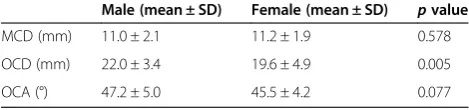
OCA, OCD, and MCD measurements were analyzed, and the results are summarized in Table 2. The mean MCDs were 11.0 and 11.2 mm in males and females, re-spectively. The mean OCDs were 22.0 mm (male) and 19.6 mm (female), and the OCAs were 47.2° (male) and 45.5° (female). No significant differences in OCA and MCD were observed between the genders (p>0.01), but a significant difference in the OCD was observed (p<0.01).
Discussion
In this study, we have determined reference values for estimating the occipitocervical neutral position using three key measurements. A comprehensive evaluation using all three parameters should provide an accurate estimation of the neutral position.
Conceptually, the occipitocervical neutral position is the functional and balanced position of the head on the cervical spine. A person feels most comfortable in this position. Radiologically, occipitocervical neutral is de-fined as the position in which the subject looks straight ahead during a standard lateral cervical radiograph; in this position, the mandible should not overlap C2or C3[12].
All these criteria are subjective, and they are not practic-able for patients with injuries or during an operation. Harris et al. and Uno et al. studied the relationship be-tween the occiput and the axis in normal and pathologic states to identify occipitovertebral dissociation [13,14], but they did not establish a measurement to define the occipi-tocervical neutral position.
The motion characteristics of the occipitocervical seg-ment are obviously different from those of other cervical
parts because of the occipitocervical segment’s specific anatomic features. The three-dimensional movements of the occipitoatlantal junction and the atlantoaxial junc-tion meet the requirements for fine movements of the head; at the same time, these movements make it chal-lenging to define the occipitocervical neutral position. Wills et al. used an individualized occipitocervical angle to evaluate the change in lordosis at the occipitocervical junction in patients treated with posterior occipitocervical arthrodesis [15]. This angle can only be used for compari-sons in the same person because it is involved in the entry point of the occipital wire and the anteriorinferior corner of the lowest fused vertebral body, which varies in pa-tients. The O-C2 angle, created by McGregor’s line and
the inferior surface of the axis, has been commonly used in the literature [10,11]. This method can be used to evaluate the outcome of surgical treatment. However, the O-C2 angle is quite sharp, and it is not convenient to
obtain precise measurements during surgery. The OCA and the OCD proposed by Phillips in 1999 were of clin-ical significance [8]; however, the OCA is still not easy to measure during surgery, and the OCD varies greatly in different people.
In this paper, we introduced the MCD measurement to make the evaluation process quicker. The MCD is more sensitive than the OCD because the distance from the occipitoatlantal junction (the axis of nodding) to the angle of the mandible is longer than the distance be-tween the occipitoatlantal junction and the occipital pro-tuberance. That is to say, the motion of the head affects the MCD much more than it affects the OCD. We believe that the MCD is more reliable than the OCD because morphologic variations of the C2 spinous process are more common than variations of the mandibular angle. Because the normal value of the MCD ranges from ap-proximately 9 to 13 mm on the basis of our measurements and was approximately half the sagittal length of the C3 vertebral body, it was convenient, in clinical practice, to roughly judge the position by eye based on the ratio of the MCD/sagittal length of the C3 body, which made hard-copy films unnecessary during surgery. For these rea-sons, we used the MCD as a quick screening method for judging the occipitocervical neutral position in the op-erating theater.
In our study, we emphasize that the head rotation should be corrected, and the tube should be vertical to the sagittal plane of the head during radiography. On a Table 1 Inter- and intraobserver agreement for the MCD, OCD, and OCA
MCD OCD OCA
Intraobserver Interobserver Intraobserver Interobserver Intraobserver Interobserver
Fvalues 0.007 0.001 0.340 0.267 0.050 0.024
pvalues 0.993 0.999 0.712 0.766 0.951 0.976
Table 2 Final results of the measurements of the MCD, OCD, and OCA
Male (mean ± SD) Female (mean ± SD) pvalue
MCD (mm) 11.0 ± 2.1 11.2 ± 1.9 0.578
OCD (mm) 22.0 ± 3.4 19.6 ± 4.9 0.005
standard lateral radiograph in neutral rotation, the two mandibular angles are separated from each other by less than 1 cm. However, slight rotation of the head, which is common in clinical practice, can significantly affect the positions of the mandibular angles. We select the mid-point between the two mandibular angles to eliminate this variation (Figure 2a, b).
The OCD revealed significant differences between males and females, whereas the OCA and the MCD did not. Although variations of the C2spinous process may
affect the OCD value, we found that variations exist in both males and females and have a tendency to balance. The differences in atlas heights between males and fe-males should be taken into consideration to explain the different OCDs. A conclusion can be made only when the
atlas heights in a large sample are measured and statistical comparisons are made. In addition, the tube-film distance in our study was 1.8 m, in contrast to 2.0 m in Phillips’ study. Although the OCD result reported by our group is similar to that found by Phillips’group, they are not com-parable because of the different tube-film distances.
The OCA, OCD, and MCD values correlate with each other during the motion of the occipitocervical junction. For example, in flexion of the head, the OCA and the MCD decrease, while the OCD increases. Although each parameter can define the neutral position to some ex-tent, we emphasize performing a comprehensive evalu-ation using the three parameters. The OCA is the most accurate parameter, the MCD has the highest sensitivity, and the OCD is a good assistant for the MCD.
Figure 2The MCDs were less affected when the head was slightly rotated.The figure shows the head in neutral position(a). The head turned right(b), and the head turned left(c).
It should be noted that the subjects in our study were people with normal cervical spines, just like those in Phillips’group. For patients with severe occipitocervical de-formity, compensatory morphological changes in the lower cervical spine should be considered together with the occipitocervical position. Figure 3a shows a patient with chronic atlantoaxial dislocation secondary to occipitaliza-tion. The small OCA was caused by cervical hyperlordosis and anterior rotation of the head. Although anterior trans-lation of the atlas should increase the MCD, anterior rotation of the head counteracted anterior translation, resulting in a short MCD. After surgery (Figure 3b), we achieved an OCA within the normal range (53°), and the curve of the cervical spine was improved at the same time. The MCD was longer than normal, which might have been caused by incomplete reduction of the atlas and decreased cervical lordosis. The OCD was obviously shortened, mainly because of the poorly developed atlas. Therefore, we con-cluded that the occipitocervical neutral position varies in se-vere pathological conditions, and the values offered in this paper should be used conditionally for such cases. Whether the values can be restored to within a normal range de-pends on correcting the individual patient’s basic pathology. It would be poor practice to pursue normal measurement values without correcting the primary pathology.
Another limitation of this study is that some of the demographic characteristics of the study sample (e.g., body size, weight) were not included due to imperfections in the clinical database. As such, prospective studies are still needed to obtain more reliable measurements.
Conclusions
This study offers reference values for OCA and OCD for the estimation of the occipitocervical neutral position. The introduction of the MCD made the evaluation more direct and more comprehensive. The long-term results of occipitocervical fusion are expected to be improved when the OCA, OCD, and MCD are adjusted according to the lateral radiograph during surgery. However, con-sidering the complicated anatomic characteristics of the investigated region, it is more justified to say that the method used in this study serves as a proposed approach rather than a universal treatment regimen.
Abbreviations
BCR:Body/canal ratio; MRI: Magnetic resonance imaging; CT: Computed tomography; OCA: The occipitocervical angle; OCD: The occipitocervical distance; MCD: Mandible cervical distance.
Competing interests
The authors declare that they have no competing interests.
Authors’contributions
GL conceived of the study, participated in its design and coordination, and helped to draft the manuscript. JT participated in the design of the study and drafted the manuscript. SL collected the data and performed the statistical analysis. All authors read and approved the final manuscript.
Acknowledgments
The authors thank all colleagues in the Department of Orthopedic Surgery of Yantaishan Hospital for their assistance and support during this work.
Author details 1
Department of Orthopedic Surgery, Yantai Affiliated Hospital of Binzhou Medical University, Yantai 264000, People’s Republic of China.2Department of Orthopedic Surgery, Yantaishan Hospital, No. 91 Jiefang Road, Zhifu district, Yantai 264000, People’s Republic of China.
Received: 13 May 2014 Accepted: 16 September 2014
References
1. Lee SC, Chen JF, Lee ST:Clinical experience with rigid occipitocervical fusion in the management of traumatic upper cervical spinal instability. J Clin Neurosci2006,13:193–198.
2. Shin H, Barrenechea IJ, Lesser J, Sen C, Perin NI:Occipitocervical fusion after resection of craniovertebral junction tumors.J Neurosurg Spine2006, 4:137–144.
3. Nakagawa T, Yone K, Sakou T, Yanase M:Occipitocervical fusion with C1 laminectomy in children.Spine1997,22:1209–1214.
4. Rodgers WB, Coran DL, Emans JB, Hresko MT, Hall JE:Occipitocervical fusions in children. Retrospective analysis and technical considerations. Clin Orthop Relat Res1999,364:125–133.
5. Kim LJ, Rekate HL, Klopfenstein JD, Sonntag VK:Treatment of basilar invagination associated with Chiari I malformations in the pediatric population: cervical reduction and posterior occipitocervical fusion. J Neurosurg2004,101(Suppl 2):189–195.
6. Matsunaga S, Ijiri K, Koga H:Results of a longer than 10-year follow-up of patients with rheumatoid arthritis treated by occipitocervical fusion. Spine2000,25:1749–1753.
7. Matsunaga S, Sakou T, Onishi T, Hayashi K, Taketomi E, Sunahara N, Komiya S: Prognosis of patients with upper cervical lesions caused by rheumatoid arthritis: comparison of occipitocervical fusion between c1 laminectomy and nonsurgical management.Spine2003,28:1581–1587.
8. Toyama Y, Matsumoto M, Chiba K, Asazuma T, Suzuki N, Fujimura Y, Hirabayashi K:Realignment of postoperative cervical kyphosis in children by vertebral remodeling.Spine1994,19:2565–2570.
9. Matsunaga S, Onishi T, Sakou T:Significance of occipitoaxial angle in subaxial lesion after occipitocervical fusion.Spine2001,26:161–165. 10. Yoshida M, Neo M, Fujibayashi S, Nakamura T:Upper-airway obstruction
after short posterior occipitocervical fusion in a flexed position. Spine2007,32:267–270.
11. Phillips FM, Phillips CS, Wetzel FT, Gelinas C:Occipitocervical neutral position.Spine1999,24:775–778.
12. Wholey MH, Bruwer AJ, Baker HL:The lateral roentgenogram of the neck; with comments on the atlanto-odontoid-basion relationship. Radiology1958,71:350–356.
13. Harris JH, Carson GC, Wagner LK:Radiologic diagnosis of traumatic occipitovertebral dissociation: 1. Normal occipitovertebral relationships on lateral radiographs of supine subjects.Am J Roentgenol1994,162:881–886. 14. Uno K, Kataoka O, Shiba R:Occipitoatlantal and occipitoaxial
hypermobility in Down syndrome.Spine1996,21:1430–1434.
15. Wills BP, Auerbach JD, Glotzbecker MP, Drummond DS, Dormans JP:Change in lordosis at the occipitocervical junction following posterior occipitocervical fusion in skeletally immature children.Spine2006,31:2304–2309.
doi:10.1186/s13018-014-0087-2