R E S E A R C H
Open Access
Prediction of response to therapy with ezatiostat
in lower risk myelodysplastic syndrome
Naomi Galili
1*, Pablo Tamayo
2, Olga B Botvinnik
2, Jill P Mesirov
2, Margarita R Brooks
1, Gail Brown
3and Azra Raza
1Abstract
Background:Approximately 70% of all patients with myelodysplastic syndrome (MDS) present with lower-risk disease. Some of these patients will initially respond to treatment with growth factors to improve anemia but will eventually cease to respond, while others will be resistant to growth factor therapy. Eventually, all lower-risk MDS patients require multiple transfusions and long-term therapy. While some patients may respond briefly to
hypomethylating agents or lenalidomide, the majority will not, and new therapeutic options are needed for these lower-risk patients. Our previous clinical trials with ezatiostat (ezatiostat hydrochloride, Telentra®, TLK199), a glutathione S-transferase P1-1 inhibitor in clinical development for the treatment of low- to intermediate-risk MDS, have shown significant clinical activity, including multilineage responses as well as durable red-blood-cell
transfusion independence. It would be of significant clinical benefit to be able to identify patients most likely to respond to ezatiostat before therapy is initiated. We have previously shown that by using gene expression profiling and grouping by response, it is possible to construct a predictive score that indicates the likelihood that patients without deletion 5q will respond to lenalidomide. The success of that study was based in part on the fact that the profile for response was linked to the biology of the disease.
Methods:RNA was available on 30 patients enrolled in the trial and analyzed for gene expression on the Illumina HT12v4 whole genome array according to the manufacturer’s protocol. Gene marker analysis was performed. The selection of genes associated with the responders (R) vs. non-responders (NR) phenotype was obtained using a normalized and rescaled mutual information score (NMI).
Conclusions:We have shown that an ezatiostat response profile contains two miRNAs that regulate expression of genes known to be implicated in MDS disease pathology. Remarkably, pathway analysis of the response profile revealed that the genes comprising the jun-N-terminal kinase/c-Jun molecular pathway, which is known to be activated by ezatiostat, are under-expressed in patients who respond and over-expressed in patients who were non-responders to the drug, suggesting that both the biology of the disease and the molecular mechanism of action of the drug are positively correlated.
Background
Myelodysplastic syndrome (MDS) is a clonal stem cell dis-order resulting in bone marrow failure and variable cytope-nias. Development of new treatment strategies has greatly improved the outlook for patients with MDS. There are three FDA-approved drugs for therapy of patients who have become transfusion-dependent, including two hypo-methylating drugs (HMAs), azacitidine and decitabine,
and the thalidomide derivative lenalidomide. Patients with higher-risk disease have been shown to benefit from HMA therapy [1,2], while patients with lower-risk disease with a karyotype of clonally restricted deletion of the long arm of chromosome 5 (deletion 5q or del[5q]) are highly responsive to lenalidomide [3,4]. Only 26% of transfusion-dependent lower-risk patients without del(5q) will also become transfusion-independent while on treatment [5], but the FDA has not approved lenalidomide for these patients. There are few treatment options for the major-ity of transfusion-dependent MDS patients with lower-risk disease. This situation represents a significant unmet medical need. Once disease-modifying therapy is * Correspondence:ng2368@columbia.edu
1Department of Medicine, Division of Hematology and Oncology, Columbia
University Medical Center and New York Presbyterian Hospital, 177 Fort Washington Ave., New York, NY 10032, USA
Full list of author information is available at the end of the article
required by the patient, it is a challenge for the treating physician to decide which drug will best benefit the indi-vidual patient, as only a subset responds to any given agent.
Ezatiostat (ezatiostat hydrochloride, TelintraW, TLK199), a glutathione analog inhibitor of the enzyme glutathione S-transferase P1-1 (GSTP1-1), causes dissociation of the enzyme from the jun-N-terminal kinase/c-Jun (JNK/JUN) complex, leading to JNK activation by phosphorylation. Activated JNK phosphorylates c-JUN, which ultimately results in the stimulation of all myeloid lineages hemato poietic progenitor’s proliferation and maturation. In addition, subsequent activation of the caspase-dependent apoptotic pathway increases reactive oxygen species in human leukemia blast cells. This cascade can trigger apoptosis. In other words, the therapeutic action of eza-tiostat appears to include both proliferation of normal myeloid progenitors as well as apoptosis of the malig-nant clone.
Our previous phase 2 study of ezatiostat demonstrated that this drug can elicit a therapeutic response in a pro-portion of patients with lower-risk MDS [6]. Trilineage responses were observed in 4 of 16 patients (25%) with trilineage cytopenia. Hematologic Improvement-Erythroid (E) was observed in 9 of 38 patients (24%), Neutrophil (N) in 11 of 26 patients (42%), and HI-Platelet (HI-P) in 12 of 24 patients (50%). In a subgroup of 9 patients who were red-blood-cell (RBC)-transfusion-dependent and HMA-naïve, a 47% HI-E rate was observed. Three (16%) of these patients achieved complete RBC-transfusion independence, and 3 of 9 (33%) reported multi-lineage responses. While the responses seen in the lower-risk patients resulted in hematologic improvement with clinically significant reductions in RBC-transfusion require-ments, and in some cases transfusion-independence, it is clear that in this heterogeneous disease it would be advanta-geous if a diagnostic predictor of response could be devel-oped to optimize treatment outcomes.
Gene expression profiling studies can define signatures that are capable of improving existing classification and prognosis of multiple diseases, especially malignancies which tend to be heterogeneous or of unknown or uncer-tain origin. MDS is a group of hematopoietic stem cell dis-orders that pose a unique challenge for gene expression profiling by virtue of their inherent heterogeneity. How-ever, we have previously shown that profiling can generate distinct expression signatures based on the uniform group-ing of patient response to a specific drug therapy [7]. In an attempt to identify the subset of lower-risk patients likely to benefit from therapy with ezatiostat, we exam-ined pre-therapy marrow cells from ezatiostat-treated MDS patients by gene expression profiling in order to identify signatures which differentiate responders from non-responders.
Methods
Patient samples
A separate research protocol was submitted to the in-stitutional review boards (IRBs) at the University of Massachusetts Memorial Medical Center, Worcester, MA, and at Saint Vincent’s Comprehensive Cancer Center, New York, NY, seeking permission to perform the micro-array analysis as described below. Once the research proto-col was approved by the respective IRBs and informed consent was obtained from each patient, samples from lower-risk MDS patients treated with ezatiostat in the phase 2 clinical trial at those institutions were obtained. Mononuclear cells from pre-therapy bone marrow aspi-rates were stored in Trizol at °C.
All patients had low- or intermediate-1-risk MDS as determined by the International Prognostic Scoring System (IPSS) and had not received growth factors for 4 weeks prior to study enrollment. Hematological improvement re-sponse was based on International Working Group (IWG) 2006 criteria [8,9].
Microarrays
Total RNA was purified from 5-10 × 106mononuclear cells using Trizol (Invitrogen) and analyzed for gene expression on the Illumina HT12v4 whole genome array according to the manufacturer’s protocol. RNA was available on 30 patients enrolled in the trial at the two institutions.
Gene marker analysis
score. Because in our study the number of samples is small, we opted for not performing the permutation test and focused instead on analyzing the 100 genes with the highest (50) and lowest (50) NMI scores.
Gene set/pathway analysis
To project the gene profiles into the space of pathways, we used a single-sample Gene Set Enrichment Analysis (ssGSEA) [12-17]. The gene-expression values were first rank-normalized and sorted independently, sample per sample. Then a per-gene enrichment score for each gene set/pathway was computed based on the integrated dif-ference between the empirical cumulative distribution functions of: i) the genes in the gene set vs. ii) the genes not in the set. This procedure is similar to the computa-tion of standard Gene Set Enrichment Analysis [13], but it is based on absolute rather than differential expression.
The selection of gene sets/pathways more associated with the responders vs. non-responders phenotype was obtained using an NMI as was done with the gene profiles (see above paragraph). The sources of gene sets/pathways were: i) the C2 sub-collection of curated and functional gene sets from the Molecular Signatures Database (MSigDB) release 2.5 (www.broadinstitute.org/msigdb); ii) an internal data-base of signatures of oncogene activation containing over 300 gene sets defined from data generated in our labora-tory, from GEO datasets, and from the biomedical litera-ture; and iii) gene sets representing hematopoietic cell populations [18]. We considered a total of 2776 gene sets. The selection analysis was restricted to the 60 gene sets/ pathways with the 30 highest and 30 lowest NMI scores.
Results and discussion
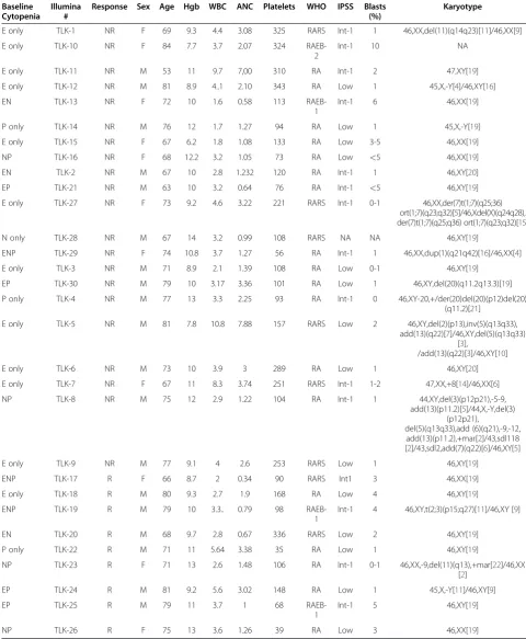
In order to identify an expression signature of ezatiostat response, prior to therapy with the drug, the genome-wide gene expression profiles of bone marrow aspirate mono-nuclear cells were obtained from patients with MDS. Samples of nine responders and 21 non-responders were available for analysis. The nine responders included one with a baseline single erythroid cytopenia, one with a single platelet cytopenia, one with erythroid-neutrophil cytopenias, two with erythroid-platelet cytopenias, two with neutrophil-platelet cytopenias and two with triline-age cytopenia (Table 1). The non-responders included 11 patients with a single erythroid cytopenia, one with single platelet cytopenia, one with single netrophil cyto-penia, two with erythroid-platelet cytopenias, two with erythroid-neutrophil cytopenias, and one with trilineage cytopenias. There were 18 patients with refractory anemia (RA); eight with RA with ringed sideroblasts (RARS); three with RA with excess blasts, type 1 (RAEB-1); and one with RAEB-2. Patient samples had similar representation in both the responder and the non-responder groups (Table 1).
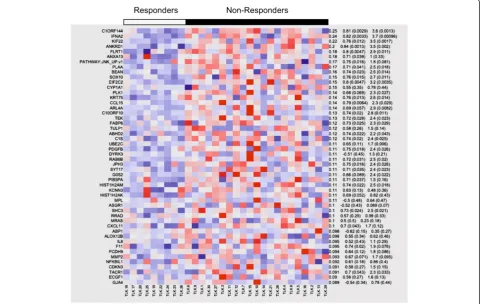
We compared the gene expression profiles of responders and non-responders to identify genes that correlate with ezatiostat response. The top 100 marker genes (50 under-expressed and 50 over-under-expressed in the responders) were identified using a sensitive metric based on the normal mu-tual information (NMI; see Methods) (Figure 1A and B). A majority of the top genes in both profiles are tran-scripts of as-yet unknown function. Most notably, how-ever, there are two microRNA (miR) genes that are differentially expressed. Responders under-express miR-129 and over-express miR-155. miRNAs are small non-coding RNAs of 18–25 nucleotides that bind the 3’UTR of mRNA, resulting in suppressed translation or mRNA degradation. This post-transcriptional control has been found to be perturbed in a wide variety of tumors, where it has been shown to have both oncogenic and tumor-suppressor activities [19]. Surprisingly, both miRNAs have been shown to mediate control of molecular path-ways associated with the pathophysiology of MDS.
Reduced expression of miR-129 has been found in a variety of primary solid tumors and has been shown to reduce proliferation by targeting the G!S cell cycle kinase CDK6 in lung epithelial-derived cells [20]. Inter-estingly, one of the direct targets of miR-129 is the onco-gene SOX4, a member of the SRY-related high mobility group box family of transcription factors [21]. Over-expression of SOX4 has been demonstrated in pros-tate, liver, lung, bladder, and medulloblastoma cancers exhibiting poor prognosis [21]. SOX4 has also been shown to target growth factor receptors that when stimu-lated increase proliferation as well as inhibit differentiation via suppression of other transcription factors [22]. Sup-porting the role of SOX4 in myeloid cells are the in vitro studies showing that over-expression of SOX4 in 32D cells resulted in the suppression of cytokine-induced granulo-cyte differentiation [23]. Other predicted target genes are components of the RISC complex that processes miRNAs from their precursor molecules [22]. Thus the low expres-sion of miR129 seen in responders would be expected to aberrantly affect proliferation and differentiation and, through dysregulated miRNA processing, to participate in oncogenic transformation. These are precisely the path-ways that are associated with the evolution of MDS.
Table 1 Myelodysplastic syndrome disease characteristics of patients treated with ezatiostat and analyzed by Illumina expression arrays
Baseline Cytopenia
Illumina #
Response Sex Age Hgb WBC ANC Platelets WHO IPSS Blasts (%)
Karyotype
E only TLK-1 NR F 69 9.3 4.4 3.08 325 RARS Int-1 1 46,XX,del(11)(q14q23)[11]/46,XX[9]
E only TLK-10 NR F 84 7.7 3.7 2.07 324
RAEB-2
Int-1 10 NA
E only TLK-11 NR M 53 11 9.7 7,00 310 RA Int-1 2 47,XY[19]
E only TLK-12 NR M 81 8.9 4..1 2.10 343 RA Low 1 45,X,-Y[4]/46,XY[16]
EN TLK-13 NR F 72 10 1.6 0.58 113
RAEB-1
Int-1 6 46,XX[19]
P only TLK-14 NR M 76 12 1.7 1.27 94 RA Low 1 45,X,-Y[19]
E only TLK-15 NR F 67 6.2 1.8 1.08 133 RA Low 3-5 46,XX[19]
NP TLK-16 NR F 68 12.2 3.2 1.05 73 RA Low <5 46,XX[19]
EN TLK-2 NR M 67 10 2.8 1.232 120 RA Int-1 1 46,XY[20]
EP TLK-21 NR M 63 10 3.2 0.64 76 RA Int-1 <5 46,XY[19]
E only TLK-27 NR F 73 9.2 4.6 3.22 221 RARS Int-1 0-1 46,XX,der(7)t(1;7)(q25;36)
ort(1;7)(q23;q32)[5]/46,Xdel(X)(q24q28), der(7)t(1;7)(q25;q36) ort(1;7)(q23;q32)[15]
N only TLK-28 NR M 67 14 3.2 0.99 108 RARS NA NA 46,XY[19]
ENP TLK-29 NR F 74 10.8 3.7 1.27 56 RA Int-1 1 46,XX,dup(1)(q21q42)[16]/46,XX[4]
E only TLK-3 NR M 71 8.9 2.1 1.39 108 RA Low 0-1 46,XY[19]
EP TLK-30 NR M 79 10 3.17 3.36 101 RA Low 1 46,XY,del(20)(q11.2q13.3)[19]
P only TLK-4 NR M 77 13 3.3 2.25 93 RA Int-1 0 46,XY-20,+/der(20)del(20)(p12)del(20)
(q11.2)[21]
E only TLK-5 NR M 81 7.8 10.8 7.88 157 RARS Low 2 46,XY,del(2)(p13),inv(5)(q13q33),
add(13)(q22)[7]/46,XY,del(5)(q13q33) [3],
/add(13)(q22)[3]/46,XY[10]
E only TLK-6 NR M 73 10 3.9 3 289 RA Low 1 46,XY[20]
E only TLK-7 NR F 67 11 8.3 3.74 251 RARS Int-1 1-2 47,XX,+8[14]/46,XX[6]
NP TLK-8 NR M 75 12 2.9 1.22 104 RA Int-1 1 44,XY,del(3)(p12p21),-5-9,
add(13)(p11.2)[5]/44,X,-Y,del(3) (p12p21),
del(5)(q13q33),add (6)(q21),-9,-12, add(13)(p11.2),+mar[2]/43,sdl118 [2]/43,sdl2,add(7)(q22)[6]/46,XY[5]
E only TLK-9 NR M 77 9.1 4 2.6 253 RARS Low 1 46,XY[19]
ENP TLK-17 R F 66 8.7 2 0.34 90 RARS Int1 3 46,XX[19]
E only TLK-18 R M 80 9.3 2.7 1.9 168 RA Low 4 46,XY[19]
ENP TLK-19 R M 79 10 3.3.. 0.79 98
RAEB-1
Int-1 4 46,XY,t(2;3)(p15;q27)[11]/46,XY [9]
EN TLK-20 R M 68 9.7 2.8 0.67 336 RARS Low 2 46,XY[19]
P only TLK-22 R M 71 11 5.64 3.38 35 RA Low 1 46,XY[19]
NP TLK-23 R F 71 13 2.6 1.48 106 RA Int-1 0-1 46,XX,-9,del(11)(q13),+mar[22]/46,XX
[2]
EP TLK-24 R M 81 9.2 5.6 3.02 148 RA Low 1 45,X,-Y[11]/46,XY[9]
EP TLK-25 R M 79 11 3.7 1 68
RAEB-1
Int-1 5 46,XY[19]
NP TLK-26 R F 75 13 3.6 1.26 39 RA Low 3 46,XX[19]
in granulocyte/monocyte expansion, with these cells hav-ing dysplastic features [24]. This proliferation was accom-panied by decreased erythrocytes, megakaryocytes, and lymphocytes in the marrow. In addition, when expression analysis was performed on the marrow cells, genes known to be important for normal hematopoiesis were found to be down-regulated.
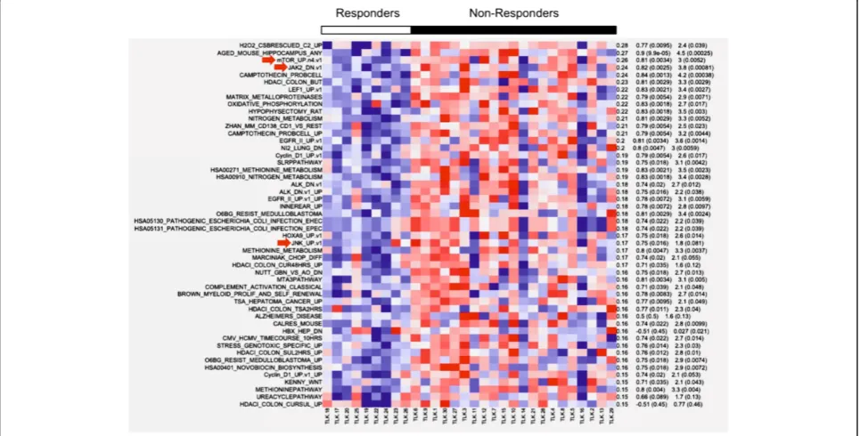
Single-sample Gene Set Enrichment Analysis was per-formed to find the most salient differences in terms of pathways and biological processes between responders and non-responders. Most notably, three pathways, mTOR, JAK2 and JNK, were all found to be under-expressed in the responders (Figure 2). All three have significant impli-cations in the process of hematopoiesis.
Figure 1Patients who responded to ezatiostat under-expressed miR-129 (A) and over-expressed miR-155 (B).
The serine/threonine kinase Akt is the upstream regula-tor of mTOR and functions as an antiapoptotic kinase. AKT is the major downstream target of PI3K (phophoino-sitide-3 kinase), which may be activated by receptor tyro-sine kinases (RTKs), including epidermal growth factor receptor (EGFR), insulin-like growth factor-1 receptor (IGF-1R), and G protein-coupled receptors (GPCRs). It has been shown that the PI3K/Akt/mTOR pathway is acti-vated in high-risk MDS, when compared to lower risk or healthy controls [26]. In addition, mTOR was specifically shown to be upregulated in the myeloid progenitors of high-risk MDS. These results suggest that this pathway participates in the evolution of MDS and that patients with low expression of these genes may respond to ezatiostat. JAK2 (tyrosine Janus Kinase-2) is an important regulator of erythropoiesis. When erythropoietin binds to its recep-tor on progenirecep-tor cells, the receprecep-tor forms homodimers that physically associate with JAK2, resulting in phosphor-ylation and activation. The activated tyrosine residues then associate with multiple downstream adaptors and effec-tors, including PI3K and JNK [27,28]. The resulting effects are promotion of erythroid differentiation and the synthe-sis of hemoglobin. As with the mTOR pathway, those patients able to respond to ezatiostat appear to be those who under-express genes of the JAK2 activation pathway.
Lastly, and most striking, was the finding that the JNK/ JUN pathway, which has been shown to be central to ezatiostat’s molecular mechanism of action, is also under-expressed in responding patients. This gene set, as defined by the GEO dataset GDS2081, was derived from expres-sion studies in primary cultured human epidermal kerati-nocytes, with activated JNK/JUN exposed to the JNK inhibitor drug SP600125 and analyzed on Affymetrix HGU95Av2 arrays [29]. A heatmap of responders/ non-responders was derived from the combined enrich-ment score of the top/bottom 200 genes, of which the top expressing genes are shown in Figure 3. Most notably, the gene-set profile of the JNK-inhibited keratinocytes is highly similar to the gene-set profile of patients who respond to ezatiostat. In other words, the profile is the same when the JNK pathway is dysregulated in vitro by the drug or patho-logically, as in some MDS patients. Ezatiostat has been shown to activate the JNK/JUN pathway; thus it is reason-able to expect that patients whose pre-treatment marrow cells show low expression will respond to ezatiostat ther-apy. In contrast, we show here that patients whose cells do not under-express the JNK/JUN pathway are not likely to benefit from additional activation by ezatiostat.
In conclusion, a bedside-to-bench strategy correlating MDS patient pre-treatment genomic data with clinical
response to ezatiostat has yielded positive markers for this investigational drug’s clinical efficacy. These signature genes and signaling pathways positively correlate with the known mechanism of action of ezatiostat. The gen-omic signature reported herein that distinguishes responders from non-responders among MDS patients treated with ezatiostat may enable the future selection of patients who are most likely to positively benefit from ezatiostat treatment. These markers could potentially be developed into a clinical diagnostic test for MDS patient sensitivity to ezatiostat treatment.
Competing interests
Gail Brown is an employee of Telik, Inc. Azra Raza has received honoraria from Celgene Corporation to serve on their speakers’bureau. Naomi Galili, Pablo Tamayo, Olga B Botvinnik, Jill P Mesirov and Margarita Roserika Brooks declare that they have no competing interests.
Author details
1Department of Medicine, Division of Hematology and Oncology, Columbia
University Medical Center and New York Presbyterian Hospital, 177 Fort Washington Ave., New York, NY 10032, USA.2The Eli and Edythe L. Broad
Institute of Massachusetts Institute of Technology and Harvard University, 301 Binney Street, Cambridge, Massachusetts 02142, USA.3Telik, Inc, 300 Hanson
Way, Palo Alto, CA 94304, USA.
Authors’contributions
NG conceived the study, obtained the Illumina results, and wrote the manuscript. PT, OBB and JPM did the computational analysis and generated the figures. MRB coolected the clinical characteristics and made Table 1. GB and AR conceived the study, assigned the clinical response criteria and participated in the writing of the manuscript. All authors read and approved the final manuscript.
Received: 28 February 2012 Accepted: 6 May 2012 Published: 6 May 2012
References
1. Silverman LR, Demakos EP, Peterson BL, Kornblith AB, Holland JC, Odchimar-Reissig R, Stone RM, Nelson D, Powell BL, DeCastro CM, Ellerton J, Larson RA, Schiffer CA, Holland JF:Randomized controlled trial of azacitidine in patients with the myelodysplastic syndrome: a study of the cancer and leukemia group B.J Clin Oncol2002,20:2429–2440.
2. Kantarjian H, Issa JP, Rosenfeld CS, Bennett JM, Albitar M, DiPersio J, Klimek V, Slack J, de Castro C, Ravandi F, Helmer R III, Shen L, Nimer SD, Leavitt R, Raza A, Saba H:Decitabine improves patient outcomes in myelodysplastic syndromes: results of a phase III randomized study.Cancer2006,106:1794–1803. 3. Dewald G, Bennett J, Giagounidis A, Raza A, Feldman E, Powell B, Greenberg
P, Thomas D, Stone R, Reeder C, Wride K, Patin J, Schmidt M, Zeldis J, Knight R:Myelodysplastic syndrome-003 study investigators. Lenalidomide in the myelodysplastic syndrome with chromosome 5q deletion.N Engl J Med2006,355(14):1456–1465.
4. Fenaux P, Giagounidis A, Selleslag D, Beyne-Rauzy O, Mufti G, Mittelman M, Muus P, Te Boekhorst P, Sanz G, Del Cañizo C, Guerci-Bresler A, Nilsson L, Platzbecker U, Lübbert M, Quesnel B, Cazzola M, Ganser A, Bowen D, Schlegelberger B, Aul C, Knight R, Francis J, Fu T, Hellström-Lindberg E: MDS-004 Lenalidomide del5q Study Group. A randomized phase 3 study of lenalidomide versus placebo in RBC transfusion-dependent patients with Low-/Intermediate-1-risk myelodysplastic syndromes with del5q.
Blood2011,118(14):3765–3776.
5. Raza A, Reeves JA, Feldman EJ, Dewald GW, Bennett JM, Deeg HJ, Dreisbach L, Schiffer CA, Stone RM, Greenberg PL, Curtin PT, Klimek VM, Shammo JM, Thomas D, Knight RD, Schmidt M, Wride K, Zeldis JB, List AF:Phase 2 study of lenalidomide in transfusion-dependent, low-risk, and intermediate-1 risk myelodysplastic syndromes with karyotypes other than deletion 5q.
Blood2008,111(1):86–93.
6. Raza A, Galili N, Smith SE, Godwin J, Boccia RV, Myint H, Mahadevan D, Mulford D, Rarick M, Brown GL, Schaar D, Faderl S, Komrokji RS, List AF,
Sekeres M:A phase 2 randomized multicenter study of 2 extended dosing schedules of oral ezatiostat in low to intermediate-1 risk myelodysplastic syndrome.Cancer2012 Apr 15,118(8):2138–2147. 7. Ebert BL, Galili N, Tamayo P, Bosco J, Mak R, Pretz J, Tanguturi S, Ladd-Acosta C,
Stone R, Golub TR, Raza A:An erythroid differentiation signature predicts response to lenalidomide in myelodysplastic syndrome.PLoS Med2008, 5(2):e35.
8. Greenberg P, Cox C, LeBeau MM, Fenaux P, Morel P, Sanz G, Sanz M, Vallespi T, Hamblin T, Oscier D, Ohyashiki K, Toyama K, Aul C, Mufti G, Bennett J: International scoring system for evaluating prognosis in myelodysplastic syndromes.Blood1997,89(6):2079–2088.
9. Cheson BD, Greenberg PL, Bennett JM, Lowenberg B, Wijermans PW, Nimer SD, Pinto A, Beran M, de Witte TM, Stone RM, Mittelman M, Sanz GF, Gore SD, Schiffer CA, Kantarjian H:Clinical application and proposal for modification of the International Working Group (IWG) response criteria in myelodysplasia.Blood2006,108:419–425.
10. Cover T, Thomas J:Elements of Information Theory, 2nd. Ed.: Wiley Series in Telecommunications and Signal Processing. Los Angeles: IEEE Computer Society Press; 2006.
11. Li M, Chen X, Li X, Ma B, Paul MB, Vitányi MB:The similarity metric.IEEE Trans Inf Theory2004,50(12):3250–3264.
12. Barbie DA, Tamayo P, Boehm JS, Kim SY, Moody SE, Dunn IF, Schinzel AC, Sandy P, Meylan E, Scholl C, Fröhling S, Chan EM, Sos ML, Michel K, Mermel C, Silver SJ, Weir BA, Reiling JH, Sheng Q, Gupta PB, Wadlow RC, Le H, Hoersch S, Wittner BS, Ramaswamy S, Livingston DM, Sabatini DM, Meyerson M, Thomas RK, Lander ES, Mesirov JP, Root DE, Gilliland DG, Jacks T, Hahn WC:Systematic RNA interference reveals that oncogenic KRAS-driven cancers require TBK1.Nature
2009,462(7269):108–112.
13. Subramanian A, Tamayo P, Mootha VK, Mukherjee S, Ebert BL, Gillette MA, Paulovich A, Pomeroy SL, Golub TR, Lander ES, Mesirov JP:Gene set enrichment analysis: a knowledge-based approach for interpreting genome-wide expression profiles.Proc Natl Acad Sci USA
2005,102(43):15545–15550.
14. Jagani Z, Mora-Blanco EL, Sansam CG, McKenna ES, Wilson B, Chen D, Klekota J, Tamayo P, Nguyen PT, Tolstorukov M, Park PJ, Cho YJ, Hsiao K, Buonamici S, Pomeroy SL, Mesirov JP, Ruffner H, Bouwmeester T, Luchansky SJ, Murtie J, Kelleher JF, Warmuth M, Sellers WR, Roberts CW:Dorsch M Loss of the tumor suppressor Snf5 leads to aberrant activation of the Hedgehog-Gli pathway.Nat Med2010,16:1429–1433.
15. Wolfer A, Wittner BS, Irimia D, Flavin RJ, Lupien M, Gunawardane RN, Meyer CA, Lightcap ES, Tamayo P, Mesirov JP, Liu XS, Shioda T, Toner M, Loda M, Brown M, Brugge JS, Ramaswamy S:MYC regulation of a “poor-prognosis”metastatic cancer cell state.Proc Natl Acad Sci USA
2010,107(8):3698–3703.
16. Cho YJ, Tsherniak A, Tamayo P, Santagata S, Ligon A, Greulich H, Berhoukim R, Amani V, Goumnerova L, Eberhart CG, Lau CC, Olson JM, Gilbertson RJ, Gajjar A, Delattre O, Kool M, Ligon K, Meyerson M, Mesirov JP, Pomeroy SL:Integrative genomic analysis of Medulloblastoma identifies a molecular subgroup that drives poor clinical outcome.J Clin Oncol2011,29(11):1424–1430.
17. Tamayo P, Cho YJ, Tsherniak A, Greulich H, Ambrogio L, Schouten-van Meeteren N, Zhou T, Buxton A, Kool M, Meyerson M, Pomeroy SL, Mesirov JP:Predicting relapse in patients with medulloblastoma by integrating evidence from clinical and genomic features.J Clin Oncol
2011,29(11):1415–1423.
18. Novershtern N, Subramanian A, Lawton LN, Mak RH, Haining WN, McConkey ME, Habib N, Yosef N, Chang CY, Shay T, Frampton GM, Drake AC, Leskov I, Nilsson B, Preffer F, Dombkowski D, Evans JW, Liefeld T, Smutko JS, Chen J, Friedman N, Young RA, Golub TR, Regev A, Ebert BL:Densely interconnected transcriptional circuits control cell states in human hematopoiesis.Cell2011, 144(2):296–309.
19. Esquela-Kerscher A, Slack FJ:Oncomirs - microRNAs with a role in cancer.Nat Rev Cancer2006,6(4):259–269.
20. Wu J, Qian J, Li C, Kwok L, Cheng F, Liu P, Perdomo C, Kotton D, Vaziri C, Anderlind C, Spira A, Cardoso WV, Lü J:miR-129 regulates cell proliferation by downregulating Cdk6 expression.Cell Cycle2010, 9(9):1809–1818.
21. Huang YW, Liu JC, Deatherage DE, Luo J, Mutch DG, Goodfellow PJ, Miller DS, Huang TH:Epigenetic repression of microRNA-129-2 leads to overexpression of SOX4 oncogene in endometrial cancer.Cancer Res
22. Scharer CD, McCabe CD, Ali-Seyed M, Berger MF, Bulyk ML, Moreno CS: Genome-wide promoter analysis of the SOX4 transcriptional network in prostate cancer cells.Cancer Res2009,69(2):709–717.
23. Boyd KE, Xiao YY, Fan K, Poholek A, Copeland NG, Jenkins NA, Perkins AS: Sox4 cooperates with Evi1 in AKXD-23 myeloid tumors via
transactivation of proviral LTR.Blood2006,107(2):733–741. 24. O’Connell RM, Rao DS, Chaudhuri AA, Boldin MP, Taganov KD, Nicoll J,
Paquette RL, Baltimore D:Sustained expression of microRNA-155 in hematopoietic stem cells causes a myeloproliferative disorder.J Exp Med
2008,205(3):585–594.
25. Vargova K, Curik N, Burda P, Basova P, Kulvait V, Pospisil V, Savvulidi F, Kokavec J, Necas E, Berkova A, Obrtlikova P, Karban J, Mraz M, Pospisilova S, Mayer J, Trneny M, Zavadil J, Stopka T:MYB transcriptionally regulates the miR-155 host gene in chronic lymphocytic leukemia.Blood2011,117(14):3816–3825. 26. Follo MY, Mongiorgi S, Bosi C, Cappellini A, Finelli C, Chiarini F, Papa V, Libra
M, Martinelli G, Cocco L, Martelli AM:The Akt/mammalian target of rapamycin signal transduction pathway is activated in high-risk myelodysplastic syndromes and influences cell survival and proliferation.
Cancer Res2007,67(9):4287–4294.
27. Tong W, Zhang J, Lodish HF:Lnk inhibits erythropoiesis and Epo-dependent JAK2 activation and downstream signaling pathways.Blood2005, 105(12):4604–4612. Epub 2005.
28. Arcasoy MO, Jiang X:Co-operative signalling mechanisms required for erythroid precursor expansion in response to erythropoietin and stem cell factor.Br J Haematol2005,130(1):121–129.
29. Gazel A, Banno T, Walsh R, Blumenberg M:Inhibition of JNK promotes differentiation of epidermal keratinocytes.J Biol Chem
2006,281(29):20530–20541.
doi:10.1186/1756-8722-5-20
Cite this article as:Galiliet al.:Prediction of response to therapy with ezatiostat in lower risk myelodysplastic syndrome.Journal of Hematology & Oncology20125:20.
Submit your next manuscript to BioMed Central and take full advantage of:
• Convenient online submission
• Thorough peer review
• No space constraints or color figure charges
• Immediate publication on acceptance
• Inclusion in PubMed, CAS, Scopus and Google Scholar
• Research which is freely available for redistribution


