R E S E A R C H
Open Access
Development and characterization of a
high-throughput in vitro cord formation model
insensitive to VEGF inhibition
Beverly L Falcon
1, Belinda O
’
Clair
4, Don McClure
2, Glenn F Evans
1, Julie Stewart
3, Michelle L Swearingen
1,
Yuefeng Chen
1, Kevin Allard
4, Linda N Lee
1, Kuldeep Neote
1, Dyke P McEwen
4, Mark T Uhlik
1and
Sudhakar Chintharlapalli
1*Abstract
Background:Anti-VEGF therapy reduces tumor blood vessels, however, some vessels always remain. These VEGF insensitive vessels may help support continued tumor growth and metastases. Manyin vitroassays examining multiple steps of the angiogenic process have been described, but the majority of these assays are sensitive to VEGF inhibition. There has been little focus on the development of high-throughput,in vitroassays to model the vessels that are insensitive to VEGF inhibition.
Methods:Here, we describe a fixed end-point and kinetic, high-throughput stem cell co-culture model of cord formation.
Results:In this system, cords develop within 24 hours, at which point they begin to lose sensitivity to VEGF inhibitors, bevacizumab, and ramucirumab. Consistent with the hypothesis that other angiogenic factors maintain VEGF-independent vessels, pharmacologic intervention with a broad spectrum anti-angiogenic antagonist (suramin), a vascular disrupting agent (combretastatin), or a combination of VEGF and Notch pathway inhibitors reduced the established networks. In addition, we used ourin vitroapproach to develop anin vivoco-implant vasculogenesis model that connects with the endogenous vasculature to form functional blood vessels. Similar to thein vitro system, over time these vessels become insensitive to VEGF inhibition.
Conclusion:Together, these models may be used to identify novel drugs targeting tumor vessels that are not sensitive to VEGF inhibition.
Keywords:Angiogenesis, Vascular endothelial growth factor (VEGF), Adipose derived stem cells (ADSC), Endothelial colony forming cells (ECFC)
Background
Preclinical studies indicate that most solid tumors require angiogenesis, the formation of new blood vessels from existing vessels, for growth, survival, and metastasis. While many factors regulate tumor angiogenesis, vascular endothelial growth factor (VEGF) appears to have a dom-inant role, inducing vascular permeability, endothelial cell proliferation and migration, and new blood vessel growth. Numerous drugs have been developed to target the VEGF
pathway with receptor tyrosine kinase inhibitors, with sol-uble decoy receptors, or with antibodies targeting the VEGF ligand or receptor. Inhibition of VEGF signaling re-duces tumor growth in many preclinical models [1,2], however, the benefits of targeting VEGF in mouse models have not completely translated to the clinic. While the FDA has approved multiple VEGF pathway inhibitors for clinical treatment of certain cancers, not all patients bene-fit from these treatments. Some tumors may initially re-spond but eventually become refractory, while others show no clinical benefit of inhibiting the VEGF pathway [3]. Some preclinical models have even shown resistance
* Correspondence:[email protected]
1
Eli Lilly and Company, Department of Cancer Angiogenesis, Lilly Corporate Center, Indianapolis, IN 46285, USA
Full list of author information is available at the end of the article
and increased metastatic spread associated with VEGF in-hibition [4-6].
Both preclinical and clinical studies have shown that despite significant reductions in tumor blood vessels with VEGF signaling blockade, some tumor blood vessels remain [7,8]. The blood vessels that remain have a dis-tinct phenotype typically associated with more pericyte coverage [8-13]. There are a number of possible explana-tions for this effect. First, the initial reduction in tumor blood vessels leads to tumor cell hypoxia, which, in turn, can cause tumor cells to either secrete more VEGF to overcome the anti-VEGF therapy or stimulate the release
of other pro-angiogenic cytokines [4,14-16]. “Vascular
normalization” may also play a role in VEGF resistant
tumor vessels. VEGF inhibitors can transiently improve pericyte and basement membrane coverage, decrease tumor vessel tortuosity and hyperpermeability, and in-crease oxygen and drug delivery [10,17,18]. These vessels may be formed via normalization of the atypical pheno-type associated with tumor vessels or a pruning of the abnormal vessels leaving behind pre-existing vessels that
have a more normal phenotype. Studies by Hal Dvorak’s
group indicate that tumor blood vessels are heterogeneous consisting of at least six distinctly different blood vessel types: (1)“mother”vessels, (2) glomeruloid microvascular proliferations, (3) vascular malformations, (4) capillaries, (5) feeder arteries (6) and draining veins [19,20]. Interest-ingly, only subpopulations of these vessels are sensitive to
VEGF inhibition. Immunodeficient mice expressing
VEGF-A164initially form vessel subtypes such as“mother” vessels and GMPs that are sensitive to VEGF inhibitors while later stage vessels are VEGF-independent [20,21]. Thus, the developmental stage of tumor vasculature is critical to anti-VEGF therapy sensitivity and the lack of good in vitroresistance models has slowed the develop-ment of non-VEGF anti-angiogenic therapies. In particu-lar, studies should be developed to identify novel ways of targeting the tumor blood vessels that remain or are in-sensitive to VEGF inhibition.
Many in vitro assays have been developed that examine multiple steps in the angiogenic process. These assays inter-rogate sprouting and tip formation, migration and prolifera-tion, lumen formaprolifera-tion, and tube or cord formation.In vivo assays also look at many of these similar processes. The ma-jority of these assays, however, are driven by the addition of VEGF or other growth factors to the system and remain sensitive to VEGF inhibition [22-25]. Disrupting established vessels, cords, or tubes which may be insensitive to VEGF inhibitors, however, has not been a major focus ofin vitro orin vivoapproaches. Here, we describe anin vitrocord for-mation assay that demonstrates insensitivity to VEGF inhib-ition. Similar to what is seen in vivo, resistance to VEGF inhibition is associated with cord maturity and pericyte asso-ciation. The advantage of this approach is its increased
throughput and ability to identify novel anti-angiogenic agents that can inhibit VEGF-independent vessels. Finally, we show the translatability of thisin vitroapproach using an in vivomodel of vasculogenesis to validate the effectiveness of novel treatments on the ability to decrease blood vessels that are insensitive to VEGF inhibition.
Results
Characterization of multiplein vitroangiogenesis models
Multiplein vitromodels of angiogenesis or cord formation were examined (Figure 1). Traditionally, co-cultures of HUVECs and NHDFs have been used to analyze and quan-tify growth factor and drug effects on angiogenesis [26]. Recently, a co-culture model of ECFCs and ADSCs, which has a shorter experimental duration and presence of pericyte biology, has been described [22]. In all of the models examined, cord formation occurred in the controls with increased cord formation induced by 20 ng/mL VEGF (Figure 1a). We observed a 44% increase in cords in the NHDF/HUVEC co-culture model while there was a 76% increase in cords in the ADSC/ECFC co-culture model at this VEGF concentration (Figure 1a). The optimized media used for these assays, however, contain serum and angio-genesis related growth factors such as epidermal growth factor (EGF) and basic fibroblast growth factor (FGF). In order to reduce background cord formation and increase responsiveness to exogenously added angiogenic growth factors, a basal media (BM) was developed which lacks serum and any additional growth factors. When the ADSC/ECFC co-culture was run in BM, the background cord formation decreased by 68% and there was a 194% in-crease in cord formation with the addition of VEGF (Figure 1a). Immunocytochemical characterization showed that cords formed in the ADSC/ECFC co-cultures express
multiple markers common to the in vivo vasculature
[27-29] (Figure 1b). CD31 (PECAM-1), VEGFR-2, and VE-cadherin were expressed by the endothelial cells forming the cords (Figure 1b). In addition, only ADSCs that were in close proximity with endothelial cells differentiated into
cells expressing SMA and PDGFR-β, indicative of a
pericyte-like phenotype [28] (Figure 1b, arrows). These pericyte markers were not expressed in the ADSC feeder layer found away from the cords. Finally, vascular base-ment membrane markers, such as nidogen and type IV col-lagen, were expressed and associated with the cords in this co-culture system (Figure 1b). In contrast, in the NHDF/ HUVEC co-culture model, the cords expressed endothelial and basement membrane markers, but pericyte markers were not expressed (data not shown).
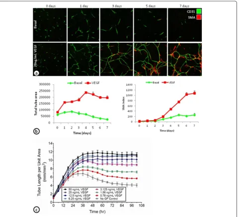
Time course of ADSC/ECFC cord formation
To further characterize the development of basal and VEGF-induced cords and its associated SMA cells,
(Figure 2a and b). After 1 day, many of the basal and VEGF-induced cords have already formed. The basal cords were only stable for a few days, before reductions in total tube area were seen beginning around day 3. VEGF-induced cords were stable over the 7 day time course with slight increases in total tube area after the first 24 hours (Figure 2a and b). The increase in SMA index was not observed until day 3, and increased
dramatically over the next 48 hours (Figure 2a and b). These results indicate that VEGF-induced cords are ini-tially formed within the first day. After the first day, however, the cords appear to remodel and may become more stable by the differentiation of the ADSCs into SMA expressing pericyte-like cells.
Continuous monitoring of cord formation using GFP-expressing ECFCs cultured with ADSCs demonstrated
concentration dependent increases in VEGF induced cord formation (Figure 2c). Similar to the fixed endpoint studies, the increased VEGF induced cord formation was
found in the first 24–36 hours. After 36 hours, the
higher concentrations of VEGF-induced cords were stable over the next 3 days (Figure 2c).
Targeting the components of the cord formation system
To determine whether VEGF-induced cords can be targeted with anti-VEGF therapy, cords were treated with a receptor tyrosine kinase inhibitor targeting the VEGF
receptors among others (sunitinib), an antibody targeting the VEGF-A ligand (avastin; bevacizumab), or an antibody targeting VEGFR-2 (IMC-1121B; ramucirumab) (Figure 3a). Blocking VEGF signaling with sunitinib, bevacizumab, or
ramucirumab all concentration-dependently reduced
VEGF-driven cord formation in the ADSC/ECFC
co-culture system. Sunitinib (0.2 μM) maximally decreased
VEGF-induced total tube area by 89% (EC50= 0.027 μM),
bevacizumab (20 μg/mL) by 65% (EC50= 0.174 μM), and
ramucirumab (20 μg/mL) by 80% (EC50= 0.623 μM)
(Figure 3a and b). In addition to VEGF-induced cords,
sunitinib and ramucirumab decreased basal total tube area by 75% and 72% respectively, while bevacizumab only decreased basal cords by 10% (Figure 3a and b). While the inhibitors of VEGF signaling decreased cord formation in a concentration-dependent manner, even at the highest concentrations, cords were not completely eliminated. This may indicate that the remaining cords are dependent on other growth factors secreted by the
feeder layer of ADSCs. In fact, examination of cords indicate that the basal cords formed in the ADSC/ECFC co-culture system are also dependent on HGF [22] (Additional file 1: Figure S1).
The ability to target SMA positive pericyte differenti-ation was also examined. Previous studies indicate that PDGF expression and stimulation of its receptor,
PDGFR-β, are important for pericyte recruitment [30-32]. Here,
cords were allowed to form for 4 days to allow for some pericyte differentiation. After 4 days, the cords were treated with 20μg/mL IgG1or a PDGFR-βblocking
anti-body (IMC-2C5; [33]). Inhibition of PDGFR-β signaling
with IMC-2C5 decreased the SMA index induced by VEGF by 79%, indicating a reduction in pericyte coverage, but the total tube area was not affected (Figure 3c and b).
Development of VEGF insensitive cords
To determine whether cords remain sensitive to inhibi-tors of VEGF signaling, VEGF-induced cords were allowed to establish for 0, 1, 2, or 4 days prior to addition of bevacizumab or ramucirumab. Bevacizumab
(20μg/mL) decreased total tube area by 64% when given
on day 0, by 25% on day 1, 13% on day 2, and 16% on day 4 (Figure 4a). Cord formation was reduced by 75%
when ramucirumab (20μg/mL) was given on day 0, 33%
on day 1, 21% on day 2, and 18% on day 4 (Figure 4a). These results indicate that once established, cords become increasingly resistant to VEGF inhibition. To further characterize this insensitivity to VEGF inhibition, continuous live-cell monitoring of the cords after ramucirumab or bevacizumab treatment at day 0 (neoangiogenic mode) or day 4 (established mode) was
compared (Figure 4b and c). Bevacizumab (25μg/mL)
de-creased neoangiogenic cord formation by 70%, but only
reduced established cord formation by 20% (Figure 4b).
Likewise, ramucirumab treatment (10 μg/mL) decreased
neoangiogenic cord formation by 90% and only decreased established cord formation by 30% (Figure 4c).
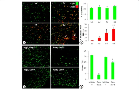
In vivomodel of VEGF insensitive vessels
A limitation of anyin vitromodel is its translatability to in vivo biology. Here, to investigate whether similar
VEGF-independent vessels can be established in vivo, a
co-implant model ofin vivovasculogenesis with ADSCs
and ECFCs was performed. Injection of a mixture of ADSCs and ECFCs in Matrigel develop blood vessels within 3 days. Evidence of blood perfusion (identified with an erythrocyte marker, TER119) in the vessels was seen beginning at 4 days and increased with additional time (Figure 5a and b). At 6 days, however, evidence of hemorrhage, or TER119 not associated with blood vessels became evident (Figure 5a, arrows). Four-day
treatment with ramucirumab beginning on day 0 de-creased the percent area of CD31 by 81%. However, when ramucirumab treatment was given for 4 days beginning on day 4, the percent area of CD31 was only reduced by 24% (Figure 5c and d); recapitulating the in vitroobservations.
Targeting VEGF-independent cords
A number of different mechanisms, including the upregulation of other angiogenic pathways such as the Notch pathway, have been described to play a role in the development of tumor vessels that are insensitive to anti-VEGF therapy [4,14,15,34]. To determine whether other classes of anti-vascular therapy can reduce cords that are insensitive to VEGF inhibition, a broad spectrum anti-angiogenic antagonist (suramin), a vascu-lar disrupting agent (combretastatin), and a combination therapy of a VEGFR-2 inhibitor (ramucirumab) and a
gamma secretase inhibitor (GSI; LY411575 [35,36]) were tested on established cords (Figure 6). VEGF-induced cords were allowed to establish for 4-days prior to addition of sura-min, combretastatin, or the ramucirumab/GSI (LY411575)
combination. Suramin treatment (100 μM) decreased
VEGF-established cords by 90% (Figure 6a), combretastatin (11 nM) decreased cords by 100% (Figure 6b), and the com-bination of ramucirumab (10μg/mL) and a GSI (LY411575; 10 nM) decreased established cords by 50% (Figure 6c). The reduction in established cords with the combination of ramucirumab and a GSI (LY411575) was greater than either
of the drugs alone (Figure 6c). Similarly, in the co-implant
model of in vivo vasculogenesis, ramucirumab or the GSI
(LY411575) alone led to slight reductions in vessels that were allowed to establish for 4 days prior to treatment. However, the combination of ramucirumab and the GSI (LY411575) almost completely eliminated the vessels (Figure 6d).
Discussion
Results from this study indicate that a co-culture system of progenitor cells and endothelial cells can create a cord network with components found in the vasculature:
endothelial cells, pericytes, and basement membrane. Exogenously added VEGF can stimulate cord formation and the cords that develop become insensitive to VEGF
inhibition as the cords mature. An in vivo co-implant
model of vasculogenesis, using the same progenitor and endothelial cells as thein vitroapproach, develops func-tional blood vessels that anastamose with the host vascu-lature. These vessels also become insensitive to VEGF pathway inhibition with time. Together, these studies
in-dicate that the combined use of an in vitro
high-throughput established cord formation assay and an
established in vivo co-implant model of vasculogenesis
can be used to identify novel drugs that can target VEGF-independent blood vessels.
Here, we investigate two different in vitro models of
angiogenesis. The first model uses co-cultures of human umbilical vein endothelial cells (HUVEC) with normal human dermal fibroblasts (NHDF) and the other uses co-cultures of endothelial colony forming cells (ECFC) with adipose-derived stem cells (ADSC). When seeded with NHDF, the HUVECs recapitulate the major phases of the angiogenic process, initially proliferating and migrat-ing into endothelial clusters followed by differentiation and branching into complex networks over the 7–10 day assay. Under basal conditions, little tube formation occurs,
while VEGF addition on day 3–4 stimulates tube
forma-tion in a concentraforma-tion-dependent manner [37-39]. Fur-ther characterization of the HUVEC/NHDF approach shows that the pharmacological and physiological effects on endothelial cell biology are highly translatable to previ-ousin vivocharacterizations, exemplified by DLL4/Notch inhibition resulting in increased branch point formation late in the angiogenic process [39-41]. One of the major advantages of the ADSC and ECFC co-culture model is that the process of developing cords occurs quickly and incorporates a pericyte-like biology associated with the cords. Unlike the NHDF/HUVEC model and other similar models, in the ADSC/ECFC model, the majority of VEGF-driven cords are formed within the first 24 hours [23,24,38,42]. Additional stimulation allowed for further remodeling of the vessels and differentiation of the
ADSCs into SMA or PDGFR-βexpressing pericyte-like
cells. Characterization of these SMA associated cords indicates that drugs targeting VEGF or PDGF can in-hibit the cords or the pericytes, respectively, similar to
what has previously been shownin vivo[8,28]. Further,
VEGF is not the only growth factor that induces rapid cord formation. Other pro-angiogenic growth factors, including FGF and EGF, also induce cord formation in a concentration-dependent manner and exhibit differ-ent phenotypes and kinetics (manuscript under prepar-ation). In addition to the assay duration and the cord
similarities to in vivo vascular structure, the use of
basal media in the ADSC/ECFC co-culture assay has
dramatically increased the response window for drug screening purposes.
Interestingly, we found that inhibition of the VEGF re-ceptor with sunitinib or ramucirumab leads to decreases in basal and VEGF driven cord formation, but inhibition of the ligand with bevacizumab only affected the VEGF driven cords. There may be several explanations for this effect. Endothelial cells can make VEGF and signal in an autocrine fashion. In fact, previous studies using endothe-lial cell specific knockout of VEGF indicates that autocrine VEGF signaling is required for the homeostasis of blood vessels [43]. It is also feasible that ligand-independent mechanisms or signaling through heterodimerization of VEGFR2 with other receptors may play an important role in basal cord formation [44]. Internalization of the recep-tor with ramucirumab or the multi-targeted nature of Sunitinib may play a role as well. Finally, VEGF secreted in the co-culture system may get bound to the extracellu-lar matrix, where it may not be accessible to VEGF anti-bodies, but may still be able to be affected by receptor inhibition. These possibilities require further exploration as it is unclear what mechanism or mechanisms are in-volved in the ECFC/ADSC co-culture assay.
Unlike most tube and cord formation assays, established cords in the ADSC and ECFC co-culture system lose their dependence on VEGF once the cords are developed. Even after 1 day of establishment, the cords are less sensitive to multiple inhibitors of the VEGF signaling pathway. The mechanism of this VEGF-independence is not clear. Pericyte coverage is thought to make vessels insensitive to VEGF inhibition, but in this assay, we see increased VEGF independence after 1 day even though the SMA differenti-ation does not occur until day 3. In addition, inhibition of the PDGFR after 4 days of establishment decreased the SMA index but did not significantly alter total tube area. While it is still possible that pericytes play an important role in maintenance of established vessels, in this assay system other growth factors secreted by the ADSC feeder layer likely play a major role in maintaining the cords once they have been created. Clearly, there are some cords that can form without the addition of VEGF. Previous studies and our data indicate that HGF is highly expressed by the
ADSCs and contributes to basal cord formation [22]. In
vivo studies show that inhibition of VEGF and the HGF
other growth factors secreted from the ECFCs or ADSC feeder layer may maintain cords following the initial VEGF stimulation.
Using an in vivo co-implant model of vasculogenesis
with ADSCs and ECFCs, functional vessels can form after anastamosing with the host vasculature. These ves-sels form over 3 days and blood cells labeled with TER119 can be seen beginning 4 days after the cells are injected into the flank. Treatment of the vasculogenic plugs with a VEGF inhibitor dramatically decreases blood vessel formation if given at the beginning of the assay. If, however, VEGFR signaling was not inhibited until the vessels have established and have blood flow (at day 4), there is little effect. While the mechanism of this insensitivity is not know, it would be interesting to characterize our VEGF independent vessels to determine whether they have similar phenotypes as those described by the Dvorak laboratory [20,21]. Nonetheless, these
re-sults are consistent with our high-throughput in vitro
assay and provide a uniquein vivomodel to examine the effects of novel drugs on vessels that are insensitive to VEGF inhibition.
As proof of principle, a broad-spectrum anti-angiogenic in-hibitor (suramin), a vascular disrupting agent (combretastatin), and a combination of a gamma secretase and VEGF in-hibitor were tested on VEGF established cords. Suramin blocks a variety of growth factors including many angiogenesis-related factors. One of the proposed mecha-nisms of VEGF-independent tumor vessels is that inhib-ition of VEGF leads to induction of other proangiogenic factors [4,14,15,34]. Suramin is likely able to block many of these other proangiogenic factors to reduce the cords in our established cord assay system.
The vascular disrupting agent combretastatin is a microtubule-depolymerizing agent which binds to tubulin dimers to prevent microtubule polymerization. This re-sults in mitotic arrest and apoptosis of endothelial cells. In addition, combretastatin disrupts the endothelial cell junc-tion molecule (VE-cadherin) leading to vascular collapse [45] andin vivo studies show that combretastatin is able to reduce immature vessels [45,46]. We observed reduc-tions in established cords with combretastatin treatment. Clearly, while combretastatin may not reduce all mature vesselsin vivo, it is able to target a unique population of vessels or cords that are insensitive to VEGF inhibition. In fact, preclinical and clinical studies indicate that combin-ing combretastatin with bevacizumab is more efficacious than either inhibitor alone [47,48].
A recentin vivostudy indicates that VEGF-independent vessels are driven by DLL4-Notch signaling and are sensi-tive to gamma secretase inhibition [34]. Consistent with this novel strategy to overcome anti-angiogenic resistance, a gamma secretase inhibitor was tested in ourin vitroand in vivomodels alone or in combination with inhibition of
VEGF signaling. In thein vitrosystem, treatment with ei-ther compound alone prevented a slight increase in cords associated with feeding the cells with fresh VEGF, but did not disrupt established networks. However, when VEGF and gamma secretase inhibitors were combined, there was a reduction in the number of cords. Similarly, in the in vivo co-implant model, ramucirumab or the gamma secretase inhibitor alone elicited a slight reduction in the vessels, but the combination reduced the vessels signifi-cantly more. These results indicate that our established cord assays may be used to identify new pathways in-volved in anti-VEGF/VEGFR directed therapy resistance and potential combinatorial strategies.
Many current angiogenesis assays used to screen anti-angiogenic agents are highly VEGF dependent. However, from preclinical and clinical analysis, there clearly exists a population of tumor vessels that are insensitive to VEGF inhibition. Thus, angiogenic assays are needed in which novel agents can be tested for their effectiveness on vessels which are not dependent on VEGF. The ECFC/ADSC assay is high throughput and relatively quick. Results can be obtained in approximately a week and can be run in 96-well and 384-well formats and similar co-culture approaches have previously been used in high-throughput drug discovery [24,49]. In addition, labeling the ECFCs with GFP is a feasible approach to monitor cord formation and effects on established cords using continuous live-cell monitoring. Together, these results indicate that a co-culture cord formation system with ADSCs and ECFCs is a useful method to identify and characterize novel drugs on VEGF-independent cords. It would be interesting to identify selective markers on tumor vessels that remain after VEGF ther-apy and determine if the same markers exist in this co-culture system. If so, these in vitro and in vivo systems would be conducive to interrogate the mechanisms by which vessels become insensitive to VEGF inhibition though use of shRNA/siRNA knockdowns. With more and more studies being published regarding mechanisms of VEGF resistance, additional targets should be tested in thisin vitroco-culture system.
Conclusions
Despite in vivo evidence that VEGF independent vessels exist, the majority of the in vitro assays used are dependent on VEGF. We described an in vitro cord for-mation assay that shows insensitivities to inhibition of the VEGF pathway. In addition, we were able to show the translatability of this assay using an in vivo model of vasculogenesis. Together, the combined use of this in vitro high-throughput established cord formation
assay and an established in vivo co-implant model of
Methods
Cell lines and media
Human adipose derived stem cells (ADSCs) isolated from lipoaspirates collected during surgical liposuction procedures were purchased from Lonza (Allendale, NJ). Cells were grown in EGM2-MV media (Cambrex; Walkersville, MD) and used at passage 4–6. Endothelial colony forming cells (ECFCs) isolated from cord-blood derived endothelial cells were grown on Collagen I coated flasks in EGM2-MV media supplemented with an
additional 5% FBS and used at passage 7–10 (Lonza).
For studies examining cord formation over time with continuous live-cell monitoring, ECFCs were lentivirally transduced to express CytoLight Green, a soluble variant of GFP, and optimized for imaging in the IncuCyte™ im-aging system. Human umbilical vein endothelial cells
(HUVECs) and normal human dermal fibroblast
(NHDF) cells and media were purchased from Cambrex. HUVECs were grown in EGM media with 10% FBS and NHDF cells were maintained in EGM-2 media.
Co-culture assay of endothelial cells and fibroblasts
HUVEC and NHDF co-culture cord formation assays were performed with AngioKit optimized media (TCS Cellworks, Birmingham, UK) as previously described
[26,37-39]. Briefly, 20K NHDF cells in 100 μL of media
were plated in each well of a 96-well plate and incubated
overnight at 37°C, 5% CO2.The next day, HUVECs were
added on top of the NHDF cells at 1800 cells/well in
100 μL and incubated overnight at 37°C, 5% CO2. On
the third day and every subsequent third day, the media was changed to optimized media containing 20 ng/mL VEGF (R&D Systems). On day 10, the co-culture was fixed, stained, and imaged as described below.
Neoangiogenic ADSC and ECFC co-culture cord formation assay
ADSC and ECFC co-culture assays were performed with AngioKit optimized media, basal media (MCDB-131
medium with 30 μg/mL L-ascorbic acid 2-phosphate,
1 μM dexamethasone, 50μg/mL tobramycin, 10 μg/mL
r-transferrin AF, and 10 μg/mL insulin) or basal media
plus (MCDB-131 medium with 0.3% FBS, 30 μg/mL
L-ascorbic acid 2-phosphate, 50μg/mL tobramycin, 10μg/
mL r-transferrin AF, and 10μg/mL insulin). ADSCs were
plated in 96-well plates at 40–50K cells per well in
100 μL and incubated overnight at 37°C, 5% CO2. The
next day, the media was removed and 4–5K ECFCs per
well in 50–100 μL of media was plated on top of the
ADSC monolayer and incubated at 37°C, 5% CO2for 3–
6 hours before the addition of growth factors and inhibi-tors. After the ECFCs attach, growth factors and test agents were added to the 50–100μL of media at 2–5× to achieve the final concentrations as indicated. Co-cultures
were grown for 0–7 days at which time the cells were
fixed, stained, and imaged as described below.
Established ADSC and ECFC co-culture cord formation assay
Established ADSC and ECFC co-culture assays were plated as described above for the neoangiogenesis assay. After the ECFCs were allowed to attach, 20 ng/mL VEGF was use to stimulate and establish the cord network. After 1–4 days the media was changed to contain fresh VEGF in the presence or absence of inhibitors at the indicated con-centrations. After addition of the inhibitors, cultures were allowed to grow an additional 3–4 days before the cells were fixed, stained, and imaged as described below to in-vestigate network disruption or cord regression.
Fixation and staining of fixed endpoint cords
At the completion of the assay, ADSC/ECFC cords were fixed and permeabilized with either 70% ice cold ethanol
for 20–30 minutes or 3% paraformaldehyde for 10
mi-nutes followed by 70% ice cold ethanol for 20 mimi-nutes. Cells were blocked with PBS + 1% bovine serum albu-min (BSA) for 30 albu-minutes at room temperature. Primary antibodies were diluted in PBS + 1% BSA and stained ei-ther sequentially or in combination for >90 minutes at 37°C. Endothelial cells were identified with sheep anti-CD31 (PECAM-1; Sigma; 1:200), rabbit anti-VEGFR-2 (55B11; Cell Signaling; 1:50), or goat anti-VE-cadherin (Santa Cruz; 1:50) antibodies. Cy3 conjugated mouse anti-smooth muscle actin (SMA; Sigma; 1:200) and rabbit anti-platelet-derived growth factor receptor beta
(PDGFR-β; Y92; LifeSpan; 1:50) antibodies identified
pericytes associated with the cords. Vascular basement membranes were identified with goat anti-Nidogen (R&D Systems; 1:50) and goat anti-type IV collagen (Millipore; 1:50) antibodies. After a brief wash, second-ary AlexaFluor 488- and 555-conjugated donkey anti-sheep, donkey anti-rabbit, donkey anti-goat secondary antibodies (Invitrogen; 1:400) were incubated for ~60 minutes at room temperature. Nuclei were identified with Hoechst 33342 (Invitrogen; 1:1000) for 5 minutes at room temperature. After Hoechst staining, the cells were washed and imaged as described below.
Fixed endpoint imaging and quantification
Cord formation images were captured using a Cellomics Arrayscan VTI and analyzed with the Tube Formation bio-application reading at a magnification of 5×. Objects were identified using an algorithm to detect CD31 stain-ing of cords. Total tube area was calculated from 9 fields
for each well with 3–4 wells for each treatment. SMA
Continuous monitoring of cord formation
ADSCs and ECFCs transduced with CytoLight Green were seeded for the assay as described above. After 3–4 hours at 37°C, the cells were treated with test reagents (growth factors ± compounds or antibodies), placed into the IncuCyte FLR for imaging, and allowed to form net-works over the course of the 4 day experiment. If run-ning the assay in neoangiogenic mode, looking at the inhibition of tube formation, the assay was terminated at the 96 hour time point. If studying tube regression was desired, the assay was run in established mode. To do this, growth factor-driven networks were formed over the first 96 hours of the assay. At this point, a full media replacement occurred including fresh growth factor in the presence or absence of test agent. The assay plate was then placed back in the IncuCyte FLR and imaged over the desired time frame to quantify regression of established networks.
For imaging and quantification, phase-contrast and fluorescent images were automatically collected every 6 hours in the IncuCyte FLR to detect network formation using the Tiled Field of View (FOV) mosaic imaging mode. The integrated Angiogenesis Analysis Module was used to identify the fluorescent signal from back-ground in order to quantify multiple assay metrics, such as tube length and branch formation, for each time point. In the first step of the process, the angiogenesis algorithm analyzed each fluorescent image and assigned a segmentation mask that closely resembles thein vitro network. From here, the mask was refined and filtered to exclude non-tube forming events, specifically measur-ing angiogenesis over time. Kinetic plots of the angio-genesis metrics was generated using the IncuCyte software, allowing for a direct comparison of test agent treatments to validated control conditions (Figure 2b).
In vivovasculogenesis assay
ADSCs and ECFCs were mixed in a ratio of 1:4 (0.5 × 10⁶/2 × 106 cells/mL) in Matrigel (BD Biosciences) and injected (0.2 mL/implant) subcutaneously into the flank of female athymic nude mice as previously described [50,51]. Three to six days following implantation, im-plants were collected and placed into zinc-tris fixative. Treatments with IgG or IMC-1121B (ramucirumab; 10 mg/kg, ip) began on day 0 or day 4. Treatments with the gamma secretase inhibitor (GSI, LY411575; 3 mg/kg QD, ip) alone or in combination with ramucirumab began on day 4. The concentration of drugs used was determined from dose response studies (data not shown). Implants were collected and fixed 4 days post treatment and analyzed using multiplexed immunohisto-chemistry of sections stained for endothelial cells with a CD31 antibody (PECAM; Bethyl; 1:50), erythrocytes with a TER-119 antibody (BD Biosciences; 1:50), and nuclei
with Hoechst 33342 (Invitrogen; 1:1000). Quantifications were made using an iCys research imaging cytometer as previously described [52].
Statistical analysis
All experiments had an n≥3 for each treatment and
similar results were seen in at least two experiments. Re-sults are expressed as means ± SEM. Statistical differ-ences were measured by ANOVA with a Tukey posthoc test using JMP software.
Additional file
Additional file 1: Figure S1.Role of HGF in basal cords CD31 stained basal cords at 3 days treated with 10μg/mL hIgG or anti-HGF antibody.
Abbreviations
VEGF:Vascular endothelial growth factor; ADSC: Adipose derived stromal cells; ECFC: Endothelial colony forming cells, HUVEC, Human umbilical vein endothelial cells; NHDF: Normal human dermal fibroblasts; EGF: Epidermal growth factor; FGF: Fibroblast growth factor; BM: Basal media; SMA: Smooth muscle actin; PDGFR-β: Platelet derived growth factor receptor beta; HGF: Hepatocyte growth factor, Bev, Bevacizumab; Ram: Ramucirumab; GSI: Gamma secretase inhibitor.
Competing interests
BLF, DM, GFE, JS, MLS, YC, LNL, KN, MU, and SC are employees of Eli Lilly and Co. BOC, KA, and DPM are employees of Essen Biosciences.
Authors’contributions
BLF played a role in conception and design of the experiments, acquisition, analysis, and interpretation of the data, and writing the manuscript. BOC and KA participated in optimization of live cell imaging and in the acquisition and analysis of the data. DM, MLS, YC, LNL, KN played a role in the conception and optimization of the cord formation assay. GFE developed, optimized, and performed the in vivo vasculogenesis assays and was involved in the design and acquisition of the data. JS was involved in the acquisition, analysis and interpretation of the in vivo vasculogenesis assay. DPM and MTU were involved in the conception and design of the experiments, analysis and interpretation of the data, and in writing the manuscript. SC played a role in conception, design and optimization of the in vitro experiments and in vivo vasculogenesis assays, analysis and interpretation of the data, and in writing the manuscript. All authors read and approved the final manuscript.
Acknowledgements
The authors would like to thank Jonathan Yingling for helpful scientific discussions, Jonathan Lee and Sarah Oliver for assay characterization and helpful discussions and Laura Benjamin and Bronek Pytowski for critical review of the manuscript.
Author details
1Eli Lilly and Company, Department of Cancer Angiogenesis, Lilly Corporate Center, Indianapolis, IN 46285, USA.2Eli Lilly and Company, Department of BioTDR, Lilly Corporate Center, Indianapolis, IN 46285, USA.3Eli Lilly and Company, Department of Tailored Therapeutics, Lilly Corporate Center, Indianapolis, IN 46285, USA.4Essen BioScience, Inc., 300 West Morgan Road, Ann Arbor, MI 48108, USA.
Received: 28 February 2013 Accepted: 23 April 2013 Published: 27 April 2013
References
coopted vessels in a model of neuroblastoma.Proc Natl Acad Sci U S A
2002,99:11399–11404.
2. Prewett M, Huber J, Li Y, Santiago A, O’Connor W, King K, Overholser J, Hooper A, Pytowski B, Witte L,et al:Antivascular endothelial growth factor receptor (fetal liver kinase 1) monoclonal antibody inhibits tumor angiogenesis and growth of several mouse and human tumors.
Cancer Res1999,59:5209–5218.
3. Ellis LM, Hicklin DJ:VEGF-targeted therapy: mechanisms of anti-tumour activity.Nat Rev Cancer2008,8:579–591.
4. Casanovas O, Hicklin DJ, Bergers G, Hanahan D:Drug resistance by evasion of antiangiogenic targeting of VEGF signaling in late-stage pancreatic islet tumors.Cancer Cell2005,8:299–309.
5. Ebos JM, Lee CR, Cruz-Munoz W, Bjarnason GA, Christensen JG, Kerbel RS:
Accelerated metastasis after short-term treatment with a potent inhibitor of tumor angiogenesis.Cancer Cell2009,15:232–239. 6. Paez-Ribes M, Allen E, Hudock J, Takeda T, Okuyama H, Vinals F, Inoue M,
Bergers G, Hanahan D, Casanovas O:Antiangiogenic therapy elicits malignant progression of tumors to increased local invasion and distant metastasis.Cancer Cell2009,15:220–231.
7. Griffioen AW, Mans LA, de Graaf AM, Nowak-Sliwinska P, de Hoog CL, de Jong TA, Vyth-Dreese FA, van Beijnum JR, Bex A, Jonasch E:Rapid angiogenesis onset after discontinuation of sunitinib treatment of renal cell carcinoma patients.Clin Cancer Res2012,18:3961–3971.
8. Inai T, Mancuso M, Hashizume H, Baffert F, Haskell A, Baluk P, Hu-Lowe DD, Shalinsky DR, Thurston G, Yancopoulos GD, McDonald DM:Inhibition of vascular endothelial growth factor (VEGF) signaling in cancer causes loss of endothelial fenestrations, regression of tumor vessels, and appearance of basement membrane ghosts.Am J Pathol2004,165:35–52. 9. Batchelor TT, Sorensen AG, di Tomaso E, Zhang WT, Duda DG, Cohen KS,
Kozak KR, Cahill DP, Chen PJ, Zhu M,et al:AZD2171, a pan-VEGF receptor tyrosine kinase inhibitor, normalizes tumor vasculature and alleviates edema in glioblastoma patients.Cancer Cell2007,11:83–95.
10. Winkler F, Kozin SV, Tong RT, Chae SS, Booth MF, Garkavtsev I, Xu L, Hicklin DJ, Fukumura D, di Tomaso E,et al:Kinetics of vascular normalization by VEGFR2 blockade governs brain tumor response to radiation: role of oxygenation, angiopoietin-1, and matrix metalloproteinases.Cancer Cell
2004,6:553–563.
11. Benjamin LE, Golijanin D, Itin A, Pode D, Keshet E:Selective ablation of immature blood vessels in established human tumors follows vascular endothelial growth factor withdrawal.J Clin Invest1999,103:159–165. 12. Benjamin LE, Hemo I, Keshet E:A plasticity window for blood vessel
remodelling is defined by pericyte coverage of the preformed endothelial network and is regulated by PDGF-B and VEGF.
Development1998,125:1591–1598.
13. Bergers G, Song S, Meyer-Morse N, Bergsland E, Hanahan D:Benefits of targeting both pericytes and endothelial cells in the tumor vasculature with kinase inhibitors.J Clin Invest2003,111:1287–1295.
14. Bergers G, Hanahan D:Modes of resistance to anti-angiogenic therapy.
Nat Rev Cancer2008,8:592–603.
15. Cascone T, Herynk MH, Xu L, Du Z, Kadara H, Nilsson MB, Oborn CJ, Park YY, Erez B, Jacoby JJ,et al:Upregulated stromal EGFR and vascular remodeling in mouse xenograft models of angiogenesis inhibitor-resistant human lung adenocarcinoma.J Clin Invest2011,
121:1313–1328.
16. Takeda T, Okuyama H, Nishizawa Y, Tomita S, Inoue M:Hypoxia inducible factor-1alpha is necessary for invasive phenotype in Vegf-deleted islet cell tumors.Sci Rep2012,2:494.
17. Jain RK:Normalization of tumor vasculature: an emerging concept in antiangiogenic therapy.Science2005,307:58–62.
18. Tong RT, Boucher Y, Kozin SV, Winkler F, Hicklin DJ, Jain RK:Vascular normalization by vascular endothelial growth factor receptor 2 blockade induces a pressure gradient across the vasculature and improves drug penetration in tumors.Cancer Res2004,64:3731–3736.
19. Nagy JA, Chang SH, Shih SC, Dvorak AM, Dvorak HF:Heterogeneity of the tumor vasculature.Semin Thromb Hemost2010,36:321–331.
20. Nagy JA, Dvorak HF:Heterogeneity of the tumor vasculature: the need for new tumor blood vessel type-specific targets.Clin Exp Metastasis2012,
29:657–662.
21. Sitohy B, Nagy JA, Jaminet SC, Dvorak HF:Tumor-surrogate blood vessel subtypes exhibit differential susceptibility to anti-VEGF therapy.
Cancer Res2011,71:7021–7028.
22. Merfeld-Clauss S, Gollahalli N, March KL, Traktuev DO:Adipose tissue progenitor cells directly interact with endothelial cells to induce vascular network formation.Tissue Eng Part A2010,16:2953–2966.
23. Evensen L, Link W, Lorens JB:Imaged-based high-throughput screening for anti-angiogenic drug discovery.Curr Pharm Des2010,16:3958–3963. 24. Evensen L, Micklem DR, Link W, Lorens JB:A novel imaging-based
high-throughput screening approach to anti-angiogenic drug discovery.
Cytometry A2010,77:41–51.
25. Prigozhina NL, Heisel A, Wei K, Noberini R, Hunter EA, Calzolari D, Seldeen JR, Pasquale EB, Ruiz-Lozano P, Mercola M, Price JH:Characterization of a novel angiogenic model based on stable, fluorescently labelled endothelial cell lines amenable to scale-up for high content screening.
Biol Cell2011,103:467–481.
26. Hetheridge C, Mavria G, Mellor H:Uses of the in vitro endothelial-fibroblast organotypic co-culture assay in angiogenesis research.
Biochem Soc Trans2011,39:1597–1600.
27. Falcon BL, Hashizume H, Koumoutsakos P, Chou J, Bready JV, Coxon A, Oliner JD, McDonald DM:Contrasting actions of selective inhibitors of angiopoietin-1 and angiopoietin-2 on the normalization of tumor blood vessels.Am J Pathol2009,175:2159–2170.
28. Sennino B, Falcon BL, McCauley D, Le T, McCauley T, Kurz JC, Haskell A, Epstein DM, McDonald DM:Sequential loss of tumor vessel pericytes and endothelial cells after inhibition of platelet-derived growth factor B by selective aptamer AX102.Cancer Res2007,67:7358–7367.
29. You WK, Sennino B, Williamson CW, Falcon B, Hashizume H, Yao LC, Aftab DT, McDonald DM:VEGF and c-Met blockade amplify angiogenesis inhibition in pancreatic islet cancer.Cancer Res2011,71:4758–4768. 30. Armulik A, Abramsson A, Betsholtz C:Endothelial/pericyte interactions.Circ
Res2005,97:512–523.
31. Betsholtz C, Lindblom P, Gerhardt H:Role of pericytes in vascular morphogenesis.EXS2005,94:115–125.
32. Enge M, Bjarnegard M, Gerhardt H, Gustafsson E, Kalen M, Asker N, Hammes HP, Shani M, Fassler R, Betsholtz C:Endothelium-specific platelet-derived growth factor-B ablation mimics diabetic retinopathy.EMBO J2002,
21:4307–4316.
33. Shen J, Vil MD, Prewett M, Damoci C, Zhang H, Li H, Jimenez X, Deevi DS, Iacolina M, Kayas A,et al:Development of a fully human anti-PDGFRbeta antibody that suppresses growth of human tumor xenografts and enhances antitumor activity of an anti-VEGFR2 antibody.Neoplasia2009,11:594–604. 34. Li JL, Sainson RC, Oon CE, Turley H, Leek R, Sheldon H, Bridges E, Shi W,
Snell C, Bowden ET,et al:DLL4-Notch signaling mediates tumor resistance to anti-VEGF therapy in vivo.Cancer Res2011,71:6073–6083. 35. Lanz TA, Hosley JD, Adams WJ, Merchant KM:Studies of Abeta
pharmacodynamics in the brain, cerebrospinal fluid, and plasma in young (plaque-free) Tg2576 mice using the gamma-secretase inhibitor N2-[(2S)-2-(3,5-difluorophenyl)-2-hydroxyethanoyl]-N1-[(7S)-5-methyl-6-oxo-6,7-di hydro-5H-dibenzo[b,d]azepin-7-yl]-L-alaninamide (LY-411575).
J Pharmacol Exp Ther2004,309:49–55.
36. Wong GT, Manfra D, Poulet FM, Zhang Q, Josien H, Bara T, Engstrom L, Pinzon-Ortiz M, Fine JS, Lee HJ,et al:Chronic treatment with the gamma-secretase inhibitor LY-411,575 inhibits beta-amyloid peptide production and alters lymphopoiesis and intestinal cell differentiation.J Biol Chem
2004,279:12876–12882.
37. Bishop ET, Bell GT, Bloor S, Broom IJ, Hendry NF, Wheatley DN:An in vitro model of angiogenesis: basic features.Angiogenesis1999,3:335–344. 38. Chen Y, Wei T, Yan L, Lawrence F, Qian HR, Burkholder TP, Starling JJ, Yingling
JM, Shou J:Developing and applying a gene functional association network for anti-angiogenic kinase inhibitor activity assessment in an angiogenesis co-culture model.BMC Genomics2008,9:264.
39. Donovan D, Brown NJ, Bishop ET, Lewis CE:Comparison of three in vitro human‘angiogenesis’assays with capillaries formed in vivo.
Angiogenesis2001,4:113–121.
40. Noguera-Troise I, Daly C, Papadopoulos NJ, Coetzee S, Boland P, Gale NW, Lin HC, Yancopoulos GD, Thurston G:Blockade of Dll4 inhibits tumour growth by promoting non-productive angiogenesis.Nature2006,444:1032–1037. 41. Ridgway J, Zhang G, Wu Y, Stawicki S, Liang WC, Chanthery Y, Kowalski J, Watts
RJ, Callahan C, Kasman I,et al:Inhibition of Dll4 signalling inhibits tumour growth by deregulating angiogenesis.Nature2006,444:1083–1087. 42. Evensen L, Micklem DR, Blois A, Berge SV, Aarsaether N, Littlewood-Evans A,
43. Lee S, Chen TT, Barber CL, Jordan MC, Murdock J, Desai S, Ferrara N, Nagy A, Roos KP, Iruela-Arispe ML:Autocrine VEGF signaling is required for vascular homeostasis.Cell2007,130:691–703.
44. Olsson AK, Dimberg A, Kreuger J, Claesson-Welsh L:VEGF receptor signalling—in control of vascular function.Nat Rev Mol Cell Biol2006,
7:359–371.
45. Vincent L, Kermani P, Young LM, Cheng J, Zhang F, Shido K, Lam G, Bompais-Vincent H, Zhu Z, Hicklin DJ,et al:Combretastatin A4 phosphate induces rapid regression of tumor neovessels and growth through interference with vascular endothelial-cadherin signaling.J Clin Invest
2005,115:2992–3006.
46. Jockovich ME, Bajenaru ML, Pina Y, Suarez F, Feuer W, Fini ME, Murray TG:
Retinoblastoma tumor vessel maturation impacts efficacy of vessel targeting in the LH(BETA)T(AG) mouse model.Invest Ophthalmol Vis Sci
2007,48:2476–2482.
47. Nathan P, Zweifel M, Padhani AR, Koh DM, Ng M, Collins DJ, Harris A, Carden C, Smythe J, Fisher N,et al:Phase I trial of combretastatin A4 phosphate (CA4P) in combination with bevacizumab in patients with advanced cancer.Clin Cancer Res2012,18:3428–3439.
48. Siemann DW, Shi W:Dual targeting of tumor vasculature: combining Avastin and vascular disrupting agents (CA4P or OXi4503).Anticancer Res
2008,28:2027–2031.
49. Lee JA, Uhlik MT, Moxham CM, Tomandl D, Sall DJ:Modern phenotypic drug discovery is a viable, neoclassic pharma strategy.J Med Chem2012,
55:4527–4538.
50. Meier T, Uhlik M, Chintharlapalli S, Dowless M, Van Horn R, Stewart J, Blosser W, Cook J, Young D, Ye X,et al:Tasisulam sodium, an antitumor agent that inhibits mitotic progression and induces vascular normalization.
Mol Cancer Ther2011,10:2168–2178.
51. Melero-Martin JM, Bischoff J:Chapter 13. An in vivo experimental model for postnatal vasculogenesis.Methods Enzymol2008,445:303–329. 52. Falcon BL, Stewart J, Ezell S, Hanson J, Wijsman J, Ye X, Westin E, Donoho G,
Credille K, Uhlik MT:High-content multiplexed tissue imaging and quantification for cancer drug discovery.Drug Discov Today2012. Epub ahead of print.
doi:10.1186/1756-8722-6-31
Cite this article as:Falconet al.:Development and characterization of a high-throughput in vitro cord formation model insensitive to VEGF inhibition.Journal of Hematology & Oncology20136:31.
Submit your next manuscript to BioMed Central and take full advantage of:
• Convenient online submission
• Thorough peer review
• No space constraints or color figure charges
• Immediate publication on acceptance
• Inclusion in PubMed, CAS, Scopus and Google Scholar
• Research which is freely available for redistribution