R E S E A R C H
Open Access
Combined aberrant expression of E-cadherin and
S100A4, but not
β
-catenin is associated with
disease-free survival and overall survival in
colorectal cancer patients
Sang-Jeon Lee
1, Song Yi Choi
2, Wun-Jae Kim
3, Meiying Ji
4, Taek-Gu Lee
1, Bo-Ra Son
5, Soon Man Yoon
4,
Rohyun Sung
2, Eun Jeoung Lee
4, Sei Jin Youn
4and Seon Mee Park
4*Abstract
Background/Aims:Epithelial-to-mesenchymal transition (EMT) in cancers is related to metastasis, recurrence, and poor prognosis. We evaluated whether EMT-related proteins can act as prognostic biomarkers in colorectal cancer (CRC) patients.
Methods:We evaluated the expression of E-cadherin,β-catenin, and S100A4 by immunohistochemistry (IHC) in 333 CRC tissues from the tumor center and invasive margin. Tumor budding, cell grade, tumor stage, type of tumor growth, peritumoral lymphocyte infiltration (TLI), and perineural- or lymphovascular invasion were evaluated as pathological parameters. mRNA levels of E-cadherin, N-cadherin,β-catenin, and S100A4 from 68 specimens from the same set were analyzed by real time quantitative RT-PCR.
Results:Loss of E-cadherin, nuclearβ-catenin, and gain of S100A4 were higher in the invasive margin than in the tumor center. Loss of E-cadherin was associated with cell grade, macroscopic type, perineural invasion, and tumor budding, β-catenin with microsatellite instability and tumor site, and S100A4 with growth type, macroscopic type, AJCC stage, lymphovascular invasion, and perineural invasion. The aberrant expression of E-cadherin and S100A4 notβ-catenin in the invasive margin was a significant and independent risk factor for disease-free and overall-survival by multivariate analysis, along with AJCC stage and perineural invasion. mRNA levels ofβ-catenin and S100A4 were correlated with the IHC findings at the tumor invasive margin. E-cadherin and N-cadherin showed a weak inverse correlation. Conclusions:The combination of loss of E-cadherin and gain of S100A4 in the tumor invasive margin can be used to stratify patients with the same AJCC stage into different survival groups.
Virtual slides:The virtual slides for this article can be found here: http://www.diagnosticpathology.diagnomx.eu/ vs/9398289629244673
Keywords:Epithelial to mesenchymal transition, E-cadherin,β-catenin, S100A4, Tumor budding, Colorectal cancer
Background
The incidence of colorectal cancers (CRC) has been in-creasing in Korea since 1999. In 2009, CRC was the fourth most fatal cancer [1]. Although the 5-year survival rate of CRC overall has been reported to be as high as 71.3% [1], the survival rate in patients with recurrence is only 40%
[2]. The recurrence rate of stage I - III CRC patients who received curative resection has been reported to be 27.3% [2]. In addition to American Joint Committee on Cancer (AJCC) stage, biomarkers to predict recurrence are needed to select those patients who should be treated more aggres-sively. The growth pattern of the invasive margin, tumor budding, tumor grade, perineural invasion, and lympho-vascular invasion have been reported to predict a poor prognosis [3,4]. Tumor buds are thought to be responsible for the subsequent steps in invasion and metastasis [5]. * Correspondence:smpark@chungbuk.ac.kr
4
Department of Internal Medicine, Chungbuk National University College of Medicine and Medical Research Institute, Cheongju, Republic of Korea Full list of author information is available at the end of the article
They are considered the histological hallmark of the epithe-lial to mesenchymal transition (EMT) [6].
EMT is the process by which mature epithelial cells change in appearance and lose cell–cell contacts and epi-thelial protein expression while at the same time acquiring the phenotypic characteristics of mesenchymal cells [7]. Many different EMT-related proteins and transcriptional factors that promote tumor progression and local or dis-tant metastasis have been reported. Immunohistochemical staining (IHC) of human tissues obtained from patients with CRC demonstrated that the loss or attenuation of epithelial marker expression and the gain of mesenchymal marker expression are closely related to tumor progres-sion and poor prognosis.
The initial step in tumor invasion and metastasis is the break-up of adhesion junctions mediated by E-cadherin, resulting in extension of the tumor cells into the stroma and their attachment to the extracellular matrix. Loss of E-cadherin in CRC correlates with clinicopathologic fea-tures of aggressive CRC and predicts poor prognosis [8]. Dysfunction of the Wnt-signaling pathway plays an im-portant role in colorectal carcinogenesis and Wnt signaling dysfunction leads to the nuclear accumulation of ß-catenin [9]. Nuclear translocation of β-catenin triggers an EMT and a proinvasive gene expression [10]. Nuclearβ-catenin expression has been observed in advanced CRC, but the prognostic significance was not clarified; it was related to poor prognosis [11], no effect [9,12] or even favorable prognosis [13]. S100A4 is directly involved in the forma-tion of metastasis from several different tumor types via increased cell motility and invasion [14]. In CRC, nu-clear expression of this protein is related to advanced tumor stage [15] and poor metastasis-free and overall survival [16]. EMT-related proteins such as E-cadherin,
β-catenin, and S100A4 are known to be related to carcino-genesis and tumor progression, but the relation of these protein expressions and whether these proteins can serve as prognostic biomarkers of CRC were not clarified.
The aim in this study was to evaluate whether EMT-related protein expression and clinicopathological features of CRC are useful prognostic predictors or not. We com-pared the patterns of EMT protein expression in the tumor center and invasive margin and determined if there were correlations between the IHC findings and mRNA expres-sion levels of various EMT-related genes.
Methods
Patients and tissue samples
Paraffin-embedded tissues were obtained from the de-partment of pathology and fresh frozen specimens were provided by the National Biobank of Korea, with the approval of the Ethics Committee of Chungbuk National University Hospital. Three hundred thirty-three CRC pa-tients (male:female, 189:144), who underwent complete
resection (R0) and were followed-up for more than 5 years were enrolled in this study. The patients did not receive any chemotherapy or radiation therapy before surgery. Among the enrolled patients, 68 fresh-frozen specimens were obtained for real time RT-PCR. At the time of sur-gery, tumor tissues and matched normal tissues were immediately sampled from the resected colorectal speci-men by pathologists. The tissue was frozen in liquid nitro-gen, and kept at−80°C.
Histopathology
Each tumor was re-evaluated by analysis of the medical records and the tissue slide files by one pathologist. Tumor location (right side or left side), the degree of differentiation (well, moderately, and poorly), depth of tumor invasion, lymph node or distant metastasis, and microsatellite status (characterized in 166 cases) were assessed. The stages were defined according to the TNM staging system of AJCC. We also reviewed macroscopic type (polypoid, ulcerofungating, and ulceroinfiltrative), invasion to the lymphatics or vessels, and perineural invasion. Tumor growth was classified as expanding- or infiltrative-type and tumor budding was evaluated by cytokeratin staining. The number of tumor buds was counted in five regions of clusters of tumor cells comprising less than 5 cells at 200x magnification. We divided cases into those with low (median <10) or high
budding (median≥10) (Figure 1).
Tissue microarray (TMA) construction and immunohistochemistry (IHC)
After all slide reviews, 3-mm sized TMAs were constructed. Areas from the center and the invasive margin of the tumor with the lowest degree of differentiation but abundant in cells with high mitotic activity were chosen from the ori-ginal blocks. These representative areas were marked by an experienced pathologist on hematoxylin and eosin (H&E)-stained slides from selected paraffin blocks. Serial 4-um sections were then cut from the TMA paraffin blocks. The sections were mounted on Capillary Gap plus Slides. Before IHC staining, all sections were deparaffinized and heated in citrate buffer (10 mM/L citric acid, pH 6.0) in a microwave oven. After inactivation by exposure to 3% H2O2for 10 min, the sections were incubated with blocking
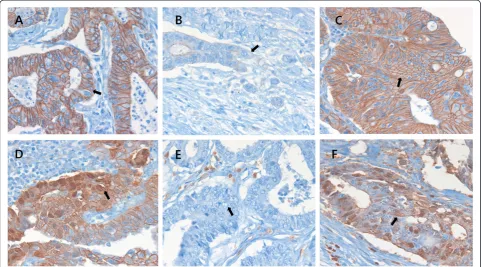
served as a negative control. The slides were then coun-terstained with hematoxylin. The results were assessed by two pathologists who were blinded to the patients’details. E-cadherin immunostaining was evaluated according to a method described previously [17]. E-cadherin staining was classified as grade 0 (preserved) when more than 90% of the CRC cells on a section showed strong membranous staining, or grade 1 (reduced or lost) when less than 90% of the CRC cells showed positive membranous staining. Forβ-catenin, the percentage of tumor cells with nuclear staining was evaluated and scored as grade 0 (0-30%) or grade 1 (30-100%). To evaluate S100A4 expression, stain-ing intensity was recorded as 0 (no stainstain-ing), 1 (weak staining), 2 (intermediate staining), or 3 (strong staining). The proportion of stained cells was expressed as a per-centage. The staining score was obtained by multiplying the intensity and percentage of stained cells and classified
as grade 0 (low expression) and grade 1 (high expression) (Figure 2).
Real time quantitative RT-PCR of EMT related genes Tumor and matched normal tissues were homogenized and total RNA was isolated using an RNeasy Mini kit (Qiagen, Tokyo, Japan) following the manufacturer’s in-structions. All samples were treated with RNase-free DNase (Qiagen). RNA quality control and quantification were car-ried out on an Agilent 2100 Bioanalyzer using a RNA 6000 Pico LabChip (Agilent Technologies, Tokyo, Japan). First-strand complementary DNA was made from total RNA using the Prime Script RT-reagent kit (Amersham Biosci-ences Europe GmbH, Freiburg, Germany) according to the manufacturer’s instructions. To quantify the
expres-sion levels of E-cadherin, β-catenin, N-cadherin, and
S100A4, real-time PCR amplification was performed
with a Rotor Gene 6000 instrument (Corbett Research, Mortlake, Australia). Real-time PCR assays using SYBR Premix EX Taq (TAKARA BIO INC., Otsu, Japan) were carried out in micro-reaction tubes (Corbett Research, Mortlake, Australia). The PCR reaction was performed in a final volume of 10μl, consisting of 5μl of 2x SYBR premix EX Taq buffer, 0.5 μl of each 5’- and 3’- primer
(10 pmol/μl), and 1 μl of sample cDNA. The product
was 10-fold serially diluted from 100 pg/μl to 0.1 pg/μl. Dilution series of PCR products were used to establish standard curves for real-time PCR. Spectral data were cap-tured by using Rotor-Gene Real-Time Analysis Software 6.0 Build 14 (Corbett Research, Mortlake, Australia). All samples were run in triplicate. Glyceraldehyde-3-phosphate dehydrogenase (GAPDH) was used as an endogenous RNA reference gene. Gene expression was
normalized to the expression of GAPDH. Primers and product sizes for E-cadherin, N-cadherin,β-catenin, S100A4, and GAPDH are summarized in Table 1.
Statistical analysis
Differences were compared using Fisher’s exact test or Pearson’s test for qualitative variables and Student’s t-test or analysis of variance for continuous variables. Prognosis was determined by disease-free survival and overall sur-vival. Prognostic factors were examined by univariate and multivariate analyses (Cox proportional hazards model). All statistical tests were two sided, and statistical signifi-cance was accepted at the P < 0.05 level. All analyses were performed using SPSS version 12.0 (SPSS Inc., Chicago, IL, USA).
Figure 2Immunohistochemical staining for E-cadherin,β-catenin, and S100A4 in colorectal cancers (×400).E-cadherin, membranous staining at the center of the tumor (A, arrow) and loss or attenuation of expression at the invasive margin of tumor buds (B, arrow).β-catenin, membranous staining (C, arrow) and nuclear staining at the invasive margin of tumor buds (D, arrow). Negative (E, arrow) and positive (F, arrow) staining for S100A4.
Table 1 Sequences of primers and product size used in real time RT-PCR
Gene Forward Backward size
E-cadherin AGTCACGCTGAATACAGTGG CATTTTCTGGGCAGCTGATG 161
N-cadherin CAGTGCAGTCTTATCGAAGG GAAAGCTTCTCACGGCATAC 158
β-catenin AATGGCAGTGCGTTTAGCTG ATAGCACCTTCAGCACTCTG 233
S100A4 CACAAGTACTCGGGCAAAGA TACACATCATGGCGATGCAG 211
Results
Clinicopathologic parameters related to EMT-related protein expression
Clinicopathologic characteristics of the patients showed in Table 2. The relation between clinicopathological pa-rameters and aberrant expression of E-cadherin,β-catenin and S100A4 in the invasive margin was observed. Loss of E-cadherin was related with cell grade (p = 0.050), macro-scopic type (p = 0.014), perineural invasion (p = 0.037) and high tumor budding (p = 0.010). Nuclearβ-catenin expres-sion was higher in microsatellite stable (MSS) than micro-satellite instable (MSI) tumors (p = 0.004) and left CRCs than in right CRCs (p = 0.037). S100A4 expression was increased in infiltrative growth (p = 0.017), macroscopic type (p = 0.024), T-stage (p = 0.010), nodal stage (p = 0.002), AJCC stage (p = 0.001), ratio of metastatic lymph nodes (p = 0.019), lymphovascular invasion (p = 0.034), and peri-neural invasion (p = 0.050). It was also related with TLI (p = 0.001) (Table 3).
Relation of E-cadherin,β-catenin and S100A4 expression The aberrant expression of EMT-related proteins was higher in the invasive margin than in the tumor center; loss of E-cadherin, 17.9% versus 31.8% (p < 0.0001), nuclear expression of β-catenin, 10.6% versus 12.5% (p < 0.0001), and gain of S100A4, 10.2% versus 15.5% (p < 0.0001), respectively. Aberrant expression of each protein was related to the others: S100A4 versus E-cadherin (r = 0.312, p = 0.050), S100A4 versus β-catenin (r = 0.166, p = 0.004) and E-cadherin versusβ-catenin (r = 0.152, p = 0.009).
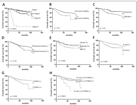
Prognostic factors of disease-free survival and overall survival Disease-free survival was associated with AJCC stage (p < 0.0001, Figure 3A), tumor budding (p = 0.007, Figure 3B), tumor growth type (p = 0.003, Figure 3C), and perineural invasion (p = 0.004, Figure 3D). Aberrant expression of the E-cadherin (p = 0.007, Figure 3E), S100A4 (p = 0.004, Figure 3F), and combination of both proteins in the in-vasive margin were related to disease free survival (p = 0.005, Figure 3G), whereas β-catenin was inversely related (p = 0.032, Figure 3H). Multivariate Cox analysis showed that combination of E-cadherin and S100A4 ex-pressions in the invasive margin and the AJCC stage are independent risk factor for disease-free survival (Table 4). Overall survival was associated with AJCC stage (p < 0.0001, Figure 4A), tumor budding (p = 0.01, Figure 4B), tumor growth type (p = 0.01, Figure 4C), perineural invasion (p = 0.02, Figure 4D), and TLI (p = 0.005, Figure 4E). Ab-errant expression of E-cadherin (p = 0.002, Figure 4F), S100A4 (p = 0.003, Figure 4G), and combination of both proteins in the invasive margin were associated with overall survival time (p = 0.0002, Figure 4H), whereas
β-catenin was not. Cox regression analysis showed that
the combination of E-cadherin and S100A4 in the
Table 2 Clinical and pathological characteristics of 333 patients with colorectal cancers
Characteristics N (%)
Age (years), Mean ± SD (range) 63.6 ± 11.1 (25-86)
Gender
Male 189 (56.8)
Female 144 (43.2)
Tumor Grade
Well 77 (23.1)
Moderately 236 (70.9)
Poorly 11 (3.3)
Undetermined 9 (2.7)
Depth of Invasion
T1 13 (3.9)
T2 43 (12.9)
T3 210 (63.1)
T4 67 (20.1)
Nodal Stages
N0 195 (58.6)
N1(a, b) 86 (25.8)
N2(a, b) 52 (15.6)
AJCC stage
I 44 (13.2)
II 143 (42.9)
III 119 (35.7)
IV 27 (8.1)
Lymphovascular Invasion
Negative 240 (72.1)
Positive 93 (27.9)
Perineural invasion
Negative 306 (91.9)
Positive 27 (8.1)
Lymphocyte infiltration
Mild 112 (33.6)
Moderate 170 (51.1)
Severe 45 (13.5)
Type of Tumor Growth
Expanding 118 (36.3)
Infiltrative 207 (62.2)
Undetermined 8 (2.4)
Tumor budding
Yes 44 (13.2)
Table 3 Relation between clinico-pathological parameters and immunohistochemistry of E-cadherin,β-cadherin, and S100A4 at invasive margin in colorectal cancers (n = 305)
Parameters E-cadherin P β-cadherin P S100A4 P
Strong (n = 208)
Weak or Loss (n = 97)
Nuclear, Low (n = 267)
Nuclear, High (n = 38)
Low (n = 267)
High (n = 267)
Preoperative CEA (ng/ml) 7.0 ± 22.5 6.5 ± 12.4 0.811 6.5 ± 19.8 8.4 ± 18.3 0.573 5.3 ± 13.1 12.5 ± 35.7 0.014
MSI status (n = 166) 0.167 0.004 0.386
MSI-H (n = 8) 3(37.2) 5(62.5) 8(100.0) 0(0.0) 7(87.5) 1(12.5)
MSI-L (n = 6) 3(50.0) 3(50.0) 4(66.7) 2(33.3) 5(83.3) 1(16.7)
MSS (n = 135) 91(67.4) 44(32.6) 107(81.1) 25(18.9) 107(80.5) 26(19.5)
Tumor Location 0.455 0.037 0.704
Right (n = 66) 48(72.7) 18(27.3) *6.5 ± 18.7 60(87.0) 9(13.0)
Left (n = 236) 158(66.9) 78(33.1) *12.2 ± 22.7 194(84.0) 37(16.0)
Tumor Budding 0.011 0.799 0.480
Low (<10) (n = 263) 188(70.9) 77(29.1) 230(87.1) 34(12.9) 224(85.2) 39(14.8)
High (≥10) (n = 40) 20(50.0) 20(50.0) 37(90.2) 4(9.8) 32(80.0) 8(20.0)
Macroscopic Tumor Type 0.616 0.909 0.038
Polypoid (n = 21) 17(77.3) 5(22.7) 18(85.7) 3(14.3) 21(100.0) 0(0.0)
Ulcerofungating (n = 160) 109(66.9) 54(33.1) 143(88.3) 19(11.7) 138(86.3) 22(13.8)
Ulceroinfiltrative (n = 122) 82(68.3) 38(31.7 106(86.9) 16(13.1) 97(79.5) 25(20.5)
Tumor Grade 0.735 0.817 0.577
Well (n = 73) 53(72.6) 20(27.4) 64(86.5) 10(13.5) 60(82.2) 13(17.8)
Moderate or Poor (n = 227) 151(66.5) 73(34.0) 196(87.5) 28(12.5) 191(85.3) 33(14.7)
Ratio of Metastatic Lymph Node 0.11 ± 0.19 0.13 ± 0.21 0.567 0.12 ± 0.20 0.09 ± 0.19 0.317 0.11 ± 0.19 0.18 ± 0.23 0.019
Nodal Stages 0.567 0.288 0.002
N0 (n = 178) 128(70.3) 54(29.7) 155(85.2) 27(14.8) 161(90.4) 17(9.6)
N1 (n = 77) 49(63.6) 28(36.4) 69(92.0) 6(8.0) 57(74.0) 20(26.0)
N2 (n = 48) 31(67.4) 15(32.6) 43(89.6) 5(10.4) 38(79.2) 10(20.8)
Depth of Invasion 0.513 1.000 0.010
T1, T2 (n = 50) 38(73.1) 14(26.9) 44(88.0) 6(12.0) 48(96.0) 2(4.0)
T3, T4 (n = 253) 170(67.2) 83(32.8) 223(87.5) 32(12.5) 208(82.2) 45(17.8)
AJCC stage 0.272 0.669 0.001
I (n = 39) 27(67.5) 13(32.5) 35(87.5) 5(12.5) 37(94.9) 2(5.1)
II (n = 133) 96(71.1) 39(28.9) 114(85.1) 20(14.9) 119(89.5) 14(10.5)
III (n = 110) 74(68.5) 34(31.5) 97(89.8) 11(10.2) 81(73.6) 29(26.4)
IV (n = 21) 11(50.0) 11(50.0) 21(91.3) 2(8.7) 19(90.5) 2(9.5)
Lymphovascular Invasion 0.891 0.341 0.050
Negative 150(68.5) 69(31.5) 195(88.6) 25(11.4) 189(87.1) 28(12.9)
Positive 57(67.1) 28(32.9) 72(84.7) 13(15.3) 66(77.6) 19(22.4)
Neural Invasion 0.037 0.507 0.050
Negative 196(69.8) 85(30.2) 248(87.9) 34(12.1) 240(85.7) 40(14.3)
Positive 11(47.8) 12(52.2) 19(82.6) 4(17.4) 15(68.2) 7(31.8)
Lymphocyte infiltration 0.325 0.821 < 0.0001
Mild 70(72.9) 26(27.1) 88(88.9) 11(11.1) 70(71.4) 28(28.6)
Moderate 108(67.1) 53(32.9) 138(86.3) 22(13.8) 144(89.4) 17(10.6)
invasive margin, the AJCC stage and perineural inva-sion were independent risk factors of overall survival time (Table 5).
Association between mRNA expression and IHC findings Transcript levels ofβ-catenin and S100A4 were correlated with IHC findings at the tumor invasive margin;β-catenin (r = 0.369, p = 0.003) and S100A4 (r = 0.504, p < 0.0001). However, there was no relation between the RT-PCR data and the IHC findings for E-cadherin. E-cadherin and N-cadherin showed a weak inverse relation without statistical
significance (r = -0.169, p = 0.084). mRNA levels of N-cadherin were higher in recurrent or mortality cases (p = 0.040 and p = 0.042, respectively).
Discussion
In this study, we demonstrated that the combination of the loss of E-cadherin and gain of S100A4 in the invasive margin of CRC is an independent prognostic factor of a poorer outcome, along with AJCC stage and perineural invasion. Nuclear expression ofβ-catenin was not related to patient survival.
Table 3 Relation between clinico-pathological parameters and immunohistochemistry of E-cadherin,β-cadherin, and S100A4 at invasive margin in colorectal cancers (n = 305)(Continued)
Type of Tumor Growth 0.159 1.000 0.044
Expanding 79(73.1) 29(26.9) 93(87.7) 13(12.3) 96(90.6) 10(9.4)
Infiltrative 125(65.1) 67(34.9) 168(87.0) 25(13.0) 156(81.3) 36(18.8)
Lymph Node Ratio, number of metastatic lymph nodes / number of harvested lymph nodes;CEAcarcinoembryonic antigen serum level, *, percentage of β-catenin nuclear expression at invasive margin.
When tumor cells start to invade and metastasize, ad-hesion molecules undergo alterations. Down-regulation of E-cadherin in CRC is associated with malignant fea-tures. Loss of E-cadherin has been shown to be associated with tumor budding [18] and lymph node metastasis in CRC [19] and to predict disease recurrence and long-term
survival in CRC [8,20,21]. In this study, loss of E-cadherin at the invasive margin of CRCs was associated with high tumor budding, perineural invasion, and a poor prognosis. S100A4 is localized in the nucleus, cytoplasm, and extra-cellular space and has a wide range of biological functions ranging from regulation of angiogenesis to cell survival, Table 4 Cox regression analysis for disease free survival
time in colorectal cancers
Parameters P value Hazard Ratio 95% CI
*EMT, invasive front 0.026 1.60 1.01-2.43
Lymphovascular invasion 0.625 1.15 0.90-4.28
Perineural invasion 0.092 1.96 0.90-4.28
Lymphocyte infiltration 0.987 1.00 0.66-1.52
Tumor growth type 0.306 1.43 0.72-2.85
Tumor budding (≥10/x200HPF) 0.168 1.61 0.82-3.15
AJCC stage 0.000 3.03 2.03-4.52
*EMT included the expressions of E-cadherin and S100A4.
Figure 4Analysis of overall survival time according to histopathologic parameters and EMT related protein expression.AJCC stage (A), tumor budding (B), type of tumor growth (C), Perineural invasion (D), Peritumoral lymphocytic infiltration (TLI) (E), E-cadherin expression (F), S100A4 expression (G), and combination of E-cadherin and S100A4 (H).
Table 5 Cox multivariate analysis for overall survival time in colorectal cancers
Parameters P value Hazard Ratio 95% CI
*EMT, invasive front 0.036 1.54 1.03-2.31
Lymphovascular invasion 0.963 1.01 0.58-1.78
Perineural invasion 0.010 2.58 1.25-5.34
Lymphocyte infiltration 0.696 0.92 0.60-1.41
Tumor growth type 0.119 1.73 0.87-3.44
Tumor budding (≥10/x200HPF) 0.778 0.90 0.60-1.41
AJCC stage 0.001 1.84 1.26-2.68
motility, and invasion [14]. We found that S100A4 expres-sion in the invasive margin was increased in infiltrative tumors, those with a lymph node metastasis, advanced AJCC stage, or lymphovascular- and perineural invasion, which are all parameters representing tumor aggressive-ness. We demonstrated that high expression of S100A4 is associated with recurrence and mortality. These re-sults were consistent to the previous studies [15,22,23], in which S100A4 is related to a poor prognosis. One study reported that S100A4 is overexpressed in cell pop-ulations enriched for stem-like cells, which is associated
with Wnt/APC/β-catenin signaling pathway and S100A4
worked as β-catenin/TCF target gene [24]. Inconsistently other results, S100A4 was also related to lymphocyte infiltration, which protects tumor progression and de-stroys tumor budding. These findings were resulted from that S100A4 expressing fibroblasts, monocytes, macro-phages, T lymphocytes, neutrophilic granulocytes, or endo-thelial cells may be misinterpreted as S100A4 expressing cancer cells [14].
β-catenin plays to maintain cell-to-cell adhesion along with E-cadherin. However, β-catenin also acts as a tran-scription factor in the Wnt signal transduction pathway and Wnt signaling dysfunction leads to the nuclear accu-mulation of ß-catenin [9]. Several studies have reported that nuclear ß-catenin expression in the invasive margin is associated with high tumor budding and poor prognosis [11], whereas other studies did not find such a relationship [9,12,20]. In addition, recent study showed that aberrant
β-catenin expression was related to favorable prognosis [13]. We did not find any association between β-catenin expression and tumor budding or overall survival in our patient cohort. These inconsistencies may result from dif-ferent CRC subtypes, the existence of more than one type of CRC in a study [25], or different evaluation methods [3]. We showed thatβ-catenin nuclear expression was in-creased in MSS tumors compared to MSI tumors and that it was higher in left CRCs than right CRCs. These results are consistent with those of a previous study [26], which
reported that MSI-high,CpG island methylator phenotype
(CIMP)-high, and BRAF mutations, which are characteris-tics of right CRCs, showed an inverse association with
cytoplasmic and nuclear β-catenin expression. MSS and
MSI colorectal cancers are increasingly being recognized as distinct entities with significantly different pathological characteristics, behaviors, and prognoses [27]. MSI is asso-ciated with significantly lower levels of nuclear ß-catenin and impaired EMT than MSS [27]. In agreement with these reports, we found that 44.4% of MSI-H cases versus 72.4% of MSS or MSI-L cases were tumor budding positive. Aberrant expression of E-cadherin,β-catenin, and S100A4 showed parallel patterns each other. These results sug-gested that overexpression of S100A4 inhibits E-cadherin expression andβ-catenin plays a role in driving
S100A4-dependent EMT induction [14]. Although individual change of E-cadherin and S100A4 was related to patients’prognosis in univariate survival analysis, multivariate Cox analysis re-vealed that the combination of E-cadherin loss and S100A4 overexpression was the only prognostic factor in addition to AJCC stage, which is still the most important prognostic factor in CRC. These results highlight the fact that multi-marker phenotypes rather than single protein are needed in IHC biomarker investigations [3].
Because the tumor biology of the invasive margin is different than that of the tumor center, the patterns of EMT protein expression are expected to be different at the two sites. In this study, EMT-related changes in the expression of E-cadherin,β-catenin and S100A4 were more severe in the invasive margin than in the tumor center and EMT changes in the invasive margin, but not the tumor center, had prognostic significance.
Tumor budding is a putative hallmark of CRC cell inva-sion and has previously been shown to be associated with various clinicopathological parameters, including lymph node metastasis, vascular and lymphatic invasion, distant metastasis, local recurrence, and poor outcomes [28]. In this study, high tumor budding was associated with the ra-tio of metastatic lymph nodes, type of tumor growth, and perineural invasion (data not shown). It has also been classified as an additional prognostic factor. However, our results suggest that the combination of E-cadherin and S100A4 expression at the invasive margin of CRC is su-perior to tumor budding for predicting prognosis.
The mRNA levels of E-cadherin,β-catenin, and S100A4
were not found to be associated with histopathological parameters or prognosis in this study. Forβ-catenin and S100A4, mRNA levels reflected that of the encoded pro-tein, but this was not the case for E-cadherin. However, the switch in expression from E-cadherin to N-cadherin and the higher expression of N-N-cadherin in patients with a poor prognosis were demonstrated in this study. Recent study also showed that N-cadherin was highly expressed in type II papillary renal cell carcinomas than type I cancers and type II cancers were related to poor prognosis [29].
In conclusion, our results suggest that aberrant expres-sion of E-cadherin and S100A4 in the invasive margin was well related with clinicopathological parameters and IHC of both proteins is useful marker to predict progno-sis in CRC.
Abbreviations
AJCC:American Joint Committee on Cancer; CRC: Colorectal cancer; EMT: Epithelial-to-mesenchymal transition; GAPDH: Glyceraldehyde-3-phosphate dehydrogenase; IHC: Immunohistochemistry; H&E: Hematoxylin and eosin; MSI: Microsatellite instability; MSS: Microsatellite stable; TLI: Peritumoral lymphocyte infiltration; TMA: Tissue microarray.
Competing interests
Authors’contributions
S-JL contstructed the manuscript. SYC and RS carried out pathologic study. W-JK checked mRNA levels. MJ constructed tissue microarray. T-GL, B-RS, SMY, and SJY were responsible for clinical data. SMP designed and constructed manuscript. All authors read and approved the final manuscript.
Acknowledgements
This study was supported by a grant from the National R&D Program for Cancer Control, Ministry of Health and Welfare, Republic of Korea (1120330).
Author details
1Department of Surgery, Chungbuk National University College of Medicine and Medical Research Institute, Cheongju, Chungbuk 361-763, Republic of Korea.2Department of Pathology, Chungbuk National University College of Medicine and Medical Research Institute, Cheongju, Republic of Korea. 3Department of Urology, Chungbuk National University College of Medicine and Medical Research Institute, Cheongju, Republic of Korea.4Department of Internal Medicine, Chungbuk National University College of Medicine and Medical Research Institute, Cheongju, Republic of Korea.5Department of Laboratory Medicine, Chungbuk National University College of Medicine and Medical Research Institute, Cheongju, Republic of Korea.
Received: 20 February 2013 Accepted: 21 May 2013 Published: 19 June 2013
References
1. Jung KW, Park S, Kong HJ, Won YJ, Lee JY, Seo HG, Lee JS:Cancer statistics in Korea: incidence, mortality, survival, and prevalence in 2009.
Cancer Res Treat2012,44:11–24.
2. Tsai HL, Chu KS, Huang YH, Su YC, Wu JY, Kuo CH, Chen CW, Wang JY: Predictive factors of early relapse in UICC stage I-III colorectal cancer patients after curative resection.J Surg Oncol2009,100:736–743. 3. Zlobec I, Lugli A:Prognostic and predictive factors in colorectal cancer.
J Clin Pathol2008,61:561–569.
4. Zlobec I, Molinari F, Martin V, Mazzucchelli L, Saletti P, Trezzi R, De Dosso S, Vlajnic T, Frattini M, Lugli A:Tumor budding predicts response to anti-EGFR therapies in metastatic colorectal cancer patients.World J Gastroenterol2010,16:4823–4831.
5. Prall F:Tumour budding in colorectal carcinoma.Histopathology2007, 50:151–162.
6. Zlobec I, Lugli A:Epithelial mesenchymal transition and tumor budding in aggressive colorectal cancer: tumor budding as oncotarget.Oncotarget 2010,1:651–661.
7. Kalluri R, Neilson EG:Epithelial-mesenchymal transition and its implications for fibrosis.J Clin Invest2003,112:1776–1784. 8. Elzagheid A, Buhmeida A, Laato M, El-Faitori O, Syrjänen K, Collan Y,
Pyrhönen S:Loss of E-cadherin expression predicts disease recurrence and shorter survival in colorectal carcinoma.APMIS2012,120:539–548. 9. Brabletz T, Jung A, Hermann K, Gu¨nther K, Hohenberger W, Kirchner T:
Nuclear overexpression of the oncoprotein b-catenin in colorectal cancer is localized predominantly at the invasion front.Pathol Res Pract 1998,194:701–704.
10. Sánchez-Tilló E, De Barrios O, Siles L, Cuatrecasas M, Castells A, Postigo A: β-catenin/TCF4 complex induces the epithelial-to-mesenchymal transition (EMT)-activator ZEB1 to regulate tumor invasiveness.Proc Natl Acad Sci U S A2011,108:19204–19209.
11. Baldus SE, Mönig SP, Huxel S, Landsberg S, Hanisch FG, Engelmann K, Schneider PM, Thiele J, Hölscher AH, Dienes HP:MUC1 and nuclear ß-catenin are coexpressed at the invasion front of colorectal carcinomas and are both correlated with tumor prognosis.Clin Cancer Res2004, 10:2790–2796.
12. Horkko TT, Klintrup K, Makinen JM, Napankangas JB, Tuominen HJ, Makela J, Karttunen TJ, Makinen MJ:Budding invasive margin and prognosis in colorectal cancer-no direct association with beta-catenin expression.
Eur J Cancer2006,42:964–971.
13. Wangefjord S, Brändstedt J, Ericson Lindquist K, Nodin B, Jirström K, Eberhard J:Associations of beta-catenin alterations and MSI screening status with expression of key cell cycle regulating proteins and survival from colorectal cancer.Diagn Pathol2013,8:10.
14. Boye K, Maelandsmo GM:S100A4 and metastasis: a small actor playing many roles.Am J Pathol2010,176:528–535.
15. Flatmark K, Pedersen KB, Nesland JM, Rasmussen K, Aamodt G, Mikalsen S-O, Bjørnland K, Fodstad Ø, Mælandsmo GM:Nuclear localization of the metastasis-related protein S100A4 correlates with tumour stage in colorectal cancer.J Pathol2003,200:589–595.
16. Gongoll S, Peters G, Mengel M, Piso P, Klempnauer J, Kreipe H, Von Wasielewski R:Prognostic significance of calcium binding protein S100A4 in colorectal cancer.Gastroenterology2002,123:1478–1484.
17. Asayama Y, Taguchi Ki K, Aishima Si S, Nishi H, Masuda K, Tsuneyoshi M: The mode of tumour progression in combined hepatocellular carcinoma and cholangiocarcinoma: an immunohistochemical analysis of E-cadherin, alpha-catenin and beta-catenin.Liver2002,22:43–50. 18. Zlobec I, Lugli A, Baker K, Roth S, Minoo P, Hayashi S, Terracciano L, Jass JR:
Role of APAF-1, and peritumoral lymphocytic infiltration in tumour budding in colorectal cancer.J Pathol2007,212:260–268.
19. Karamitopoulou E, Zlobec I, Patsouris E, Peros G, Lugli A:Loss of E-cadherin independently predicts the lymph node status in colorectal cancer.
Pathology2011,43:133–137.
20. Roca F, Mauro LV, Morandi A, Bonadeo F, Vaccaro C, Quintana GO, Specterman S, De Kier Joffé EB, Pallotta MG, Puricelli LI, Lastiri J:Prognostic value of E-cadherin, beta-catenin, MMPs (7 and 9) and TIMPs (1 and 2) in patients with colorectal carcinoma.J Surg Oncol2006,93:151–160. 21. Ngan CY, Yamamoto H, Seshimo I, Ezumi K, Terayama M, Hemmi H,
Takemasa I, Ikeda M, Sekimoto M, Monden M:A multivariate analysis of adhesion molecules expression in assessment of colorectal cancer.J Surg Oncol2007,95:652–662.
22. Boye K, Nesland JM, Sandstad B, Mælandsmo GM, Flatmark K:Nuclear S100A4 is a novel prognostic marker in colorectal cancer.Eur J Cancer 2010,46:2919–2925.
23. Kho PS, Jankova L, Fung CL, Chan C, Clarke C, Lin BP, Robertson G, Molloy M, Chapuis PH, Bokey EL, Dent OF, Clarke S:Overexpression of protein S100A4 is independently associated with overall survival in stage C colonic cancer but only in cytoplasm at the advancing tumour front.
Int J Colorectal Dis2012,27:1409–1417.
24. Stein U, Arlt F, Walther W, Smith J, Waldman T, Harris ED, Mertins SD, Heizmann CW, Allard D, Birchmeier W, Schlag PM, Shoemaker RH:The metastasis-associated gene S100A4 is a novel target of beta-catenin/ T-cell factor signaling in colon cancer.Gastroenterology2006, 131:1486–1500.
25. Iacopetta B:Aberrant DNA methylation: have we entered the era of more than one type of colorectal cancer ?Am J Pathol2003,162:1043–1045. 26. Kawasaki T, Nosho K, Ohnishi M, Suemoto Y, Kirkner GJ, Dehari R,
Meyerhardt JA, Fuchs CS, Ogino S:Correlation of beta-catenin localization with cyclooxygenase-2 expression and CpG island methylator phenotype (CIMP) in colorectal cancer.Neoplasia2007,9:569–577. 27. Pino MS, Kikuchi H, Zeng M, Herraiz MT, Sperduti I, Berger D, Park DY,
Iafrate AJ, Zukerberg LR, Chung DC:Epithelial to mesenchymal transition is impaired in colon cancer cells with microsatellite instability.
Gastroenterology2010,138:1406–1417.
28. Wang LM, Kevans D, Mulcahy H, O‘Sullivan J, Fennelly D, Hyland J, O‘Donoghue D, Sheahan K:Tumor budding is a strong and reproducible prognostic marker in T3N0 colorectal cancer.Am J Surg Pathol2009, 33:134–141.
29. Ludwig BC, Bernhard H, Arne S, Heinz-Joachim R, Felix B:N-cadherin is differentially expressed in histological subtypes of papillary renal cell carcinoma.Diagn Pathol2012,7:95.
doi:10.1186/1746-1596-8-99