Elif et al. World Journal of Pharmaceutical and Medical Research
CAN CAC SCORING BE USED AS A MEASURE OF THE EXTENT OF CAD IN
WOMEN?
Dr. Elif Gündoğdu1*, Elif Ergun2, Cansu Öztürk3 and Pınar Nercis Koşar2
Eskişehir Osmangazi University, Department of Radiology, Eskişehir, Turkey. Ankara Training and Research Hospital, Department of Radiology, Ankara, Turkey. Keçiören Training and Research Hospital, Department of Radiology, Ankara, Turkey.
Article Received on 25/02/2018 Article Revised on 18/03/2018 Article Accepted on 08/04/2018
INTRODUCTION
CVD (cardiovascular disease) is the primary cause of death in women. 50% of women will die of CVD compared with 4% of breast cancer.[1] Furthermore, women have a poorer prognosis with acute MI (myocardial infarction) when compared to men. They more often have no symptoms before the initial cardiac event, and nearly 40% of the initial cardiac event is fatal in women.[2] Thus, detection of individuals who carry high-risk of CAD (coronary artery disease) and diagnosis of CAD in its preclinical phase is of crucial importance enabling preventive pharmacotherapy.
Current risk stratification methods such as Framingham risk score fail to identify a sizable proportion of individuals who carry high risk of CAD.[3-6] There are dramatic differences in the prevalence and out come of risk factors in women compared with men which makes risk assessment in women more complicated.[7] In addition, traditional non-invasive testing that is used to detect CAD such as exercise stress testing and thallium sintigraphy are not as effective in women as they are in men.[8] Thus, diagnosis of CAD is often delayed with subsequent under treatment in women.[7] All forms of cardiovascular death in women show a trend toward
increasing fatality rate according to recent statistics of American Heart Association. On the contrary, substantial declination in men has been observed.[9] Therefore, it is clear that alternative ways to detect sub clinical atherosclerosis are needed and especially for women. CAC (coronary artery Ca) scoring is a promising method in this regard.
Calcification in the coronary arteries is directly related to atherosclerosis, which makes CAC a specific marker of CAD. Numerous studies have shown that CAC scoring gives prognostic information about future cardiac events and can predict the likelihood of significant stenosis of the coronary arteries. In addition it has an independent and incremental value over the traditional risk stratification methods in detecting individuals who carry risk of coronary heart disease.[10] However, several characteristics of the population such as gender, age and ethnicity have an influence on the sensitivity and specificity of CAC scoring in detecting sub clinical atherosclerosis.[11] Various studies investigated the value of CAC scoring in predicting future cardiac events as well as its correlation with the angiographic coronary artery disease in western populations however caution is
ISSN 2455-3301
WJPMR
AND MEDICAL RESEARCH
www.wjpmr.com*Corresponding Author: Dr. Elif Gündoğdu
Eskişehir Osmangazi University, Department of Radiology, Eskişehir, Turkey.
ABSTRACT
Background: To investigate the correlation between CAC (coronary artery Ca) score and the presence and extent of CAD (coronary artery disease) determined by 64-slice CT coronary angiography in a population of 568 Turkish women. Materials and Methods: 568 women, who underwent CAC scanning and coronary CTA, were included in the study. CT examinations were performed by a 64-slice CT scanner (Toshiba, Aquillon 64, Toshiba Medical Systems, Otowara, Japan.) Sensitivity and specificity of CAC scanning in detecting any degree of CAD (grade1-4) and significant CAD (grade 3-4) were calculated for various thresholds of CAC (0,100,400) for the whole study population as well as for various age groups. Correlation between age and CAC score grade, age and CAD grade and CAC score grade and CAD grade were evaluated by Spearmen test. Results: Sensitivity of CAC scoring in detecting CAD increased and the specificity decreased as the age increased. There was a statistically significant correlation between age and CAC score, between age and CAD grade, and CAC score and CAD grade.
Conclusion: CAC correlates well with the presence and extent of CAD and can be used as marker of the disease in women. However caution is required when using this test in young women.
warranted when using their data in other ethnic groups.[11]
In the present study, we aimed to investigate the correlation between CAC score and the presence and extent of CAD determined by 64-slice coronary CTA (computerized tomography angiography) in a population of 568 Turkish women. In addition, we searched for the frequency and extent of CAD in women with zero score.
MATERIALS AND METHODS
568 women, who underwent CAC scanning and coronary CTA were included in the study. They were referred to coronary CTA due to having symptoms suspected of CAD or having several risk factors of CAD. Cases with known CAD or with a prior history of coronary revascularization (by-pass graft and/ or stent placement) were excluded. Following a fasting period of 6 hours CT examinations were performed by a 64-slice CT scanner (Toshiba, Aquillon 64, Toshiba Medical Systems, Otowara, Japan.) Prior to CT scanning heart rate and arterial tension, weight and height of the patient were checked and noted by a nurse, which also filled a questionnaire that includes information about the patient’s medical history and risk factors of CAD. The characteristics of the cases are presented in Table 1.
Table 1: Patient characteristics and distribution of risk factors in the study population.
Age Range 31-86 (mean:58)
Asymptomatic 257 (45.2%) Chest pain 256 (45.1%) Palpitation 46 (8.1%) Dyspne 9 (1.5%) Family history of coronary
artery disease 35 (6.2%) Hypertension 312 (54.9%) Hypercholesterolemia 153 (26.9%) Diabetes Mellitus 106 (18.7%) Smoking 89 (15.7%)
Patients with a heart rate >65 were premedicated with oral beta blocker (Propronalol 40mg) one hour before the scan if there was no contraindication. Sublingual nitrogliserin was delivered to the patient just before the scan. A total amount of 80-85 ml contrast media with iodine concentration of ≥ 350mg/ml was injected through the right antecubital vein with a flow rate of 5ml/s. 20 ml saline was injected both prior to and following the injection of the contrast media with the same flow rate. Optimal scan timing was determined by automated bolus tracking method by placing the region of interest over the descending aorta and setting the trigger threshold to 180 HU. Scan protocols of CAC scanning and CTA are provided in table 2.
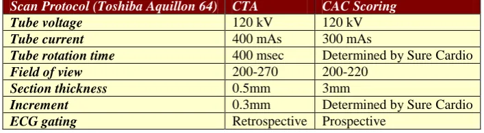
Table 2: CT Protocol of CTA (computerized tomography angiography) and CAC (coronary artery calcium) Scoring.
Scan Protocol (Toshiba Aquillon 64) CTA CAC Scoring
Tube voltage 120 kV 120 kV
Tube current 400 mAs 300 mAs
Tube rotation time 400 msec Determined by Sure Cardio
Field of view 200-270 200-220
Section thickness 0.5mm 3mm
Increment 0.3mm Determined by Sure Cardio
ECG gating Retrospective Prospective
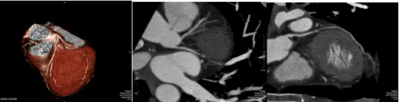
Raw CT data of CTA were reconstructed at various phases of the cardiac cycle to obtain the images without motion artifacts. Images reconstructed at 75% of the R-R interval were optimal for analysis in most of the patients.
CT data was transformed to a remote work station (Vitrea 2, Vital Images, Plymouth, Minnesota) for image analysis.
Ca score of each coronary artery and total score were calculated based on Agatston through a dedicated workstation. Total CAC score was graded using the standardized categories of Ca scoring.
Grade 0: 0 score
Grade I: Total score between 1-10 Grade II: Total score between 11-100 Grade III: Total score between 101-400 Grade V: Total score >400
In coronary CTA, wall irregularities of the coronary arteries, composition and site of the existing plaques and the degree of luminal narrowing caused by the plaque were noted. CAD was graded in to 5 groups according to the CTA findings.[12]
Grade 0: Normal.
Grade 1: Presence of a plaque that causes luminal narrowing <20% or presence of a plaque with a length < 2 cm in one or two coronary arteries.
Grade 2: Luminal narrowing of 20-49% or presence of a plaque with a length > 2cm. If 3 coronary arteries are affected by the atherosclerotic process it is also included in this group.
Grade 3: Luminal narrowing of 50-69% or presence of more than 3 arteries that is affected by the atherosclerotic process.
Cases were categorized into 5 groups according to age (< 40, 40-49, 50-59, 60-69 and > 70). Sensitivity and specificity of CAC scanning in detecting any degree of CAD (grade1-4) and significant CAD (grade 3-4) were calculated for various thresholds of CAC (0,100,400) for the whole study population as well as for each of the 5 age groups. Correlation between age and CAC score grade, age and CAD grade and CAC score grade and CAD grade were evaluated by Spearmen test.
The study was conducted in accordance with the principles of the Declaration of Helsinki.
RESULTS
Sensitivity, specificity, positive and negative predictive values of CAC score >0 in detecting any CAD were 78.4%, 99.3%, 99.08% and 83.1% respectively. When detecting grade 3-4 CAD, sensitivity and negative predictive value increased to 87.9% and 96.3% respectively, while specificity decreased to 73.10%. Sensitivity of CAC scoring in detecting CAD decreased as the CAC score increased while specificity increased with the increasing scores (Table 3). When we assessed the value of CAC in various age groups regardless of the threshold chosen, sensitivity of CAC scoring in detecting CAD increased and the specificity decreased as the age increased (Table 4-5).
Table 3: Detection of significant CAD (coronary artery disease) at various thresholds of CAC (coronary artery calcium) scores.
CAC Score Sensitivity Specificity Positive Predictivity Negative Predictivity
> 0 87,9 % 73,1% 43,1% 96,3%
> 100 52,3% 95,2% 71,8% 89,6%
> 400 16,8% 99,1% 81,8% 83,7%
Table 4: Detection of any CAD (coronary artery disease) with CAC (coronary artery calcium) score > 0 in various age groups.
Age Sensitivity Specificity Positive Predictivity Negative Predictivity
< 40 50% 100% 100% 93,75%
40 – 49 65,2% 98,8% 93,8% 91,1%
50 – 59 67,1% 100% 100% 82,3%
60 – 69 81,3% 100% 100% 75,9%
>69 92,5% 93,8% 98,4% 75%
Table 5: Detection of significant CAD(coronary artery disease) with CAC (coronary artery calcium) score > 0 in various age groups.
Age Sensitivity Specificity Positive Predictivity Negative Predictivity
40 – 49 71,4% 88,9% 31,3% 97,8%
50 – 59 80,8% 81,9% 41,2% 96,5%
60 – 69 90,7% 62,2% 44,8% 95,2%
>69 93,3% 34% 44,4% 90%
*Patients younger than 40 years of age are not presented in the table since they did not have significant CAD
Of the 568 cases 350 (61.6%) had 0 score. 246 (70%) of them were younger than 60 years of age. 59 (16.8%) of the cases without CAC had CAD (grade 1-4) while
significant CAD was detected in 13 (3.7%) of them (figure 1).
There was a statistically significant correlation between age and Ca score (r:0. 47, p<0.0001). The median CAC score of the cases younger than 60 years of age (median:0, min:0, max: 619) was found to be significantly lower than the median CAC score of the cases over 60 years of age (median: 10, min:0, max: 4953) with Mann Whitney U test (p<0.0001). Statistically significant correlation was found between age and CAD grade (r:0. 414 p<0.0001) and CAC score and CAD grade (r:0.809, p <0.0001).
DISCUSSION
Detecting CAD in its preclinical phase before the onset of symptoms is important because a significant proportion of the first CAD events are sudden cardiac death or MI.[13] Women more often than men have no symptoms before the initial cardiac event. Further more the recurrence rate of MI is higher in women and revascularization procedures have a less favorable outcome.[2] However, traditional non-invasive testing that is used to detect CAD is not as effective in women as it is in men. For instance Kwok et al.[14] showed a lower accuracy of exercise stress testing in women, artifacts caused by breast attenuation is a well known limitation of thallium scintigraphy.[8] In addition, various studies have shown that classical risk stratification models underestimate CAD risk in women and according to these models, substantial number of women with sub clinical atherosclerosis do not meet the criteria for primary prevention therapy.[1,15-17] These limitations have generated increasing interest in non-invasive measures of atherosclerosis including CAC scoring.
High correlation between CAC and total plaque burden has been shown by histopathologic studies and this correlation confines all ages and both sexes.[18,19] Coronary calcification occurs in proportion to the severity of underlying atherosclerosis.[20] Therefore, CAC score correlates with the severity of CAD and is useful in predicting significant stenosis including angiographic 3-vessel disease and/or LM disease.[21] In consistent with previous reports a significant correlation between CAC score and CAD was found in the present study. We believe CAC scoring is valuable in identifying women with CAD and is a measure of the extent of the disease.
Various authors advocate that while interpreting CAC testing clinical and demographic features of the patient population should be taken in to account.[21,22] The results of the present study indicate that sensitivity of CAC scoring in detecting CAD decreases with decreasing age. Agatston et al.[23] also showed a distinct influence of age on the relation between CAC and CAD, and stated that sensitivity improves with age while the specificity decreases. A similar observation of decreasing sensitivity of CAC in young women was made by Devries et al[24] and Eroğlu et al.[17] There fore caution is necessary when using CAC testing in young women. Although to detect the exact threshold of age when CAC testing can be used
safely is beyond the aim of our study, we believe this fact needs to be elucidated by further studies.
Even though the total CAC score is a measure of overall plaque burden consisting of both the calcific and the calcific plaque, CAC scanning fails to detect the non-calcified plaque. Numerous studies have shown that CAD may still exist in patients without CAC. The frequency of obstructive CAD with zero CAC score ranges from 0.7-32% in these studies.[17,25-28] In the present study, 16.8% of the cases with 0 score CAD (CAD1-4) while significant CAD existed in 3.7%. Thus, absence of detectable CAD did not rule out CAD. There is a debate on the clinical usefulness of detection of non-calcified plaque in patients with zero score. Some authors support that there is a modest incremental value of other diagnostic tests in patients with zero CAC score since very low cardiac event rates are reported in them and detection of non-calcified plaque in those cases may not significantly increase their low base line prognostic risk estimate.[25,27,29,30] On the other hand, in the series of Schenker et al.[22] which consists of symptomatic patient population, inducible ischemia occurred in 16% of patients with zero CAC score and cardiac event occurred in 3.9% in the 1.4 years of follow up. Additionally in the series of Greenland et al.[31] that consists of asymptomatic patients, cardiac event occurred in 4.4% of patients without CAC. Besides, it is well known that soft plaques are more susceptible to cardiac events, and they are more prone to regression by medical therapy.[26]
CONCLUSION
The results of the present study have shown that CAC correlates well with the presence and extent of CAD and can be used as marker of the disease in women. This is an important observation given the fact that other non-invasive tests, which are used to detect sub clinical atherosclerosis, do not perform well in women and risk stratification methods fail to identify a significant proportion of women who actually need preventive pharmacotherapy. In the present study sensitivity of CAC in detecting CAD decreased in young women hence we recommend caution when using this test in young women. We would also like to point out that further testing of symptomatic patients with zero CAC score is still required since CAD cannot be ruled out.
CONFLICT OF INTEREST
No conflict of interest declared
REFERENCES
1. Michos ED, Nasir K, Braunstein JB, et al. Framingham risk equation underestimates subclinical atherosclerosis risk in asymptomatic women. Atherosclerosis, 2006; 184(1): 201-6. 2. Nasir K, Raggi P, Rumberger JA, et al., Electron
beam tomography in women. Is it a valuable test? Cardiol Rev., 2005; 13(4): 174-83.
3. Akosah KO, Schaper A, Cogbill C, et al. Preventing myocardial infarction in the young adult in the first place: how do the National Cholesterol Education Panel III guidelines perform? J Am Coll Cardiol, 2003; 41: 1475–9.
4. Hecht HS, Superko R. Electron beam tomography and national cholesterol education program guidelines in asymptomatic women.J Am Coll Cardiol, 2001; 37: 1506 –11.
5. Ridker PM, Rifai N, Rose L, et al. Comparison of C-reactive protein and low-density lipoprotein cholesterol levels in the prediction of first cardiovascular events. N Engl J Med., 2002; 14: 1557-65.
6. Ridker PM, Buring JE, Shih J, et al., Prospective study of C-reactive protein and the risk of future cardiovascular events among apparently healthy women. Circulation, 1998; 98: 731-3.
7. Raggi P, Shaw LJ, Berman DS, et al. Gender-based differences in the prognostic value of coronary calcification. J Womens Health (Larchmt), 2004; 13: 273-83.
8. Budoff MJ, Shokooh S, Shavelle RM, et al. Electron beam tomography and angiography: sex differences. Am Heart J., 2002; 143: 877-82.
9. Mosca L, Banka CL, Benjamin EJ, et al. Evidence-based guidelines for cardiovascular disease prevention in women: 2007 update. J Am Coll Cardiol, 2007; 20: 1230-50.
10. Raggi P, Gongora MC, Gopal A, et al., Coronary artery calcium to predict all-cause mortality in
elderly men and women. J Am Coll Cardiol, 2008; 1: 17-23.
11. Oudkerk M, Stillman AE, Halliburton SS, et al. Coronary artery calcium screening: current status and recommendations from the European Society of Cardiac Radiology and North American Society for Cardiovascular Imaging. Eur Radiol, 2008; 18: 2785-807.
12. Kelly JL, Thickman D, Abramson SD, et al. Coronary CT angiography findings in patients without coronary calcification. AJR Am J Roentgenol, 2008; 191: 50-5.
13. O'Rourke RA, Brundage BH, Froelicher VF, et al. American College of Cardiology/American Heart Association Expert Consensus document on electron-beam computed tomography for the diagnosis and prognosis of coronary artery disease. Circulation, 2000; 4: 126-40.
14. Kwok Y, Kim C, Grady D, et al. Meta-analysis of exercise testing to detect coronary artery disease in women. Am J Cardiol, 1999; 1: 660-6.
15. Ridker PM, Rifai N, Rose L, et al. Comparison of C-Reactive Protein and Low-Density Lipoprotein Cholesterol Levels in the Prediction of First Cardiovascular Events N Engl J Med, 2002; 347: 1557-64.
16. Nasir K, Michos ED, Rumberger JA, et al. Coronary artery calcification and family history of premature coronary heart disease: sibling history is more strongly associated than parental history. Circulation, 2004; 12: 2150-6.
17. Eroğlu E, Bayrak F, Gemici G, et al. Prevalence of coronary artery disease in low to moderate-risk asymptomatic women: a multislice computed tomography study Arch Turk Soc Cardiol, 2008; 36: 439-445.
18. Sangiorgi G, Rumberger JA, Severson A, et al. Arterial calcification and not lumen stenosis is highly correlated with atherosclerotic plaque burden in humans: a histologic study of 723 coronary artery segments using nondecalcifying methodology.J Am Coll Cardiol, 1998; 31: 126-33.
19. Janowitz WR, Agatston AS, Kaplan G, et al. Differences in prevalence and extent of coronary artery calcium detected by ultrafast computed tomography in asymptomatic men and women. Am J Cardiol, 1993; 1: 247-54.
20. Arad Y, Goodman KJ, Roth M, et al. Coronary calcification, coronary disease risk factors, C-reactive protein, and atherosclerotic cardiovascular disease events: the St. Francis Heart Study.J Am Coll Cardiol, 2005; 46: 158-65.
21. Budoff MJ, Diamond GA, Raggi P, et al. Continuous probabilistic prediction of angiographically significant coronary artery disease using electron beam tomography. Circulation, 2002; 16: 1791-6.
intermediate likelihood of coronary artery disease: a combined positron emission tomography/computed tomography study. Circulation, 2008; 1: 1693-700. 23. Agatston AS, Janowitz WR, Hildner FJ, et al.
Quantification of coronary artery calcium using ultrafast computed tomography. J Am Coll Cardiol, 1990; 15: 827–832.
24. Devries S, Wolfkiel C, Fusman B, et al. Influence of age and gender on the presence of coronary calcium detected by ultrafast computed tomography. J Am Coll Cardiol, 1995; 25: 76-82.
25. Herzog C, Britten M, Balzer JO, et al. Multidetector-row cardiac CT: diagnostic value of calcium scoring and CT coronary angiography in patients with symptomatic, but atypical, chest pain. Eur Radiol, 2004; 14: 169-77.
26. Henneman MM, Schuijf JD, Pundziute G, et al. Noninvasive evaluation with multislice computed tomography in suspected acute coronary syndrome: plaque morphology on multislice computed tomography versus coronary calcium score. J Am Coll Cardiol, 2008; 15: 216-22.
27. Venkatesh V, Ellins ML, Yang S, et al. Incremental detection of coronary artery disease by assessment of non-calcified plaque on coronary CT angiography. Clin Radiol, 2009; 64: 250-5.
28. Rubinshtein R, Gaspar T, Halon DA, et al. Prevalence and extent of obstructive coronary artery disease in patients with zero or low calcium score undergoing 64-slice cardiac multidetector computed tomography for evaluation of a chest pain syndrome. Am J Cardiol, 2007; 99: 472-5.
29. HausleiterJ, Meyer T, Hadamitzky M, et al. Prevelance of non calcified coronary plaques by 64-slice computed tomography in patients with an intermediate risk for significant coronary artery disease. J Am Coll Cardiol, 2006; 48: 312-18. 30. Sarwar A, Shaw JL, Shapiro MD, et al. Diagnostic
and Prognostic Value of Absence of Coronary Artery Calcification. J Am Coll Cardiol Img, 2009; 2: 675-688.