O R I G I N A L C O N T R I B U T I O N
Open Access
Aqueous garlic extract improves renal
clearance via vasodilatory/antioxidant
mechanisms and mitigated proteinuria via
stabilization of glomerular filtration barrier
Christian Eseigbe Imafidon
1,2*, Rufus Ojo Akomolafe
2, Omotayo Alaba Eluwole
3, Isiaka Ayofe Adekunle
2and
Ruby Adebusola Agbaje
4Abstract
Background:Lead (Pb) remains an apparently indispensable material in several industrial processes. It is a potent environmental toxin with associated deleterious biological effects. The study investigated the effects of aqueous garlic extract (AGE) on renal clearance and proteinuria in Wistar rats with Pb-induced kidney injury.
Methods:Thirty male Wistar rats were divided into six groups of five rats each such that exposure to Pb (35 mg/kg i.p) for 10 consecutive days was either followed by 30 days recovery period (without treatment) or 30 days post-treatment with oral graded doses of AGE at 100, 200 and 400 mg/kg while comparisons where made against a control (2 ml/kg NORMAL SALINE) atp< 0.05. The phytochemical constituents of the extract were determined using conventional standard protocols before administration to the rats.
Results:Pb toxicity induced deleterious alterations of renal function biomarkers (creatinine, urea and total protein) in the plasma and urine, indicators of oxidative stress and lipid peroxidation (GSH, SOD, CAT and TBARS) in the kidney tissues as well as significantly lowered plasma and kidney NO level (p< 0.05) of the rats. It also significantly lowered creatinine clearance and fractional excretion of urea while urine total protein-creatinine ratio was significantly increased in the rats. Kidney histology showed evidence of Pb-induced glomerular atrophy with tubular and interstitial vacuolation. However, AGE administration was associated with significant normalization of the aforementioned biochemical parameters (p< 0.05) as well as kidney histoarchitectural improvement. The pharmacological effects of AGE were attributed to its determined phytochemical constituents.
Conclusion:AGE normalized renal clearance through vasodilatory and antioxidant mechanisms with associated mitigation of proteinuria through stabilization of glomerular filtration barrier.
Keywords:Heavy metal, Lead, Garlic, Phytochemicals, Renal clearance, Proteinuria
Background
Kidney, the principal organ of homeostasis, performs both excretory and regulatory functions [1, 2]. Renal dysfunction results in bioaccumulation of metabolic wastes which could be terminal (resulting in death), if
left unchecked [3]. While some forms of kidney
disorder are idiopathic, others have been associated with known causes namely genetic, lifestyle or chemically-induced (toxicological) [4–6]. Heavy metal toxicity, of which lead (Pb) toxicity is important, plays a significant role in the pathogenesis of kidney disorder in many population [5, 7, 8].
Lead (Pb), occurring in both organic and inorganic forms in the environment, is a toxic heavy metal of environmental and occupational concern [9,10]. Due to its unique inherent qualities namely low melting point, resistant to corrosion, malleability, ductility and softness,
© The Author(s). 2019Open AccessThis article is distributed under the terms of the Creative Commons Attribution 4.0 International License (http://creativecommons.org/licenses/by/4.0/), which permits unrestricted use, distribution, and reproduction in any medium, provided you give appropriate credit to the original author(s) and the source, provide a link to the Creative Commons license, and indicate if changes were made.
* Correspondence:staywithchris@gmail.com
1
Renal Research Laboratory, Department of Physiology, Faculty of Basic Medical and Health Sciences, Bowen University Iwo, Iwo, Osun State, Nigeria
2Department of Physiological Sciences, Faculty of Basic Medical Sciences,
Pb has found its relevance in several industrial processes such as in the production of paints, automobiles, water pipes, ceramics as well as electric storage batteries [9,
11]. Nevertheless, it is a non-biodegradable toxicant
which when absorbed into the body (either by inhalation or ingestion) has an estimated biological half-life of about 10 years, thus enhancing bioaccumulation [10,12]. Acting through disturbance of the antioxidant system, Pb-induced oxidative stress is known to occur via any of the following two pathways in order to cause toxic effects; generation of reactive oxygen species (ROS) and depletion of antioxidant reserves through the generation of ROS [13, 14]. According to literature, Pb toxicity is associated with deleterious biological effects including hemopoietic [15], nervous [12], reproductive [9], car-diovascular [16], gastrointestinal [10], hepatic [17] and kidney [18,19] dysfunctions. Since the basic mechanism of Pb-induced toxicity is disruption of the antioxidant system, therapeutic interventions are usually geared towards application of chelating agents namely dimer-caprol (BAL), Calcium Disodium EDTA (CaNa2EDTA)
and succimer (2, 3–meso-dimercaptosuccinic acid or
DMSA). These chelating agents are expensive, not readily available and sometimes burdened with un-desirable side effects [9, 20]. This study aimed at ex-ploring the therapeutic potential of a local (easily accessible) medicinal plant in an experimental model of Pb-induced kidney injury.
It was reported by WHO that, in health care aids, me-dicinal plants are used among 80% of the world’s popula-tion either as the plant extract or in the form of their active components due to their health beneficial effects [21]. Besides forming the basis for almost all medicinal therapy, herbs are contained in about 40% of prescrip-tions and interest for herbal remedies instead of syn-thetic drugs is on the increase due to their associated relatively lesser side effects [22,23].
Garlic (Allium sativum) is a common and readily access-ible plant that is cultivated both sexually and asexually [24]. It is a nutritional plant that is used as spice in food and has vast medicinal properties [8, 9]. Some of its pharmaco-logical activities namely anti-oxidant, anti-inflammatory, anti-biotic and anti-thrombotic effects have been experimen-tally demonstrated in conditions such as prostate cancer, car-diovascular disease, stroke, reproductive toxicity and kidney injury [8,9,25]. However, there is dearth of literature on the effects of garlic extract on the mechanisms of renal clearance and proteinuria in models of Pb-induced kidney injury. This study aims to bridge this gap in knowledge.
Materials and methods Metabolic cages
Metabolic cages were fabricated by Central Techno-logical Laboratory and Workshops (CTLW), Obafemi
Awolowo University (OAU), Ile-Ife, Osun state,
Nigeria [3, 26].
Plant material, lead salt, biochemical kits and chemicals Fresh garlic bulbs where purchased from a commercial supplier at Lagere market, Ile-Ife, Osun State, Nigeria and certified by a Botanist at the Department of Botany, Obafemi Awolowo University (OAU) where a voucher specimen was deposited.
Lead, in the form of lead acetate salt, was procured from Guangzhou Fischer Chemical Co., Ltd. (Guangdong, China). Assay kits for renal function tests were purchased from Randox Laboratories Ltd. (United Kindgdom) while 1, 1-Diphenyl-2-picryl-hydrazy (DPPH) was procured from Sigma Chemicals Co. (USA). All other chemicals used were of analytical grade, available commercially.
Extraction process of aqueous garlic extract (AGE) The garlic bulbs were peeled, rinsed in tap water and thereafter weighed (W1). The weighed bulbs were pulverized in distilled water with the aid of a Waring blender (Waring Commercial, Torrington, CT). With the aid of an electric shaker, the resulting mixture was subjected to constant shaking for 12 h and thereafter filtered using < 2μm pore sieve. The filtrate, without been concentrated with a rotary evaporator, was directly fixed-dried in a lyophilizer at −40 °C (Ilshin Lab. Co. Ltd., Seoul, Republic of Korea). The yield obtained (aqueous garlic extract) was weighed (W2) and there-after kept in a desiccator until when needed. The percentage yield of aqueous garlic extract (AGE) was calculated using the formula below; [3,27,28].
Percentage yield of AGEð Þ ¼%
Yield of extract in gram W2ð Þ
Weight of freshly peeled bulbs in gram W1ð Þ100%
Note:The extraction process of aqueous garlic extract (AGE) was carried out without the application of heat.
Phytochemical screening of AGE
Phytochemical screening of the extract was carried out in accordance with established standard laboratory protocols, described as follows;
The presence of alkaloids were qualitatively
deter-mined by the method of Halilu and co-workers [29]
and quantified according to the method of Harbone
[30]. Flavonoids were qualitatively determined by the
method of Halilu and co-workers [29] and quantified
by the method of Obadoni and Ochuko [31]. The
presence of tannins was determined by the method of
Halilu and co-workers [29] and thereafter quantified
of saponins was determined using Froth test as described by Benmehdi and co-workers [33] and thereafter
quanti-fied by the method of Obadoni and Ochuko [31]. The
presence of phenolics was determined and quantified by the method of Edeoga and co-workers [34]. The presence of terpenoids was qualitatively determined by the method of Benmehdi and co-workers [33]. Qualitative analysis of cardiac glycoside was by Keller-Kiliani test as described by Anjali and Sheetal [35] while the quantitative analysis was by the method of Harbone [30].
Determination of total flavonoid and total phenolic contents of AGE
Total phenolic content of AGE was determined by
the method of Singleton and Rossi [36] and as
de-scribed by Gulcin and co-workers [37] using Folin–
Ciocalteu’s phenol reagent which is an oxidizing reagent. 0.2 ml of Folin–Ciocalteu’s phenol reagent was added to a mixture of 0.1 ml of the sample and 0.9 ml of distilled water. The resulting mixture was voltexed. After 5 min of standing, 1.00 ml of 7% (w/w) Na2CO3solution was added and thereafter made up to 2.5 ml with distilled water before the resulting mixture was incubated for 90 min at room temperature. Using an ultraviolet (UV)-Vis spectro-photometer (Labtronics, India; Model LT-290), the absorbance was read at a wavelength of 750 nm against a negative control which contained 1 ml of distilled water. The gallic acid equivalent (GAE) of AGE was determined using gallic acid at 0.1 mg/ml as a standard, after preparing a calibration curve.
Total flavonoid content of AGE was determined using aluminum chloride colorimetric assay method
according to Zhilen and co-workers [38] and as
de-scribed by Miliauskas and co-workers [39]. Standard
quercetin with varying concentrations 0.1, 0.2, 0.3, 0.4 and 0.5 mg/ml was used as standard in comparison to the AGE sample. 0.4 ml of DW was added to 0.1 ml of AGE/the standard, followed by 0.1 ml of 5% so-dium nitrate solution. After 5 min, 0.1 ml of 10% aluminum chloride, and 0.2 ml of sodium hydroxide solutions were added to the resulting mixture after which the volume was made up to 2.5 ml with distilled water. The absorbance, at a wavelength of 510 nm, was
read against blank using a UV–vis spectrophotometer
(Labtronics, India; Model LT-290).
The aforementioned tests were performed in triplicate, and the final results were expressed as mean ± Standard Error of Mean in mg quercetin/GAE per gram of AGE using the formula below; [37,39].
X¼q Vð =wÞ
X = total content of flavonoid or phenolic compound in quercetin or GAE respectively;
q = concentration of quercetin or gallic acid estab-lished from the standard curve;
V = volume of AGE (ml); and. w = weight of AGE sample.
Radical scavenging activity of AGE
The extract was evaluated for radical scavenging activity
using Table 1, 1-diphenyl-2-picryl-hydrazy (DPPH)
ac-cording to the method of Blois [40]. Butylated hydroxyl anisole (BHA) solutions as well as varied concentrations of the extract (2.5, 5, 10, and 20 g/ml) were taken into different test tubes. Four millilitres (4 ml) of 0.1 mM of DPPH was added to the test tubes, voltexed and allowed to stand at 27 °C for 20 min. Absorbance of the samples was measured at 517 nm against the absorbance of the control and their radical scavenging activities were calculated as follows; [40].
Radical scavenging activityð Þ ¼% 1‐Asample Acontrol 100
Determination of acute oral lethal dose (LD50) of AGE Acute oral lethal dose (LD50) of AGE was determined by
the method of Lorke [41] as modified by Imafidon and
co-workers [27]. The modification of Lorke’s procedure was the use of 2 rats per group in the second phase of the study; Lorke proposed the use of 1 rat per group at the second phase of LD50determination.
A total of 17 adult Wistar rats were used for the
determination of LD50. In the first phase of the
Table 1Experimental protocol and dose regimen
N= 30 Group description 10 days intraperitoneal administration 30 days oral administration 30 days recovery
Group 1 (n= 5) Control NS (2 ml/kg) NS (2 ml/kg) NSa
Group 2 (n = 5) Pb Pb (35 mg/kg)a _ _
Group 3 (n = 5) Pb + Recovery Pb (35 mg/kg) _ RPa
Group 4 (n = 5) Pb + 100 mg/kg AGE Pb (35 mg/kg) 100 mg/kg AGEa _ Group 5 (n = 5) Pb + 200 mg/kg AGE Pb (35 mg/kg) 200 mg/kg AGEa _ Group 6 (n = 5) Pb + 400 mg/kg AGE Pb (35 mg/kg) 400 mg/kg AGEa _
experiment, 9 rats were divided into 3 groups of 3 rats each and thereafter received graded doses of AGE at 10 mg/kg, 100 mg/kg and 1000 mg/kg orally. The rats were observed for 24 h after which the first phase of the study was terminated.
In the second phase, 8 rats were divided into 4 groups of 2 rats each after which they received graded doses of AGE at 750 mg/kg, 1500 mg/kg, 3000 mg/kg and 6000 mg/kg orally. They were also observed for 24 h after which the second phase of the study was terminated.
Thereafter, the oral LD50 of AGE was determined using
the formula below;
LD50¼√axb
Where a = least dose that killed a rat; b = highest dose that did not kill a rat.
Preparation of stock solution of Pb and AGE
Lead (Pb), in the form of lead acetate [Pb (CH3COO)2], was used for the study. It was ensured that each 100 g rat received 0.2 ml of any of the solutions in order to avoid fluid overload.
The stock solution of Pb was prepared by dissolving 35 mg of lead acetate in 20 ml of normal saline so that each 100 g rat received intraperitoneal injection of 0.2 ml of Pb from the stock solution (35 mg/kg of Pb) for 10 consecutive days.
The stock solution for 100 mg/kg of AGE was pre-pared by dissolving 1 g of AGE in 20 ml of normal saline. The stock solutions for 200 mg/kg and 400 mg/kg of AGE were prepared by dissolving 2 g and 4 g of AGE each in 20 ml of normal saline respectively. The stock solutions for AGE were refrigerated after use while fresh samples were prepared every 48 h throughout the period of study.
Animal management and experimental protocol
The experimental protocols were in strict compliance with the guidelines for animal research, as detailed in NIH Guidelines for the Care and use of Laboratory Animals [42] and approved by local Institutional Research Committee.
Thirty male Wistar rats (aged 8–9 weeks old)
weigh-ing 120 – 150 g were used for this study. They were
purchased from the Animal Holdings Unit of the College of Health Sciences, OAU, Ile-Ife where the study was carried out. Each rat was housed in a separate metabolic cage and was allowed access to standard rat chow (ACE Feeds Plc, Osogbo, Nigeria) and water ad libitum. The rats were allowed to acclimatize to life in the metabolic cages for two weeks before the study commenced. The rats were
thereafter divided into six groups of five rats each as follows; Group 1 received normal saline (2 ml/kg) throughout the study period, after which they were sacrificed under ketamine anesthesia (65 mg/kg i.m). Group 2 received 35 mg/kg of Pb (i.p) for 10 con-secutive days after which they were euthanized under ketamine anesthesia. Group 3 were pretreated as group 2 and thereafter left for a recovery period of 30 days (without treatment) before they were also euthanized. Groups 4, 5 and 6 were pretreated as group 2 and thereafter received graded doses of AGE at 100, 200 and 400 mg/kg respectively for 30 days
before they were also euthanized (Table 1). Before the
rats were euthanized, their 24 h urine samples were collected inside the metabolic cages. Blood sample from each rat was collected by cardiac puncture (under ketamine anesthesia) into separate lithium heparinized bottles and centrifuged at 4000 rpm for 15 min using a cold centrifuge (Centrium Scientific,
model 8881) at −4 °C. The plasma obtained was
decanted into separate plain bottles using separate sterile syringes. The left kidney of each rat was excised and kept in a cooler for the preparation of homo-genates while their right kidneys were fixed in 10% formal-saline solution for histological examination.
Biochemical assays
Assessment of kidney function biomarkers, total protein concentration and plasma nitric oxide level
The biomarkers of kidney function namely creatinine and urea were assayed using Randox standard laboratory
kits (Randox, UK) according to the manufacturer’s
in-structions. However, creatinine clearance was calculated using the formula below;
Creatinine clearance mlð =minÞ ¼UcV Pc
Where Uc = urine creatinine concentration; V = urine flow rate = volume of urine/time; and. Pc = plasma creatinine concentration.
The concentration of total protein was determined by
the method of Lowry and co-workers [43] while nitric
oxide level was determined by the method of Grisham and co-workers [44].
Note: For the conversion of S.I. units from dl/ml to mg/ ml in the determination of urine total protein-creatinine ratio, the following system of conversion was used; [26].
1 dl¼100 g
FEureað Þ ¼% UureaPCr PureaUCr
100
Where Uurea= urea concentration in the urine; Purea=
urea concentration in the plasma; UCr= creatinine
concentration in the urine; and PCr= creatinine concen-tration in the plasma.
Assessment of oxidative stress status and lipid peroxidation
With the aid of an electric homogenizer (SI601001), 10% homogenate in phosphate buffer (100 mM) was prepared with the left kidney tissue at a pH of 7.4. Thereafter, the homogenate was centrifuged at 3000 rpm for 20 min and the supernatant was collected for the assessment of the following indices of oxidative stress;
Reduced glutathione (GSH) level was determined by the method of Beutler and co-workers [46], the activity of superoxide dismutase was determined by the method
of McCord and Fridovich [47], catalase (CAT) activity
was by the method of Sinha [48] while the level of
thiobartiburic acid reactive substances (TBARS) was determined according to the method of Ohkawa and co-workers [49].
Histological examination
The right kidney of each rat was fixed in 10% formal-sa-line solution. Each kidney was thereafter dehydrated in graded alcohol and embedded in paraffin wax. Sections
of about 7–8μm thick were subjected to Haematoxylin
and Eosin (H & E) staining technique for photographic assessment using Leica DM 750 camera micro-scope at × 400 magnification.
Statistical analysis
Data were analyzed by one-way analysis of variance and expressed as mean ± Standard Error of Mean. Thereafter, they were subjected to Newman Keul’s post-hoc test and the level of significance was set at p< 0.05. Differences between two variables was assessed using student’s t-test. The statistical analysis was carried out using graph pad prism 5.03 (Graph Pad Software Inc., CA, USA).
Results
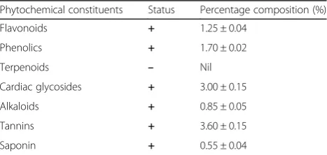
Percentage yield and phytochemical screening of AGE The percentage yield of AGE was determined to be
69.47 ± 0.51% (n= 3) (Table 2) while phytochemical
screening detected the presence of flavonoids, phenolics, cardiac glycosides, alkaloids, tannins and saponin in the extract (Table3).
Total flavonoid content, total phenolic content, radical scavenging activity and oral lethal dose of AGE
The extract was observed to have high amount of total flavonoid and phenolic contents in gram per milligram of their respective standards (Table 4) while the radical scavenging capacity was observed to increase with
in-creasing doses (Fig. 1). Oral lethal dose (LD50) of
AGE was determined to be greater than 6000 mg/kg (Table 5).
Effects of AGE on plasma and urine concentrations of creatinine, urea and total protein of Wistar rats with Pb-induced kidney injury
Pb administration was associated with a significant in-crease in the plasma creatinine concentration (mg/dl) of group 2 when compared with group 1 (p< 0.05). That of the AGE-treated groups 4, 5 and 6 were significantly lower when compared with groups 2 and 3, with no significant difference shown when compared with group 1. The urine creatinine levels of groups 2 and 3 were significantly lower than that of group 1. Groups 4, 5 and 6 showed a signifi-cantly higher level of urine creatinine when compared with group 1 (p< 0.05) (Table6).
There was a significant increase in plasma urea level (mg/dl) of groups 2 and 3 when compared with group 1 (p< 0.05). However, that of groups 4, 5 and 6 were not significantly different from group 1 (p> 0.05). On the other hand, the urine urea concentration of groups 2 and 3 were significantly lower than that of group 1 while no Table 2Percentage yield of AGE
Weight of fresh garlic bulbs (g)
Yield of AGE (g)
Percentage yield of AGE (%)
1st extraction process
500.00 342.35 68.47
2nd extraction process
500.00 350.50 70.10
3rd extraction process
500.00 349.25 69.85
The percentage yield of AGE after three separate extraction processes (n= 3) = 69.47 ± 0.51%
Table 3Qualitative and quantitative determination of phytochemicals in AGE
Phytochemical constituents Status Percentage composition (%)
Flavonoids + 1.25 ± 0.04
Phenolics + 1.70 ± 0.02
Terpenoids – Nil
Cardiac glycosides + 3.00 ± 0.15
Alkaloids + 0.85 ± 0.05
Tannins + 3.60 ± 0.15
Saponin + 0.55 ± 0.04
significant difference was recorded in that of AGE-treated groups 5 and 6 when compared with group 1 (Table6).
The plasma total protein concentration [× 10−2(mg/ml)] was significantly lowered in groups 2 and 3 when compared with group 1 (p< 0.05). That of groups 4, 5 and 6 were significantly higher than groups 2 and 3 but showed no significant difference when compared with group 1. On the other hand, the urine total protein concentration of groups 2 and 3 was significantly higher than that of group 1 (p< 0.05). However, the AGE-treated groups 5 and 6 showed no significant difference in urine total protein level when compared with group 1 (Table6).
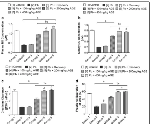
Effects of AGE plasma and kidney nitric oxide (NO) concentration, creatinine clearance and fractional excretion of urea (FEurea) of Wistar rats with Pb-induced kidney injury
The plasma NO level (μM) was significantly lowered in
groups 2 and 3 (0.39 ± 0.03 and 0.42 ± 0.03 respectively) when compared with group 1 (1.14 ± 0.04) (p< 0.05). The AGE-treated groups 4, 5 and 6 (0.94 ± 0.03; 1.12 ± 0.03 and 1.25 ± 0.02 respectively) showed significantly higher plasma NO level when compared with group 2 (0.39 ± 0.03) (p< 0.05) (Fig.2).
The kidney NO level (μM) was significantly lowered in groups 2 and 3 (0.20 ± 0.01 and 0.24 ± 0.01 respectively) when compared with group 1 (0.80 ± 0.03) (p< 0.05). The AGE-treated groups 5 and 6 (0.76 ± 0.02 and 0.75 ± 0.02 respectively) were significantly higher than groups 2 and 3 (0.20 ± 0.01 and 0.24 ± 0.01 respectively) but showed no significant difference when compared with group 1 (0.80 ± 0.03) (p< 0.05) (Fig.2).
Groups 2 and 3 (1.09 ± 0.05 and 1.13 ± 0.06 respec-tively) showed a significantly lowered creatinine
clear-ance [× 10−2 (ml/min)] when compared with group 1
(3.38 ± 0.22) (p< 0.05). However, the AGE-treated
groups 5 and 6 (3.37 ± 0.12 and 3.55 ± 0.13 respectively) showed no significant difference in creatinine clearance Table 4Total Phenolic and total flavonoid content of AGE
Total phenolic content (mg of GAE/g of AGE)
Total flavonoid content (mg of quercetin equivalent/g of AGE)
53.82 ± 1.70 69.20 ± 7.50
Each value represents Mean ± Standard Error of Mean (n= 3);GAEgallic acid equivalent
Fig. 1Radical scavenging activity of aqueous garlic extract (AGE) at different concentrations. Graph showing concentration-dependent radical scavenging activity of AGE (p< 0.0001; F = 650.85). Each value represents mean ± Standard Error of Mean (n= 3); RSA = radical scavenging activity; AGE = aqueous garlic extract; BHA = butylated hydroxyl anisole
Table 5Acute oral toxicity test (LD50) of AGE
Number of rats Dose (mg/kg) Mortality
1st PHASE
3 10 0/3
3 100 0/3
3 1000 0/3
2nd PHASE
2 750 0/2
2 1500 0/2
2 3000 0/2
2 6000 0/2
Using Lorke’s equation, LD50=√axb
a = nil; b = 6000 mg/kg. Therefore, the acute oral lethal dose of AGE is greater than 6000 mg/kg (Oral LD50of AGE > 6000 mg/kg). The adopted doses of AGE
that were used for the main study were less than 10% of the oral LD50of AGE
when compared with group 1 (3.38 ± 0.22) (p> 0.05) but were significantly higher than groups 2 and 3 (1.09 ± 0.05 and 1.13 ± 0.06 respectively) (Fig.2).
The fractional excretion of urea (%) was significantly lowered in groups 2 and 3 (19.62 ± 2.13 and 27.20 ± 1.80 respectively) when compared with group 1 (54.29 ± 1.00) (p< 0.05). Groups 5 and 6 (58.18 ± 1.91 and 59.57 ± 1.39 respectively), however, showed a significantly higher fractional excretion of urea when compared with group 1 (54.29 ± 1.00) (p< 0.05) (Fig.2).
Effects of AGE on indicators of oxidative stress (GSH, SOD, CAT) and lipid peroxidation (TBARS) of Wistar rats with Pb-induced kidney injury
Kidney GSH level (μg/mg protein) was significantly low-ered in groups 2 and 3 when compared with group 1 as well as the AGE-treated groups 4, 5 and 6 (p< 0.05). However, the AGE-treated groups 5 and 6 showed no significant difference in the GSH level when compared with group 1 (Table7).
Although the AGE-treated groups 4 and 5 showed a significantly lowered kidney SOD level (mM/mg protein) when compared with group 1 (p< 0.05), all AGE-treated groups 4, 5 and 6 showed a significantly higher SOD
level when compared with groups 2 and 3 (p< 0.05).
SOD level of groups 2 and 3 was significantly lower than group 1 (p< 0.05) (Table7).
Groups 2 and 3 had a significantly lower kidney CAT
level (μmol/min/mg protein) when compared with group
1 as well as the AGE-treated groups 4, 5 and 6 (p<
0.05). However, the AGE-treated groups 5 and 6 showed no significant difference in CAT level when compared with group 1 (p> 0.05) (Table7).
Kidney TBARS level (nmol/mg protein) was signifi-cantly higher in groups 2 and 3 when compared with group 1 as well as the AGE-treated groups 4, 5 and 6
(p< 0.05). However, the AGE-treated groups 5 and 6
showed no significant difference in TBARS level when compared with group 1 (p> 0.05) (Table7).
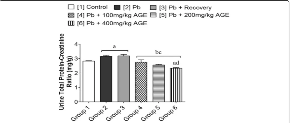
Effects of AGE on urine total protein-creatinine ratio of Wistar rats with Pb-induced kidney injury
Groups 2 and 3 (3.16 ± 0.07 and 3.18 ± 0.11 respectively) showed a significantly higher urine total protein-creatin-ine ratio (mg/g) when compared with group 1 (2.82 ± 0.04) and the AGE-treated groups 4, 5 and 6 (2.75 ± 0.16; 2.55 ± 0.04 and 2.33 ± 0.06 respectively) (p< 0.05). However, that of groups 4 and 5 (2.75 ± 0.16 and 2.55 ± 0.04 respectively) showed no significant difference when compared with group 1 (2.82 ± 0.04) (p> 0.05) while the AGE-treated group 6 (2.33 ± 0.06) had a significantly lower urine total protein-creatinine ratio when com-pared with group 1 (2.82 ± 0.04) (p< 0.05) (Fig.3).
Histological effects of AGE on the kidney of Wistar rats with Pb-induced kidney injury
Pb administration was associated with histoarchitectural distortion that was characterized by atrophied and shrunken glomerulus as well as tubular and interstitial vacuolation in groups 2. Also, the recovery group 3 showed evidence of sustained kidney histoarchitectural distortion with apparent vacuolation of renal tubules and medullary interstitium. Although representative micrograph of the AGE-treated group 4 showed evi-dence of atrophied glomerulus with mild tubular vacu-olation, those of groups 5 and 6 had similar features with the micrographic evidence of group 1 which was Table 6Effects of AGE on plasma and urine concentrations of creatinine, urea and total protein in Wistar rats with Pb-induced kidney injury
Plasma concentration Urine concentration
Groups (n = 5) Creatinine (mg/ dl)
Urea (mg/dl) Total protein [×10−2(mg/
ml)]
Creatinine (mg/ dl)
Urea (g/l) Total protein [×10−2
(mg/ml)]
[1] Control 0.60 ± 0.02 33.50 ± 0.71 36.80 ± 0.58 13.08 ± 0.28 395.70 ± 7.24 23.80 ± 0.58
[2] Pb 1.24 ± 0.05a 76.97 ± 2.25a 13.60 ± 1.03a 5.02 ± 0.54a 202.60 ± 4.57a 72.00 ± 1.64a
[3] Pb + Recovery 1.16 ± 0.06a 73.01 ± 1.48a 19.20 ± 0.66ab 5.81 ± 0.25a 230.70 ± 14.95a 69.20 ± 1.77a
[4] Pb + 100 mg/kg AGE
0.63 ± 0.02bc 58.32 ± 2.20abc 28.40 ± 0.81abc 9.06 ± 0.41abc 300.60 ± 13.22abc 29.40 ± 1.03abc
[5] Pb + 200 mg/kg AGE
0.55 ± 0.02bc 34.04 ± 0.81bcd 35.20 ± 0.74bcd 13.55 ± 0.27abc 384.30 ± 18.24bcd 22.00 ± 0.31bcd [6] Pb + 400 mg/kg
AGE
0.54 ± 0.02bc 31.09 ± 0.53bcd 35.80 ± 0.92bcd 13.98 ± 0.22abc 422.60 ± 12.33bcd 24.40 ± 0.68bcd
Each value represents mean ± Standard Error of Mean (S.E.M.) atp< 0.05
a
= significantly different from control group [1],
b
= significantly different from Pb group [2],
c
= significantly different from Pb + recovery group [3], and
d
Fig. 2Effects of AGE on plasma and kidney nitric oxide concentrations, creatinine clearance and fractional excretion of urea of Wistar rats with Pb-induced kidney injury. Each bar represents mean ± Standard Error of Mean (S.E.M.) atp< 0.05.a= significantly different from control group [1], b= significantly different from Pb group [2],c= significantly different from Pb + recovery group [3],d= significantly different from Pb + 100 mg/kg group [4], ande= significantly different from Pb + 200 mg/kg group [5]
Table 7Effects of AGE on kidney indicators of oxidative stress and lipid peroxidation in Wistar rats with Pb-induced kidney injury
Indicators of oxidative stress Indicator of lipid peroxidation
Groups (n = 5) GSH (μg/mg protein) SOD (mM/mg protein) CAT (μmol/min/mg protein) TBARS (nmol/mg protein)
[1] Control 3.36 ± 0.11 0.77 ± 0.02 2.30 ± 0.05 25.57 ± 0.88
[2] Pb 0.95 ± 0.07a 0.22 ± 0.01a 0.76 ± 0.04a 74.88 ± 1.53a
[3] Pb + Recovery 1.20 ± 0.07a 0.25 ± 0.02a 0.98 ± 0.07ab 65.24 ± 1.90ab
[4] Pb + 100 mg/kg AGE 2.44 ± 0.15abc 0.55 ± 0.02abc 1.60 ± 0.06abc 44.24 ± 1.95abc
[5] Pb + 200 mg/kg AGE 3.24 ± 0.07bcd 0.69 ± 0.02abcd 2.20 ± 0.07bcd 26.52 ± 0.36bcd
[6] Pb + 400 mg/kg AGE 3.51 ± 0.09bcd 0.76 ± 0.02bcde 2.22 ± 0.08bcd 23.08 ± 1.10bcd
Each value represents mean ± Standard Error of Mean (S.E.M.) atp< 0.05
a
= significantly different from control group [1],
b
= significantly different from Pb group [2],
c
= significantly different from Pb + recovery group [3],
d
= significantly different from Pb + 100 mg/kg group [4], and
e
characterized by an apparently normal glomerulus, renal tubules (proximal and distal) and apparently intact interstitium (Fig.4).
Discussion
The study demonstrated the therapeutic effects of aqueous garlic extract (AGE) on the renal function of Wistar rats with Pb-induced kidney injury. Worthy of note is the fact that preparation of the crude extract was without application of heat (direct lyophilizing of filtrate) in order to preserve any possible heat-labile constituent in the extract.
Abnormal levels of plasma and urine biomarkers of
renal function that were associated with Pb
administration, as shown in this study, are clear indica-tions that this heavy metal (Pb) induces both glomerular and tubular defects. This fact was well corroborated by the representative micrographic evidence which showed glomerular defects with tubular and interstitial vacuo-lation. Deleterious effects on the kidney’s filtering ability are usually associated with a significant increase in plasma levels of renal function biomarkers, hence the determination of renal clearance is essential for the assessment of glomerular filtration rate [50, 51]. This study showed that renal clearance was significantly reduced following Pb administration, with a consequent increase in the plasma level of creatinine and urea as well as significantly lowered urinary excretion of these Fig. 3Effects of AGE on urine total protein–creatinine ratio of Wistar rats with Pb-induced kidney injury. Each bar represents mean ± Standard Error of Mean (S.E.M.) atp< 0.05.a= significantly different from control group [1],b= significantly different from Pb group [2],c= significantly different from Pb + recovery group [3], andd= significantly different from Pb + 100 mg/kg group [4]
renal function biomarkers. This study, therefore, suggests that AGE has the potential to ameliorate Pb-in-duced kidney injury via modulation of glomerular and
renal tubular activities in order to bring about
normalization of renal clearance. These effects were, however, enhanced by AGE-induced increase in renal perfusion via increased secretion of vasodilatory chemo-kine(s) (NO); facts that were demonstrated in this study by the increased levels of NO and fractional excretion of urea (FEurea). While increased vasodilatory effect of AGE was characterized by increased plasma and kidney NO level, the AGE-induced increase in renal perfusion was corroborated by a significant increase in FEurea fol-lowing Pb administration. According to literature, NO is a potent vasodilator that has been reported to be essen-tial for normal kidney function due to its vital role in renal mechanisms including renin release, extracellular fluid regulation, tubulo-glomerular feedback as well as regulation of glomerular and medullary hemodynamics [52,53] while reduced FEurea level is a reflection of pre-renal effects as a result of reduced pre-renal perfusion [54, 55].
Furthermore, the Pb-induced reduction in renal perfu-sion was associated with derangements of the antioxi-dant system as demonstrated by kidney activities of GSH, SOD, CAT as well as deleterious TBARS level (an index of lipid peroxidation). This suggests a possible renal ischemia reperfusion injury. Basically, ischemia reperfusion injury refers to cell injury or damage that results from the return of blood supply after a period of inadequate blood supply to any part of the body (ischemia) [56]. Chen and co-workers [57] reported that reperfusion injury induces reactive oxygen species (ROS) generation as a result of injury or damage to mitochon-drial complexes. Since the kidney has capacity to auto regulate itself; maintaining a fairly constant blood supply despite fluctuations in arterial supply [1], it can be in-ferred that the observed significantly lowered circulating NO level must have coincided with compromised auto regulation capacity by the kidney of the Pb-treated group for renal ischemia reperfusion injury to have occurred. The findings of this study suggests that Pb administra-tion elicits deleterious effects on the renal antioxidant system through renal ischemia reperfusion injury. This mechanism apparently produced a secondary deleterious effect on renal clearance, as representative micrographic evidence showed Pb-induced kidney histoarchitectural distortions that were characterized by atrophied glo-merulus as well as tubular and interstitial vacuolation. It has been reported that the basic mechanism of Pb-in-duced deleterious biological effects is disruption of the antioxidant system via ROS generation [9]. The signifi-cantly lowered endogenous antioxidants following Pb administration can be attributed to the increased usage
of these antioxidants by the kidney to scavenge free radicals (ROS) and or reduced capacity of the renal system to replenish the used-up antioxidants in a corre-sponding rate at which they are being utilized. As predetermined in this study, AGE demonstrated a con-centration-dependent free radical scavenging capacity
(Fig. 1); making it an extract with potent antioxidant
capacity with increasing doses. The administration of AGE was found to be associated with normalization of renal clearance via improved antioxidant system and sig-nificantly lowered kidney lipid peroxidation. Apparently, this presents the extract as a potential therapeutic choice in the adjuvant treatment or management of patients with renal oxidative stress that is associated with renal ischemia reperfusion injury. The pharmacological acti-vities of the extract, as demonstrated by vasodilatory and antioxidant-boosting potentials were conferred by its important phytochemicals such as flavonoids, phenolics alkaloids and tannins (Table 3). According to literature, these phytochemicals are reputed to demonstrate anti-oxidant, anti-inflammatory and membrane-stabilizing properties bothinvivoandinvitro[58–60].
Pb administration was associated with reduced plasma total protein concentration and a significantly higher urine total protein excretion when compared with the control. It is not unlikely that the decreased plasma total protein concentration may be a direct consequence of increased urine total protein excretion; since representa-tive micrographic evidence showed apparently compro-mised glomerular filtration barrier. Nevertheless, this study demonstrated that Pb-induced kidney injury elicits decreased protein synthesis (by the liver) into the circu-lation and or increased urinary excretion of total protein. Although, under apparently normal condition, Wistar rats excrete protein in their urine [26, 61, 62], protein-uria was showed to be significantly higher in the toxic group following exposure to Pb toxicity when compared with the control. According to literature, minimal change nephropathy has been found to be associated with loss of negative charges that are normally present
in the glomerular capillary basement membrane [63].
Basically, the physiologic relevance of the negatively charged basement membrane is to repel the negatively charged plasma proteins since like-charge repels [63]. Subject to further verification, it can be inferred that Pb toxicity enhances the rapid or increased loss of normal negative charges in the basement membrane since this loss is usually associated with the unhindered passage of plasma protein through the glomerular membrane into the urine. Besides the loss of negative charge, Imafidon
and co-workers [26] reported that changes in renal
histomorphometry (Bowman’s capsular space, thickness
reflects the capacity to express proteinuria in a relation-ship that is directly proportional; that is, increased size of these features is directly proportional to increased capacity to express proteinuria. Apparently, this trans-lates to increased urine excretion of total protein due to defect(s) of the glomerular filtration barrier; apparently
lager Bowman’s capsular space. The representative
micrographic evidence of the AGE-treated groups showed apparently improved or normal kidney histoarchi-tecture when compared with both the control and toxic groups, suggesting that the extract potentially stabilizes the glomerular filtration barrier via normalization of electric charges of the basement membrane as well as histological strengthening of the filtration barrier integrity to repel plasma protein. These pharmacological effects demonstrate that the extract possesses potential stabilizing effects on the glomerular filtration barrier as its adminis-tration was associated with mitigation of the Pb-induced proteinuria; a finding that is worthy of further scientific exploration. In clinical models of kidney injury, a signifi-cantly increased urine total protein–creatinine ratio is an
indication of a compromised renal function [64, 65].
Furthermore, the urine total protein – creatinine ratio
trends with the level of proteinuria and allows for an effective monitoring of the progression of renal condition [65–67]. AGE administration significantly mitigated the
Pb-induced increase in urine total protein–creatinine
ratio. This portrays the extract as a potential choice in the adjuvant treatment or management of nephropathies that
are associated with proteinuria. Since Pb is a toxicant with a potential to bio-accumulate [10, 12], it may be inferred that a vital mechanism for the ameliorating effect of the extract is the inhibition of Pb bioaccumulation via increased (urinary) excretion from the body. This is, how-ever, subject to further scientific verification as this study did not include urinary Pb quantification to its scope.
Based on the findings of this study, a mechanistic view of Pb-induced kidney injury and the ameliorating effects
of AGE have been summarized in Fig.5.
Although this study does not indicate a clear-cut dose-dependent renal effects of aqueous garlic extract, it however demonstrates that the therapeutic effects of the extract (on Pb-induced kidney injury) increases with in-creasing doses. Additionally, at 400 mg/kg (the adopted highest dose for this study based on a pre-determined
oral LD50> 6000 mg/kg) the extract produced
thera-peutic effects on the renal function of Wistar rats. This implies that in an average human of 75 kg body weight, sub-chronic ingestion of the extract up to 30 g daily may be sufficient to produce therapeutic effects in conditions of Pb-induced kidney injury. This is, however, subject to further scientific verification and human trials.
In order to reduce the risk of sustained/irreversible kidney injury or the progression of renal dysfunction to end stage renal damage, it is recommended that Pb-ex-posed subjects should resort to prompt and efficacious treatment or management therapy as recovery period without any form of treatment was shown, by this study,
to sustain renal dysfunction. Based on the findings of this study, the consumption of garlic (to be used as spice in food or consumed raw) should be encouraged as it promotes healthy status through its beneficial biological effects. Further verification on the effects of the extract in surgically-induced renal ischemia reperfusion injury is also recommended. Although this study provides a tem-plate for further scientific exploration of effects of AGE on the glomerular filtration barrier, novel studies that takes into account the assessment or quantification of the negative charges on the glomerular basement mem-brane, special staining techniques for the assessment of the filtration membranes as well as novel appraisal of the glomerular filtration barrier using electron micros-copy are highly recommended in this scope of research on environmental toxicology and pharmacology.
Conclusion
It was concluded that aqueous garlic extract normalized renal clearance through vasodilatory and antioxidant mechanisms as well as caused the mitigation of protein-uria through stabilizing effects on the glomerular filtration barrier in Wistar rats with Pb-induced kidney injury.
Abbreviations
AGE:Aqueous garlic extract; CAT: Catalase; DPPH: 1,1-Diphenyl-2-picryl-hydrazy; FEurea: Fractional excretion of urea; GSH: Reduced glutathione; I.P.: Intraperitoneal; LD50: Lethal dose; NIH: National Institute of Health; OAU: Obafemi Awolowo University; Pb: Lead; ROS: Reactive oxygen species; RPM: Revolutions per minute; SOD: Superoxide dismutase;
TBARS: Thiobarbituric acid reactive substances; WHO: World Health Organization
Acknowledgements
The authors acknowledge members of staff of Professor Obuotor’s Laboratory, Department of Biochemistry and Molecular Biology, Obafemi Awolowo University (OAU), Ile-Ife and the Central Technological Laboratory and Workshops (CTLW), OAU, Ile-Ife, Osun State, Nigeria for their kind support and technical assistance.
Author’s contributions
ICE conceptualized the study. ICE, EOA and AIA were responsible for funding acquisition. ARO supervised and proof-read the manuscript for intellectual content. ARA and ICE carried out the biochemical analyses. All authors con-ducted the research, are responsible for data analyses, improvement of intel-lectual content and approval of final manuscript.
Funding
The authors received no form of funding support to carry out this research. The total cost of completing the research work was saddled by the authors.
Availability of data and materials Not applicable.
Ethics approval and consent to participate
This study was approved by Health Research Ethic Committee (HREC) of the Institute of Public Health, Obafemi Awolowo, University, Ile-Ife, Osun state, Nigeria.
Consent for publication Not applicable
Competing interests
The authors declare that there are no competing interests regarding the publication of this paper.
Author details
1Renal Research Laboratory, Department of Physiology, Faculty of Basic
Medical and Health Sciences, Bowen University Iwo, Iwo, Osun State, Nigeria.
2
Department of Physiological Sciences, Faculty of Basic Medical Sciences, Obafemi Awolowo University, Ile-Ife, Osun State, Nigeria.3Department of
Medical Pharmacology and Therapeutics, Faculty of Basic Medical Sciences, Obafemi Awolowo University, Ile-Ife, Osun State, Nigeria.4Department of
Biochemistry and Molecular Biology, Faculty of Sciences, Obafemi Awolowo University, Ile-Ife, Osun State, Nigeria.
Received: 29 April 2019 Accepted: 17 July 2019
References
1. Stuart IF. Human Physiology. In: McGraw-Hill. 12th ed; 2011. isbn:978-0-07-337811-4.
2. Walter FB. Medical physiology. In: a cellular and molecular approach. 1st ed, Elsevier/Saunders; 2004. isbn:978-0-8089-2449-4.
3. Adekunle IA, Imafidon CE, Oladele AA, Ayoka AO. Ginger polyphenols attenuate cyclosporine-induced disturbances in kidney function: potential application in adjuvant transplant therapy. Pathophysiol. 2018;25:101–15. 4. Bernard AM. Clinical renal toxicology. In: Sullivan JB, Krieger GR, editors.
Clinical environmental health and toxic exposures. 2nd ed. Philadelphia: Lippincott Williams & Wilkins; 2001. p. 281–9.
5. Ekong EB, Jaar BG, Weaver VM. Lead-related nephrotoxicity: a review of epidemiologic evidence. Kidney Int. 2006;70:2074–84.
6. Wedeen RP, Mallik DK, Batuman V. Detection and treatment of occupational lead nephropathy. Arch Intern Med. 1979;39:53–7.
7. Loumbourdis NS. Nephrotoxic effects of lead nitrate inRana ridibunda. Arch Toxicol. 2003;77:527–32.
8. Oladimeji ST, Omotade IO, Olusola BA. Dietary protection of garlic extract against lead-induced oxidative stress and genetic birth defects. Oxid Antioxid Med Sci. 2014;3:79–82.
9. Ayoka AO, Ademoye AK, Imafidon CE, Ojo OE, Oladele AA. Aqueous extract ofAllium sativum(Linn.) bulbs ameliorated pituitary-testicular injury and dysfunction in Wistar rats with Pb-induced reproductive disturbances. Open Access Maced J Med Sci. 2016a;4(2):200–12.
10. Nikolas CP, Eleftheria GH, Stamatis B, George NT, Aristidis MT. Lead toxicity update: a brief review. Med Sci Monit. 2005;11(10):329–36.
11. Gagan F, Deepesh G, Archana T. Toxicity of lead: a review with recent updates. Interdiscip Toxicol. 2012;5:47–58.
12. Philip AT, Gerson B. Lead poisoning–part I. Clin Lab Med. 1994a;14:423–44. 13. Patrick NDL. Lead toxicity part II: the role of free radical damage and the
use of antioxidants in the pathology and treatment of lead toxicity. Alternative Med Rev. 2006;11:114–27.
14. Sharifi AM, Ghazanfari R, Tekiyehmaroof N, Sharifi MA. Toxicology mechanisms and methods investigating the effect of lead acetate on rat bone marrow-derived mesenchymal stem cells toxicity: role of apoptosis. Toxicol Mech Methods. 2011;21:225–30.
15. Piomelli S. Childhood lead poisoning. Pediatr Clin N Am. 2002;49:1285–304. 16. Philip AT, Gerson B. Lead poisoning-part II. Effects and assay Cin Lab Med.
1994b;14:651–70.
17. Galleano M, Puntarulo S. Dietary alpha-tocopherol supplementation on antioxidant defenses after in vivo iron overload in rats. Toxicol. 1997;124:73–81.
18. Loghman-Adham M. Aminoaciduria and glycosuria following severe childhood lead poisoning. Pediatr Nephrol. 1998;12:218–21.
19. Khan N, Naqvi A, Perveen K, Rafique M. Lead induced nephrotoxicity with special reference to proximal tubule in albino rats. Pak J Pharmacol. 2008;25(1):29–35.
20. Doaa El-Nager M, Badr AA. Effect of corn oil, flax seed oil and black seed oil on testicular damage induced by lead acetate in albino mice: a histological study. Pakistan J Zool. 2013;45:1083–9.
22. Craig WJ. Health-promoting properties of common herbs. Am J Clin Nutr. 1999;70(3):491–2.
23. Shirin APR, Jamuna P. Chemical composition and antioxidant properties of ginger root (Zingiber officinale). J Med Plant Res. 2010;4(24):2674–9. 24. Block E. Garlic and other alliums: the Lore and the science. In: Royal Society
of Chemistry; 2010. isbn:0-85404-190-7.
25. Hilderbrand DC, Der R, Griffin WT, Fahim MS. Effect of lead acetate on reproduction. Am J Obstet Gynecol. 1973;115:1058–65.
26. Imafidon CE, Akomolafe RO, Oladele AA. Sexually dimorphic proteinuria in Wistar rats: relevance to clinical models. Pathophysiol. 2016;23:51–9. 27. Imafidon CE, Akomolafe RO, Sanusi AA, Ogundipe OJ, Olukiran OS, Ayowole
OA. Polyphenol-rich extract ofVernonia amygdalina(Del.) leaves ameliorated cadmium-induced alterations in feeding pattern and urine volume of male Wistar rats. J Intercult. Ethnopharmacol. 2015;4:284–92.
https://doi.org/10.5455/jice.20151107021034.
28. Online Math Learning.com. Percentage yield and percentage purity, 2015. Available from:http://www.onlinemathlearning.com/percent-yield.html. Accessed 03 Mar 2019.
29. Halilu ME, Abubakar A, Garbar MK, Isah AA. Antimicrobial and preliminary phytochemical studies of methanol extract of root bark ofCrossopteryx febrifuga(Rubiaceae). J Appl Pharm Sci. 2012;2:066–70.https://doi.org/10. 7324/japs.2012.21212.
30. Harborne JB. Phytochemical methods. 2nd ed. London: Chapman and Hall; 1980. p. 288–93.https://doi.org/10.1007/978-94-009-5921-7.
31. Obadoni BO, Ochuko PO. Phytochemical studies and comparative efficacy of the crude extracts of some haemostatic plants in Edo and Delta states of Nigeria. Glob J Pure Appl Sci. 2002;8:203–8.https://doi.org/10.4314/gjpas.v8i2.16033. 32. Allen SE, Grinshaw HM, Parkinson JA, Quarmbay C. Chemical Analysis of
Ecological Materials. 1st ed. London: Blackwell Scientific Publication; 1973. ISBN: 10: 0632003219
33. Benmehdi H, Hasnaoui O, Benali O, Salhi F. Phytochemical investigation of leaves and fruit extracts ofChamaerops humilis. Mater Environ Sci. 2012;3:320–37. 34. Edeoga HO, Okwu DE, Mbaebie BO. Phytochemical constituents of some
Nigerian medicinal plants. Afr J Biotechnol. 2005;4(7):685–8. 35. Anjali S, Sheetal S. Phytochemical analysis and free radical scavenging
potential of herbal and medicinal plant extracts. J Pharmacogn Phytochem. 2013;2:22–9.
36. Singleton VL, Rossi JA. Colorimetry of total phenolics
withphosphomolybdic-phosphotungstic acid reagents. Am J Enol Viticult. 1965;16:144–58.
37. Gulcin I, Oktay M, Kirecci E, Kufrevioglu OI. Screening of antioxidant and antimicrobial activities of anise (Pimpinella anisumL.) seed extracts. Food Chem. 2003;83:371–82.
38. Zhilen J, Mengeheng T, Jianming W. The determination of flavonoids contents in mulberry and their scavenging effects on superoxide radicals. Food Chem. 1999;64:555–9.
39. Miliauskas G, Venskutonis PR, Van Beek TA. Screening of radical scavenging activity of some medicinal and aromatic plant extracts. Food Chem. 2004;85:231–7.
40. Blois MS. Antioxidant determination by the use of stable free radical. Nature. 1958;181:1120–99.https://doi.org/10.1038/1811199a0.
41. Lorke D. A new approach to practical acute toxicity testing. Arch Toxicol. 1983;54:275–85.https://doi.org/10.1007/BF01234480.
42. NIH Guide for the Care and use of Laboratory Animals, 2011. 8th ed. Retrieved from https://grants.nih.gov/grants/./Guide-for-the-Care-and-use-of-laboratory-animals. [Accessed 01 Apr 2019].
43. Lowry OH, Nira JR, Farr LA, Rose JR. Protein measurement with the folinphenol reagent. J Biol Chem. 1951;193:265–75.
44. Grisham MB, Johnson GG, Lancaster JR. Quantitation of nitrate and nitrite in extracellular fluids. Methods Enzymol. 1996;268:237–46.
45. Carvounis CP, Nisar S, Guro-Razuman S. Significance of fractional excretion of urea in the differential diagnosis of acute renal failure. Ren Failure. 2002;62:2223–9.
46. Beutler E, Duron O, Kelly BM. Improved method for the determination of blood glutathione. J Lab Clin Med. 1963;61:882–8.
47. McCord JM, Fridovich I. Superoxide dismutase, an enzymic function for erythrocuprein (hemocuprein). J Biol Chem. 1969;244:6049–55. 48. Sinha KA. Colorimetric assay of catalase. Anal Biochem. 1971;47:389–94. 49. Ohkawa H, Ohishi N, Yagi K. Assay for lipid peroxides in animal tissues by
thiobarbituric acid reaction. Anal Biochem. 1979;95:351–8.
50. Al-Qarawi AA, Rahman HA, Mousa HM, Ali BH, El-Mougy SA. Nephroprotective action ofPhoenix dactyliferain gentamicin induced nephrotoxicity. Pharm Biol. 2008;46:227–30.
51. Valerie C, Scanlon TS. Essentials of Anatomy and Physiology. 5th ed. (Philadelphia): F.A. Davis Company; 2007. 13: 978–0–8036-1546-5 52. Oritz PA, Garvin JL. Role of nitric oxide in the regulation of nephron
transport. Am J Physiol Renal Physiol. 2002;282(5):777–84.
53. Rezaei F, Mohhamad R. Comparison of saliva nitric oxide between chronic kidney disease before and after dialysis and with control group. Open Dent J. 2018;12:213–8.
54. Carlos FV, Gustavo G, Carlos S, Griselda B. Assessment of fractional excretion of urea for early diagnosis of cardiac surgery associated acute kidney injury. Ren Fail. 2015;37(10):327–31.
55. Christos PC, Sabeeha N, Samerah GR. Significance of the fractional excretion of urea in the differential diagnosis of acute renal failure. Kidney Int. 2002;62:2223–9.
56. Christopher BA, Homer-Vanniasinkham S. Clinical implications of ischemia-reperfusion injury. Pathophysiol. 2003;9(4):229–40.
57. Chen Q, Moghaddas S, Hoppel CL, Lesnefsky EJ. Ischemia defects in the electron transport chain increase the production of reactive oxygen species from isolated rat heart mitochondria. Am J Physiol Cell Physiol. 2008;294(2):460–6.
58. Ayoka AO, Ojo EO, Imafidon CE, Ademoye AK, Oladele AA. Neuro-endocrine effects of aqueous extract ofAmaranthus viridis(Linn.) leaf in male Wistar rat model of cyclophosphamide-induced reproductive toxicity. Toxicol Rep. 2016b;3:608–19.https://doi.org/10.1016/j.toxrep.2016.07.007.
59. Health Benefits of Flavonoids, 2016. Retrieved fromhttp://www. livestrong.com/article/492244-whatare-the-health-benefits-of-flavonoids/. Accessed 01 Apr 2019.
60. Health Benefits of Plant Tannins, 2016. Retrieved fromhttp://www. medibiztv.com/articles/health-tannins. Accessed 01 Apr 2019. 61. Jeannette MA, Hackbarth H, Deerberg F, Stolte H. Proteinuria in rats in
relation to age-dependent renal changes. Lab Anim. 1980;14:95–101. 62. Perry SW. Proteinuria in the Wistar rat. J Pathol. 1965;89:729–33.
63. Ganong A, Kim EB, Susan MB, Scott B, Heddwen LB. Review of Medical Physiology. 23rd ed. New York: McGraw Hill; 2010. p. 639–85. isbn:978-0-07-160568-7.
64. Cornell University College of Veterinary Medicine, Urine Protein to Creatinine Ratio. Retreived from https://www.vet.cornell.edu/animal-health-diagnostic-center/testing/protocols/urinalysis. Accessed 22 July 2019. 65. Olaleye RO, Akomolafe RO, Imafidon CE, Ogundipe DJ, Olukiran SO, Oladele
AA. Treatment with methanolic extract ofOcimum gratissimum(Linn.) leaf reversibly normalizes urine protein-creatinine ratio in Wistar rat model of gentamicin-induced kidney injury. Int J Med Biomed Res. 2016;5(3):155–71. 66. NICE CKD Guidance (2008). Early identification and management of chronic
kidney disease in adults in primary and secondary care. Retrieved from
http://www.nice.org.uk/nicemedia/pdf/CG073NICEGuideline.pdf[Accessed 03 Apr 2019].
67. Lees GE, Brown SA, Elliot J, Graver GF, Vaden SL. Assessment and management of proteinuria in dogs and cats: 2004 ACVIM forum consensus statement (small animal). J Vet Intern Med. 2005;19:377–85.
Publisher’s Note