O R I G I N A L R E S E A R C H
Open Access
Cytomegalovirus as a cause of hypertensive
anterior uveitis in immunocompetent
patients
Jin A Choi
1, Kyu Seop Kim
2, Younhea Jung
2, Hae Young Lopilly Park
2and Chan Kee Park
2*Abstract
Background:The aims of this study are to investigate the clinical characteristics of patients with anterior hypertensive uveitis and to compare the characteristics between patients in cytomegalovirus (CMV)-positive and CMV-negative groups in their aqueous humor samples.
Immunocompetent patients (n= 42) with a history of chronic and/or recurrent hypertensive anterior uveitis
underwent ophthalmic examination and serological tests. Among the 42 patients with hypertensive anterior uveitis, aqueous humor sampling was performed in 21, and they were analyzed for viral deoxyribonucleic acids using the polymerase chain reaction (PCR).
Results:The average age of the 42 patients with hypertensive anterior uveitis was 57.6 years, and 29 (69.0 %) of the subjects were males. Of the patients, 22 (52.4 %) underwent glaucoma surgery, and the average corneal endothelial cell counts were 1908 cells/mm2. Among the 21 patients who underwent an aqueous sampling, 6 were positive for CMV-DNA, while 15 were negative. The frequency of glaucoma surgery was similar between groups (CMV positive vs. CMV negative, 66.0 vs. 66.0 %,P= 0.701). However, 66.7 % of the CMV-positive group underwent glaucoma tube shunt surgery, whereas 80 % of the CMV-negative group underwent trabeculectomy or received an ExPRESS glaucoma filtration device (Alcon, Fort Worth, TX) for glaucoma surgery (P= 0.095). The corneal endothelial cell counts were significantly lower in the CMV-positive group (CMV positive vs. CMV negative, 1245 ± 560 vs. 1981 ± 387 cells/mm2; P= 0.009).
Conclusions:CMV was found to be an etiological factor in patients with hypertensive anterior uveitis in Korea. Special caution is needed for patients with CMV-induced hypertensive anterior uveitis, considering its adverse effect on the corneal endothelium.
Keywords:Glaucoma, Uveitis, Cytomegalovirus, Endothelium, Cornea
Background
Herpes viruses are known to play a role in the idiopathic anterior uveitis associated with ocular hypertension [1–4]. There are three herpes viruses responsible for ocular inflammation; human cytomegalovirus (CMV), herpes simplex virus (HSV)-1, and varicella zoster virus (VZV). CMV has been recognized as a cause of morbidity and mortality, mostly in immunocompromised individuals [3, 4]. Recent reports have shown that CMV infection is
an emerging cause of anterior uveitis associated with ocu-lar hypertension in immunocompetent subjects [1, 2, 5, 6]. CMV is also an important cause of corneal endotheliitis, particularly in Asian populations [7–12]. However, the pathogenesis of CMV-induced anterior uveitis in im-munocompetent patients and the systemic and ocular characteristics of the disease are not well understood.
In the present study, we investigated the clinical char-acteristics of patients with anterior hypertensive uveitis and compared the ocular and systemic characteristics between CMV-positive and CMV-negative patients in their aqueous humor. Finally, we investigated the factors * Correspondence:ckpark@catholic.ac.kr
2Department of Ophthalmology and Visual Science, Seoul St. Mary’s Hospital,
College of Medicine, The Catholic University of Korea, Banpo-daero 222, Seocho-gu, Seoul 137-701, Republic of Korea
Full list of author information is available at the end of the article
associated with the corneal endothelial cell loss in the hypertensive anterior uveitis patients.
Methods
This was a retrospective review of patients with anterior hypertensive uveitis who were investigated at Seoul St. Mary’s Hospital from March 2009 to June 2014. This study was performed according to the tenets of the Declaration of Helsinki, and the study protocol was ap-proved by the institutional review/ethics boards of the Seoul St. Mary’s Hospital, the Catholic University of Korea (IRB number: KC14RISI0513). All of the patients included in this study met the following criteria: (1) an-terior uveitis with keratic precipitates (KPs) and (2) in-creased intraocular pressure (IOP). Patients with the following were excluded: (1) presence of inflammation in vitreous or retina and (2) presence of corneal endothelial changes for a known cause other than anterior uveitis.
All of the participants underwent a comprehensive ophthalmic examination, including a detailed review of medical and ocular histories, best-corrected visual acuity measurement, slit-lamp biomicroscopy, Goldmann applanation tonometry, specular microscopy using a non-contact specular microscope (Konan Noncon Robo, Konan Medical, Inc., Hyogo, Japan), dilated stereoscopic examination of the optic nerve head and fundus, stereo-scopic optic disc photography and red-free retinal nerve fiber layer (RNFL) photography (Nonmyd 7; Kowa Com-pany Ltd., Nagoya, Japan), achromatic automated perim-etry using the 24–2 Swedish Interactive Threshold Algorithm Standard program (Humphrey Visual Field (VF) Analyzer; Carl Zeiss Meditec, Inc., Dublin, CA, USA), and optical coherence tomography scans (Cirrus OCT, Carl Zeiss Meditec) to measure peripapillary RNFL thickness. Peripapillary RNFL thickness was deter-mined three times at 256 points around a set diameter (3.4 mm) circle using the fast RNFL program. Only well-focused, well-centered images without eye movement and a signal strength ≥7 were used. A global average RNFL thickness provided by the software was used for the analysis. All of the patients underwent a laboratory work-up (including complete blood count, erythrocyte sedimentation rate analysis, white cell count and differ-ential, blood chemistry) and serologic screening for IgM and IgG anti-CMV, HSV, and VZV.
Among the 42 patients with hypertensive anterior uveitis, aqueous sampling was performed in 21. Using a 30-gauge needle, 100 μL aqueous humor was aspirated under aseptic conditions and subjected to a polymerase chain reaction (PCR) assay for CMV, HSV1, and HSV2 DNA. DNA was extracted from the aqueous humor sam-ples using a QIAamp DNA minikit (Qiagen, Valencia, CA, USA). Quantitative CMV-DNA PCR testing was per-formed using an AccuPower CMV Quantitative PCR Kit
(Bioneer, Daejun, Republic of Korea). For HSV PCR, the HSV 1/2 PCR Kit (Bio-Core, Seoul, Republic of Korea) was used.
Glaucoma was defined as having glaucomatous disc appearance (thinning of neuroretinal rim, peripapillary hemorrhage, or localized pallor) associated with a typical reproducible VF defect evident on standard automated perimetry. A glaucomatous VF defect was defined as a glaucoma hemifield test result outside normal limits and the presence of at least three contiguous points in the pattern deviation plot with P values <5 %, with at least one point associated with a P value <1 % on two con-secutive reliable VF examinations.
Patients were classified according to the duration of active intraocular inflammation. Patients with at least 3 months of active intraocular inflammation were con-sidered to have chronic uveitis, and other patients to have recurrent episode of acute uveitis, which was nor-malized between attacks [2]. Glaucoma treatment was started in a step-wise manner; anti-glaucoma medication (beta-blocker, alpha-2 agonists, topical acetazolamide, and prostaglandin), and finally, glaucoma surgery. Glaucoma surgery was chosen among conventional tra-beculectomy/ExPRESS glaucoma filtration device (Alcon Laboratories, Fort Worth, TX, USA) or Ahmed glau-coma valve implant (New World Medical, Inc., Rancho Cucamonga, CA, USA) surgery, considering preoperative IOP and severity of glaucomatous optic disc damage.
Statistical analysis
For independent samples, the non-parametric Mann-Whitney U test and the χ2 test were used to compare between-group means and percentages. Multivariable ana-lysis was performed using simple and multiple linear re-gressions for corneal endothelial cell counts according to the presence of CMV-DNA in the aqueous humor. First, we adjusted for age and gender (model 1). Then we ad-justed for age, gender, lens status, and history of glaucoma surgery (model 2). APvalue <0.05 was considered to sta-tistically significant. All of the statistical analyses were per-formed using the SPSS software (ver. 14.0 for Windows; SPSS Inc.).
Results
The clinical features of our patients are summarized in Table 1. The average age of the 42 patients with hyper-tensive anterior uveitis was 57.6 years, and 29 (69.0 %) of the subjects were males. In total, 22 (52.4 %) patients underwent glaucoma surgery, and the mean corneal endothelial cell counts were 1908 cells/mm2.
CMV-negative group (CMV positive vs. CMV CMV-negative: 47.5 ± 14.8 vs. 67.6 ± 11.8 years, P= 0.006; −3.6 ± 4.2 vs. 0.0 ± 1.6 D, P= 0.031). The frequency of glaucoma surgery was similar between the groups (CMV positive vs. CMV negative, 66.0 vs. 66.0 %, P= 0.701). However, 66.7 % of CMV-positive group had an Ahmed glaucoma valve im-planted, whereas 80 % of the CMV-negative group under-went trabeculectomy or use of the ExPRESS glaucoma filtration device (P= 0.095). The corneal endothelial cell counts were significantly lower in the CMV-positive group (CMV positive vs. CMV negative: 1245 ± 560 vs. 1981 ± 387 cells/mm2;P= 0.009).
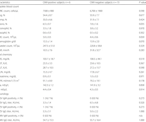
Table 3 shows the laboratory characteristics of subjects with and without CMV-PCR(+) in the aqueous humor. In the CMV-positive subjects, the percentage of mono-cytes tended to be lower than in the CMV-negative Table 1Clinical parameters of 42 patients with hypertensive
anterior uveitis
Gender (M/F) 29:13
Age (range), years 57.6 (25–88)
Spherical equivalent, D −2.6 (−11.25–0.00)
Glaucoma operation,% 22 (52.4 %)
Corneal endothelial cell count, cells/mm2 1908 (625–3067)
Unilaterality 39 (92.9 %)
KPs at baseline examination 28 (67.8 %)
Anterior chamber reaction with 1+ or less 37 (88.1 %)
Severe peripheral anterior synechiae 0 (0.0 %)
Typical feature of P-S syndrome 14 (33.3 %)
Table 2Comparisons of clinical and immunologic characteristics in subjects with or without CMV in aqueous humor
Characteristics CMV-positive subjectsn= 6 CMV-negative subjectsn= 15 Pvalue
Demographic characteristics
Male,n(%) 6 (100 %) 13 (86.7 %) 0.500
Age, years 47.5 ± 14.8 67.6 ± 11.8 0.006
Ocular characteristics
Initial BCVA 0.65 ± 0.29 0.58 ± 0.29 0.569
Final BCVA 0.47 ± 0.46 0.52 ± 0.30 0.733
Spherical equivalent, D -3.6 ± 4.2 0.0 ± 1.6 0.031
Axial length, mm 25.7 ± 1.5 24.4 ± 0.7 0.053
Glaucoma,n(%) 6 (100 %) 13 (86.7 %) 0.347
Average RNFL thickness,μm 66.2 ± 16.7 77.8 ± 19.1 0.132
Corneal endothelial cell count, mm2 1245 ± 560 1981 ± 387 0.009
Unilaterality,n(%) 83.3 80.0 0.684
KPs at baseline examination,n(%) 83.3 73.3 0.550
Presence of PAS,n(%) 4 (66.7 %) 5 (33.3 %) 0.331
Anterior chamber reaction with 1+ or less,n(%) 100.0 92.9 0.714
Lens status, phakic,n(%) 5 (83.3 %) 5 (33.3 %) 0.055
Baseline IOP, mmHg 28.83 ± 8.25 22.73 ± 7.86 0.132
Maximum IOP, mmHg 37.17 ± 8.03 39.53 ± 13.02 0.622
Final IOP, mmHg 13.3 ± 3.44 13.5 ± 5.05 0.950
Number of anti-glaucoma medication,n 2.67 ± 0.81 2.67 ± 0.81 0.970
Course of uveitis 0.544
Chronic,n(%) 4 (66.7 %) 13 (86.7 %)
Recurrent,n(%) 2 (33.3 %) 2 (13.3 %)
Glaucoma operation,n(%) 4 (66.7 %) 10 (66.7 %) 0.701
Choice of glaucoma operation 0.095
Ahmed valve,n(%) 3 (75.0 %) 2 (20.0 %)
Trabeculectomy or ExPRESS ,n(%) 1 (25.0 %) 8 (80.0 %)
HSV PCR positivity,n(%) 0 (0.0 %) 1 (6.2 %) 0.727
CMV RQ PCR, copies/mL 46,048.8 ± 91,334.5 Negative n/a
subjects, with marginal significance (P= 0.055), whereas other parameters did not differ between the groups. The presence of CMV in the aqueous humor was signifi-cantly associated with corneal endothelial cell count after controlling for age and gender (P= 0.023), which was maintained after additional adjustment for lens sta-tus and history of glaucoma surgery (P< 0.001; Table 4). The clinical information of the 6 CMV+ patients was de-scribed in the Additional file 1, and two representative cases with antiviral treatment are discussed.
Case reports Patient 1
A 68-year-old man had hypertensive anterior uveitis of his left eye with a recurrence rate of one to two times a month since 2010. On slit-lamp examination at Seoul St. Mary’s Hospital in October 2012, fine KPs in the lower part of the cornea and mild inflammatory reaction in the
Table 3Comparison of laboratory findings in subjects with or without CMV-PCR (+) in aqueous humor
Characteristics CMV-positive subjectsn= 6 CMV-negative subjectsn= 15 Pvalue
Complete blood count
WBC count, cells/μL 7300 ± 900 6,700 ± 1900 0.590
Seg, % 54.4 ± 8.7 55.9 ± 7.7 0.677
Lymp, % 35.0 ± 6.6 31.9 ± 7.1 0.424
Mono, % 6.5 ± 0.7 7.8 ± 1.6 0.055
Eosinophil, % 3.5 ± 1.8 3.8 ± 3.2 0.970
Basophil, % 0.6 ± 0.3 0.5 ± 0.2 0.302
RBC count, 106/μL 5.0 ± 0.5 4.4 ± 0.6 0.059
Hemoglobin g/dl 15.5 ± 1.4 13.9 ± 2.0 0.070
Platelet count, 103/μL 247.5 ± 51.0 220.8 ± 58.8 0.329
ESR, mm/h 10.5 ± 7.6 31.8 ± 32.7 0.283
Blood chemistry
FBS, mg/dL 103.7 ± 18.7 100.5 ± 44.1 0.519
AST, IU/L 23.3 ± 3.3 23.6 ± 10.5 0.367
ALT, IU/L 28.7 ± 10.5 27.2 ± 15.7 0.590
BUN, mg/dL 15.3 ± 4.7 17.8 ± 6.7 0.261
Creatinine, mg/dL 0.9 ± 0.1 1.0 ± 0.3 0.971
GFR, ml/min/1.73 m2 91.1 ± 8.9 79.2 ± 19.1 0.178
Na, mEq/L 142.3 ± 1.2 141.9 ± 3.2 0.858
K, mEq/L 4.4 ± 0.4 4.3 ± 0.5 0.914
Immunology
HSV IgM positivity,n(%) 1 (16.7 %) 0 (0.0 %) 0.273
HSV IgG, titer, AU/mL 3.3 ± 1.4 6.5 ± 6.8 0.320
VZV IgM positivity,n(%) 1 (16.7 %) 0 (0.0 %) 0.273
VZV IgG titer, AU/mL 5.3 ± 3.1 5.0 ± 2.2 1.000
CMV IgM positivity,n(%) 0 (0.0 %) 0 (0.0 %) n/a
CMV IgG titer, AU/mL 54.7 ± 15.3 55.7 ± 16.4 1.000
HSVherpes simplex virus,VZVvaricella zoster virus,CMVcytomegalovirus,ESRerythrocyte sedimentation rate,FBSfasting blood sugar,ASTaspartate aminotransferase, ALTalanine aminotransferase,BUNblood urea nitrogen,GFRglomerular filtration rate,n/anot applicable
Table 4Association between the presence CMV DNA in aqueous and corneal endothelial cell count in patients with anterior hypertensive uveitis
Model 1 Model 2 Model 3
Unadjusted Adjusted for age and gender
Adjusted for age, gender, lens status, and history of glaucoma surgery
B(CI) B(CI) B(CI)
CMV positivity 672.4 734.4 1034.2
(77.0–1267.9) (116.4–1352.4) (631.3–1437.1)
Pvalue 0.029 0.023 <0.001
anterior chamber were seen. The corneal endothelial cell count of the left eye was 2141 cells/mm2, and the IOP was 22 mmHg with two topical antiglaucoma medica-tions. He showed glaucomatous optic disc changes, with a mean deviation of−9.50 dB. Despite the use of topical steroids and three antiglaucoma medications for 5 months, his IOP was not controlled, and his corneal endothelial cell count in the left eye decreased to 1245 cells/mm2. A CMV-quantitative PCR analysis of aqueous humor samples for the left eye revealed 229,000 copies/mL CMV-DNA, but no HSV-DNA. Thus, an oral course of valacy-clovir (90 mg/day) was started. The ExPRESS glaucoma filtration device (P-200) was implanted under a scleral flap. Oral valacyclovir (900 mg/day) was continued for 10 days, and anterior chamber tapping revealed 66,600 copies/mL CMV-DNA. Oral valacyclovir treatment (450 mg/day) was continued for another 14 days, and re-peated anterior chamber tapping revealed 630 copies/mL CMV-DNA. After glaucoma surgery, the anterior chamber had a rare to +1 grade inflammation, and IOP was well controlled in a range of 6–9 mmHg. However, 5 months later, his corneal endothelial cell count was found to have decreased further, to 981 cells/mm2(Fig. 1).
Patient 2
A 64-year-old man had hypertensive anterior uveitis of his left eye with a recurrence rate of one to two times a month since 1991. He had been diagnosed with Posner-Schlossman syndrome, but chronic inflammation in the anterior chamber with uncontrolled IOP elevation was seen, and he was referred to Seoul St. Mary’s Hospital for glaucoma surgery. At the initial visit, mutton-fat keratic precipitates were noted in the central cornea with minimal anterior chamber reaction (Fig. 2). IOP was 21 mmHg with three antiglaucoma medications, and high pigmentation was noted in the anterior cham-ber angle. Severe glaucomatous optic disc was seen, with
a mean deviation of −18.20 dB. The corneal endothelial cell count of the left eye was 1310 cells /mm2. A CMV-quantitative PCR analysis of an aqueous humor sample for the left eye revealed 44,609 copies/mL CMV-DNA, but no HSV-DNA. Oral valacyclovir treatment (900 mg/day) was started, and an Ahmed glaucoma valve was implanted. After 14 days of oral valacyclovir treatment, the aqueous humor showed 6980 copies/mL CMV-DNA. Oral valacy-clovir was continued for 14 days, and further anterior chamber tapping showed 450 copies/mL CMV-DNA. After 1 month, the corneal endothelial cell count of the left eye was 1310 cells/mm2.
Discussions
We reported the clinical characteristics of hypertensive anterior uveitis (Table 1), which occurred unilaterally (93 %), predominantly in middle-aged (57.6 ± 17.0 years) males (69.0 %; Table 1). Despite seemingly mild intraocu-lar inflammation of the anterior chamber in the majority of patients (88.1 %) and the absence of peripheral anterior synechiae (0 %), glaucoma surgeries were required in over half of the patients (52.4 %). The male dominance in hypertensive anterior uveitis is consistent with previous results in non-Korean populations [4, 7]. Among the tients who underwent aqueous sampling, six (28.6 %) pa-tients were positive for CMV-DNA. This prevalence is similar to the previous studies, reporting 22.8 % of anter-ior uveitis associated with raised IOP [1, 4].
Notably, the corneal endothelial cell count in the CMV-positive subjects was significantly lower than in the CMV-negative subjects (P= 0.009; Table 2). This as-sociation between the presence of CMV-DNA and the corneal endothelial cell count was more evident after adjusting for age, gender, lens status, and history of glau-coma surgery (P< 0.001; Table 3). Previous studies dem-onstrated that CMV is an important pathogen for chronic corneal endotheliitis, particularly in Asian
Fig. 1Case 1: serial follow-up of specular microscopy. At the initial visit, the corneal endothelial count in the left eye was 2141 cells/mm2(a). After
topical steroid and three antiglaucoma medications for 5 months, the corneal endothelial cell count decreased to 1245 cells/mm2(b). A CMV-quantitative
populations [7–9, 13]. This study confirmed that CMV is a major cause of corneal endothelial cell loss in pa-tients with hypertensive anterior uveitis.
Evidence of valacyclovir’s efficacy was seen in our study, using serial follow-up of CMV-DNA copy number and corneal endothelial cell counts in two case patients. After 1 month of therapy, CMV-DNA copy numbers de-creased dramatically in both patients. Considering that patient 1 showed very high CMV-DNA copy numbers (44,609 copies/mL), it seems that one episode of acutely increased viral load is sufficient to cause extensive cor-neal endothelial cell loss. Consistent with our study, Kandori et al. [11] also reported a significant correlation between CMV viral load and corneal endothelial cell loss in CMV-associated uveitis.
We found that the proportion of monocytes tended to be lower in CMV-positive subjects, with marginal signifi-cance (P= 0.053; Table 2). Monocytes are the primary cell type of CMV persistence within the peripheral blood mononuclear cells [14]. CMV infection of monocytes in-duces trans-endothelial migration and monocyte to macrophage differentiation, which is productive for CMV replication [15]. Although the reason is unclear, CMV-induced differentiation of monocytes into macro-phages may be associated with the lower proportion of monocytes in CMV-positive subjects in this study.
We found that the CMV-positive subjects were signifi-cantly younger (P= 0.006, Table 2). Generally, CMV seropositivity increases with age and serves as an im-mune risk phenotype, which is associated with survival in an older population [16, 17]. The current understand-ing of the mechanisms by which CMV reactivates in relatively young immunocompetent subjects is very lim-ited. CMV is unlikely to cause clinically significant symptoms as long as it is maintained in balance by the
host immune system [18]. However, recent literature suggests that CMV is associated with the pathogenesis of cardiovascular diseases, such as atherosclerosis, auto-immune disease, and even certain cancers [19, 20]. It is known that CMV can be reactivated when circulating monocytes with latent CMV are recruited to sites of in-flammation [20]. For this reactivation, inflammatory cy-tokines, such as tumor necrosis factor and interferon, are known to play a role [21, 22]. In this regard, it is sug-gested that chronic inflammation in the anterior cham-ber may trigger CMV reactivation, which in turn, aggravates the inflammatory condition. In addition, CMV anterior uveitis is recalcitrant to topical steroid treatment [3, 4]. Recent reports show evidence that local immunosuppression by topical steroid use increases the risk of CMV infection in immunocompetent patients [23–26]. In addition, there is possibility that the frequent use of prostaglandin eyedrops have play an important role for worsening, including the uncontrolled glaucoma, considering previous reports regarding the complica-tions from glaucoma eyedrops in uveitis [27–29].
Conclusions
In conclusion, we found that CMV was a significant etiological factor in hypertensive anterior uveitis patients in Korea. In CMV-associated uveitis, extensive corneal endothelial cell damage may occur, even with effective anti-viral medication. CMV-associated hypertensive an-terior uveitis patients were younger compared to CMV-negative uveitis patients. Special caution is needed for patients with CMV-positive hypertensive anterior uveitis, given its adverse effects on the corneal endothelium.
Additional file
Additional file 1: Table S1.Demographic data and clinical manifestations
of patients with the presence of cytomegalovirus in the aqueous. (DOCX 15 kb)
Acknowledgements
Not applicable.
Funding
The authors wish to acknowledge of the financial support of the National Research Foundation of Korea Grant funded by the Korean government (MSIP) (No. NRF- 2016R1C1B1011287).
Authors’contributions
JAC and CKP contributed to the conception and design. JAC, KSK, and YHJ contributed to the acquisition of data and analysis and interpretation of data. JAC contributed in drafting the manuscript. HYLP contributed in revising the manuscript. CKP gave the final approval of the version to be published. All authors read and approved the final manuscript.
Competing interests
The authors declare that they have no competing interests.
Fig. 2Case 2: slit lamp photograph showing diffuse mutton-fat
Ethics approval
The study protocol was approved by the institutional review/ethics boards of Seoul St. Mary’s Hospital, the Catholic University of Korea (IRB number: KC14RISI0513).
Author details
1Department of Ophthalmology and Visual Science, St. Vincent’s Hospital,
College of Medicine, The Catholic University of Korea, 93-6, Ji-dong, Paldal-gu, Suwon, Kyonggi-do 442-060, Republic of Korea.2Department of
Ophthalmology and Visual Science, Seoul St. Mary’s Hospital, College of Medicine, The Catholic University of Korea, Banpo-daero 222, Seocho-gu, Seoul 137-701, Republic of Korea.
Received: 11 April 2016 Accepted: 22 August 2016
References
1. Chee SP, Bacsal K, Jap A, Se-Thoe SY, Cheng CL, Tan BH (2008) Clinical features of cytomegalovirus anterior uveitis in immunocompetent patients. Am J Ophthalmol 145:834–840
2. van Boxtel LA, van der Lelij A, van der Meer J, Los LI (2007) Cytomegalovirus as a cause of anterior uveitis in immunocompetent patients.
Ophthalmology 114:1358–1362
3. Jap A, Chee SP (2010) Emerging forms of viral uveitis in the developing world. Int Ophthalmol Clin 50:155–171
4. Jap A, Chee SP (2011) Viral anterior uveitis. Curr Opin Ophthalmol 22:483–488 5. Park SW, Yu HG (2013) Association of cytomegalovirus with idiopathic
chronic anterior uveitis with ocular hypertension in Korean patients. Ocul Immunol Inflamm 21:192–196
6. Knox DL (2008) Clinical features of cytomegalovirus anterior uveitis in immunocompetent patients. Am J Ophthalmol 146:625, author reply 625-626 7. Koizumi N, Inatomi T, Suzuki T et al (2015) Clinical features and
management of cytomegalovirus corneal endotheliitis: analysis of 106 cases from the Japan corneal endotheliitis study. Br J Ophthalmol 99:54–58 8. Kobayashi A, Yokogawa H, Higashide T, Nitta K, Sugiyama K (2012) Clinical
significance of owl eye morphologic features by in vivo laser confocal microscopy in patients with cytomegalovirus corneal endotheliitis. Am J Ophthalmol 153:445–453
9. Chee SP, Bacsal K, Jap A, Se-Thoe SY, Cheng CL, Tan BH (2007) Corneal endotheliitis associated with evidence of cytomegalovirus infection. Ophthalmology 114:798–803
10. Sungur GK, Hazirolan D, Yalvac IS, Ozer PA, Aslan BS, Duman S (2010) Incidence and prognosis of ocular hypertension secondary to viral uveitis. Int Ophthalmol 30:191–194
11. Kandori M, Miyazaki D, Yakura K, Komatsu N, Touge C, Ishikura R, Inoue Y (2013) Relationship between the number of cytomegalovirus in anterior chamber and severity of anterior segment inflammation. Jpn J Ophthalmol 57(6):497–502
12. Lewkowicz D, Willermain F, Relvas LJ et al (2015) Clinical outcome of hypertensive uveitis. J Ophthalmol 2015:974870
13. Chee SP, Jap A (2011) Immune ring formation associated with cytomegalovirus endotheliitis. Am J Ophthalmol 152:449–453, e441 14. Taylor-Wiedeman J, Sissons JG, Borysiewicz LK, Sinclair JH (1991) Monocytes
are a major site of persistence of human cytomegalovirus in peripheral blood mononuclear cells. J Gen Virol 72:2059–2064
15. Smith MS, Bentz GL, Alexander JS, Yurochko AD (2004) Human cytomegalovirus induces monocyte differentiation and migration as a strategy for dissemination and persistence. J Virol 78:4444–4453 16. Wikby A, Johansson B, Olsson J, Löfgren S, Nilsson BO, Ferguson F (2002)
Expansions of peripheral blood CD8 T-lymphocyte subpopulations and an association with cytomegalovirus seropositivity in the elderly: the Swedish NONA immune study. Exp Gerontol 37:445–453
17. Solana R, Tarazona R, Aiello AE et al (2012) CMV and immunosenescence: from basics to clinics. Immun Ageing 9:23
18. Reeves MB, MacAry PA, Lehner PJ, Sissons JG, Sinclair JH (2005) Latency, chromatin remodeling, and reactivation of human cytomegalovirus in the dendritic cells of healthy carriers. Proc Natl Acad Sci U S A 102:4140–4145 19. Halenius A, Hengel H (2014) Human cytomegalovirus and autoimmune
disease. Biomed Res Int 2014:472978
20. Söderberg-Nauclér C (2008) HCMV microinfections in inflammatory diseases and cancer. J Clin Virol 41:218–223
21. Prösch S, Heine AK, Volk HD, Krüger DH (2001) CCAAT/enhancer-binding proteins alpha and beta negatively influence the capacity of tumor necrosis factor alpha to up-regulate the human cytomegalovirus IE1/2 enhancer/ promoter by nuclear factor kappaB during monocyte differentiation. J Biol Chem 276:40712–40720
22. Prösch S, Staak K, Stein J et al (1995) Stimulation of the human cytomegalovirus IE enhancer/promoter in HL-60 cells by TNFalpha is mediated via induction of NF-kappaB. Virology 208:197–206 23. Sims JL, Chee SP (2010) Cytomegalovirus endotheliitis following
fluocinolone acetonide (Retisert) implant. Eye (Lond) 24:197–198 24. Tugal-Tutkun I, Araz B, Cagatay A (2010) CMV retinitis after intravitreal
triamcinolone acetonide injection in a patient with Behçet’s uveitis. Int Ophthalmol 30:591–593
25. Ufret-Vincenty RL, Singh RP, Lowder CY, Kaiser PK (2007) Cytomegalovirus retinitis after fluocinolone acetonide (Retisert) implant. Am J Ophthalmol 143:334–335
26. Park UC, Kim SJ, Yu HG (2011) Cytomegalovirus endotheliitis after fluocinolone acetonide (Retisert) implant in a patient with Behçet uveitis. Ocul Immunol Inflamm 19:282–283
27. Goyal R, Ram AR (2000) Brimonidine tartarate 0.2 % (Alphagan) associated granulomatous anterior uveitis. Eye (Lond) 14:908–910
28. Cates CA, Jeffrey MN (2003) Granulomatous anterior uveitis associated with 0.2 % topical brimonidine. Eye (Lond) 17:670–671
29. Warwar RE, Bullock JD, Ballal D (1998) Cystoid macular edema and anterior uveitis associated with latanoprost use. Experience and incidence in a retrospective review of 94 patients. Ophthalmology 105:263–268
Submit your manuscript to a
journal and benefi t from:
7Convenient online submission
7Rigorous peer review
7Immediate publication on acceptance
7Open access: articles freely available online
7High visibility within the fi eld
7Retaining the copyright to your article