1
Review 2
Phospholipids of Animal and Marine Origin:
3
Structure, Function, and Anti-Inflammatory
4
Properties
5
Ronan Lordan, Alexandros Tsoupras and Ioannis Zabetakis* 6
Department of Biological Sciences, University of Limerick, Limerick, Ireland;[email protected] (R.L); 7
[email protected] (A.T) 8
*Author to whom correspondence should be addressed; [email protected]; Tel.: +353-(0)-61-234-202 9
Abstract: In this review paper, the latest literature on the functional properties of phospholipids in 10
relation to inflammation and inflammation-related disorders has been critically appraised and 11
evaluated. The paper is divided into three sections: Section one addresses the relationship between 12
the anti-inflammatory bioactivities of different phospholipids in relation to their structures and 13
compositions. Sections two and three are dedicated to the structures, functions and 14
anti-inflammatory properties of dietary phospholipids from animal and marine sources. Most of 15
the dietary phospholipids of animal origin come from meat, egg and dairy products. To date, there 16
is very limited work published on meat phospholipids, undoubtedly due to the negative 17
perception that meat consumption is an unhealthy option due to its putative associations with 18
several chronic diseases. These assumptions are addressed with respect to the phospholipid 19
composition of meat products. Recent research trends indicate that dairy phospholipids possess 20
anti-inflammatory properties, which has led to an increased interest into their molecular structures 21
and reputed health benefits. Finally, the structural composition of phospholipids of marine origin 22
is discussed. Extensive research has been published in relation to ω-3 polyunsaturated fatty acids 23
(PUFAs) and inflammation, however this research has recently come under scrutiny and has 24
proved to be unreliable and controversial in terms of the therapeutic effects of ω-3 PUFA, which are 25
generally in the form of triglycerides and esters. Therefore, this review focuses on recent 26
publications concerning marine phospholipids and their structural composition and related health 27
benefits. Finally, the strong nutritional value of dietary phospholipids are highlighted with respect 28
to marine and animal origin and avenues for future research are discussed. 29
Keywords: phospholipids; atherosclerosis; inflammation; anti-inflammatory; dairy; marine; meat; 30
egg; nutrition. 31
32
1. Introduction 33
Lipids are a very heterogenic class of biomolecules with a wide range of structures and 34
functions. Lipids can be divided into two major sub-classes, neutral lipids (such as Triacylglycerol’s 35
or TAGs; waxes; terpenes), which are molecules with long hydrophobic hydrocarbon chains lacking 36
a free polar group, and polar lipids (such as phospholipids, glycolipids, etc.) that apart from their 37
hydrophobic hydrocarbon residues they also bare polar-hydrophilic group such as a 38
carbohydrate-group, or a phosphate head group with a hydrophilic residue within their structure. 39
40
1.1 Phospholipid classes and biological functions 41
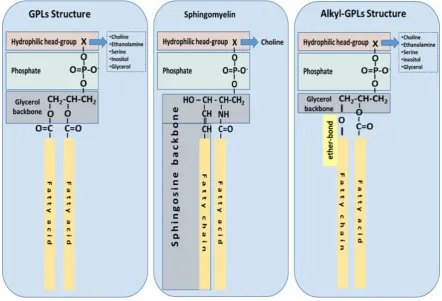
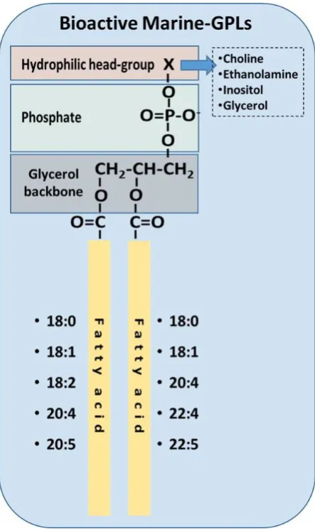
Ubiquitous to all tissues, phospholipids (PLs) are essential components of cell membranes 42
consisting of a hydrophilic head group and a hydrophobic tail giving phospholipids their 43
amphiphilic properties. Glycerophospholipids (GPLs) share a common structure consisting of two 44
fatty acid (FA) molecules esterified in the sn-1 and sn-2 positions of the glycerol moiety. This portion 45
of the molecule contributes to its hydrophobicity. The sn-3 position consists of a phosphate group 46
with a hydrophilic residue that contributes hydrophilicity (Figure 1). The simplest GPL is 47
phosphatidic acid (PA), others are named after the hydrophilic residue/group attached to the 48
phosphate group. Four main groups have been identified: ethanolamine, inositol, serine, and 49
choline. These groups form the most biologically important phospholipids, which are 50
phosphatidylethanolamine (PE), phosphatidylinositol (PI), phosphatidylserine (PS) and 51
phosphatidylcholine (PC). Lysophospholipids (Lyso-PLs) refer to phospholipids whose fatty acid 52
chain has been removed from either the sn-1 or sn-2 position. Sphingolipids (SPLs) contain the 53
long-chain amino alcohol sphingosine (instead of glycerol) esterified to a fatty acid and a phosphate 54
group. Sphingomyelin (SM) is the most representative SPL, which consists of sphingosine and bares 55
a choline molecule. SM is found in high quantities in brain and neural tissues membranes (Figure 1). 56
The biological importance of these PLs derives from their amphiphilic properties. The 57
hydrophilic head and the hydrophobic tail create a lipid bi-layer that allows for the assembly of cells’ 58
and organelles’ membranes [1-3]. These phospholipid-based bilayers form selectively permeable 59
barriers, which are essential for effective separation of a cell or organelle from its surroundings. 60
These properties allow for low membrane permeability for cellular constituents such as nutrients 61
and ions, while the organisation into a lipid bilayer provides the perfect matrix in which the 62
membrane-integral proteins are embedded. No mammalian membranes or cells are formed without 63
PLs and the integrity and function of the external (cellular) and internal (subcellular) membrane 64
systems depends on their composition and on the integrity of their phospholipid structure. Besides 65
GPLs and SPLs, biological membranes are also made up of glycolipids and cholesterol, as well as of 66
integral and peripheral membrane proteins. 67
Other forms of GPLs exist, which differ from the general structure of GPLs, such as ether-linked 68
GPLs that bare other hydrocarbon chains (saturated or unsaturated or with hydroxyl-groups, etc) 69
ether-linked to the sn-1 position of the glycerophosphate backbone, instead of a fatty acid bound by 70
ester bonds to the sn-1 position of the glycerol backbone (Figure 1c). Ether-linked GPLs can be found 71
as minor constituents of cell membranes in both prokaryotes and eukaryotes, but they are abundant 72
in archaeal organisms [4]. Some exist as bioactive molecules that seem to be maintained through 73
evolution from archaeal to eukaryotic organisms because of their lipid signalling bioactivities, 74
especially in eukaryotic organisms. One such examples includes plasmalogens and 75
platelet-activating factor, also known as PAF (1-O-alkyl-2-acetyl-sn-glyceryl-3-phosphorylcholine) 76
[5], which is potent inflammatory mediator involved in the innate immune response and chronic 77
inflammatory diseases [6,7]. 78
The lipid composition of biological membranes represents a taxonomic signature that 79
distinguishes the different kingdoms of life. Differences between ester and/or ether bonded fatty 80
acid chains at the glycerol backbone exist between different kinds of organisms [4], in addition the 81
fatty acid composition of PLs also varies depending on their origin [8]. Due to their amphipathic 82
properties, naturally occurring PLs either from plant or animal origin, generally contain an 83
unsaturated fatty acid in the sn-2 position, such as oleic acid, linoleic acid, α-linolenic acid, 84
arachidonic acid (pro-inflammatory molecule usually from animal origin) or eicosapentaenoic acid 85
(anti-inflammatory molecule usually from marine origin), whereas the sn-1 position predominantly 86
carries a saturated fatty acid (SFA), such as stearic acid or palmitic acid [9]. The correct ratio of 87
saturated to unsaturated fatty acids in the phospholipid membrane is essential to sustain the 88
membrane characteristics, since the fatty acid composition and degree of saturation directly affects 89
the fluidity of the cell membrane. Equally, the correct ratio can have a significant effect on cellular 90
with a high content of cholesterol and PLs predominantly carrying SFA, which are implicated in 92
apoptosis, cellular proliferation, and unsaturated fatty acids that act as precursors for the synthesis 93
of pro-inflammatory mediators called eicosanoids (prostaglandins - PGs, thromboxanes - TX, 94
leukotrienes - LT, lipoxins - LX) [10,11]. 95
Even though the main function of PLs is to support the formation and biofunctionality of cell 96
membranes, there are specific varied PLs that perform specialised functions in the subcellular 97
micelles and organelles. For example, PLs are structural and functional constituents of the surface 98
monolayers of lipoproteins (which transport lipids to tissues via the blood stream), the pleura and 99
alveoli of the lung and are constituents of the pericardium, joints, peritoneal and gastrointestinal 100
surfactants, while together with cholesterol and bile acids they form mixed micelles in the 101
gallbladder for fat emulsification [12]. In addition, some PLs act as lipid mediators of inflammation 102
that have the ability to influence immunological processes at the cellular level (i.e. PAF) [7]. PLs also 103
contain bound PUFAs to be released on demand as precursors of prostaglandins and other 104
eicosanoids [11], while other PLs and their metabolites are a source of secondary messengers in cell 105
signalling (e.g. diacylglycerols, phosphoinositide’s, etc.) [13] and carry out essential functions within 106
organelles such as the mitochondria [14]. Therefore, not only are PLs integral structural lipids in cell 107
membrane formation, function and integrity, but research has identified that they possess a plethora 108
of additional functions in various cell types and organisms which will be discussed further in this 109
review. 110
111
Figure 1: The most common structures of phospholipids are depicted. Phospholipids with a glycerol 112
backbone (GPLs); Sphingomyelin as a representative of a sphingosine-backbone phospholipid 113
(SPLs); Alkyl-phospholipids (Alkyl-GPLs) that have a fatty chain linked with an ether-bond at the 114
sn-1 position of the glycerol backbone. 115
1.2 Glycerophospholipid and Sphingophospholipid Biosynthesis 116
In mammalian cells, GPL synthesis requires a diacylglycerol unit, which is provided by either 117
diacylglycerol or CDP-diacylglycerol. The generation of these precursors initiates through the 118
membrane and of the endoplasmic reticulum), which links a fatty acid-CoA (generally a SFA) to the 120
sn-1 position of glycerol-3-phosphate to generate lyso-PA. Acylglycerol-3-acyltransferase is required 121
for the subsequent formation of PA in the endoplasmic reticulum, whereby it esterifies another fatty 122
acid-CoA (generally an unsaturated FA) to the sn-2 position of glycerol. PA then becomes the 123
substrate for two significant metabolic enzymatic pathways. The first pathway is controlled by a 124
cytosolic phosphatidic acid phosphatase enzyme, which takes place in the membrane of the 125
endoplasmic reticulum and produces diacylglycerols (DAG) by removing the phosphate group from 126
the sn-3 position of PA. Triacylglycerol’s (TAG) are formed by the esterification of another fatty acid 127
to the sn-3 position, and these then become the main energy source in the body. Alternatively, 128
CDP-diacylglycerol synthase, an enzyme associated primarily with the endoplasmic reticulum, 129
catalyses a reaction between CTP and PA leading to the formation CDP-diacylglycerol. In the second 130
pathway for PA synthesis, dihydroxyacetone-P is acylated to 1-acyl-dihydroxyacetone-P, which is 131
subsequently converted to lyso-PA and then PA [1,15,16]. 132
The synthesis of PC and PE occurs in the cytosol following the enzymatic addition of either a 133
choline or ethanolamine to PA [17]. The biosynthesis of PS requires the presence of PC and PE. In 134
terms of PE, PS synthesis occurs in the endoplasmic reticulum through two metabolic pathways, 135
which use differential enzymes and substrates. Initially PC exchanges a choline with a serine 136
molecule in the presence of PS synthase I, leading to the final products of PS and choline. Synthesis 137
of PS from PE follows a similar pathway where PS synthases II catalyses the substitution of an 138
ethanolamine head for a serine head, leading to the final products of PS and ethanolamine. In the 139
presence of the same enzymes, the latter reaction is unique as it is reversible, thus PS can release 140
serine and replace it with ethanolamine [1,18]. PI is also biosynthesised in the endoplasmic reticulum 141
where CDP-diacylglycerol binds to inositol, by the enzymatic actions of CDP-diacylglycerol 142
phosphatidyl transferase. These reactions result in the production of PI and cytidine 143
monophosphate (CMP). Other essential molecules often associated with the polar fraction of lipids 144
such as cardiolipin (CL) are produced through the same pathway [1,19]. 145
The synthesis of sphingomyelin starts in the endoplasmic reticulum and after a series of 146
enzymatic reactions finishes in the Golgi apparatus and the plasma membrane. Synthesis begins 147
with the condensation of serine and palmitoyl CoA by serine palmitoyltransferase forming 148
3-ketosphinganine, which is then reduced to dihydrosphingosine that is then N-acylated by one of 149
six ceramide synthases (CerS1-CerS6), each using specific acyl chains, generally with a SFA or 150
MUFA with 16-26 carbons, forming dihydroceramides that are subsequently dehydrogenated to 151
ceramides by dihydroceramide desaturase. The reaction is catalysed by the enzymes sphingomyelin 152
synthase I and sphingomyelin synthase II, which produces SM and diacylglycerols from the 153
substrates ceramide and PC [1,20]. 154
Plasmalogens are mainly synthesised in peroxisomes. They contain an aliphatic hydrocarbon 155
chain at the sn-1 position of the glycerol linked via vinyl-ether binding derived from PC and PE. 156
Generally plasmalogens are esterified with highly unsaturated fatty acids such as docosahexaenoyl 157
or arachidonoyl fatty acid at the sn-2 position of glycerol [21]. The functions of plasmalogens are not 158
yet fully understood, however it is proposed that they may act as potential biomarkers for age 159
related diseases, oxidative stress and systemic inflammation [22] 160
1.3 Inflammation and Lipid Inflammatory Mediators 161
Inflammation is a necessary protective response of the innate immune system in response to 162
physiological triggers such as pathogens or damaged cells, whereby the tissue is repaired, or the 163
pathogenic insult is eliminated. However, excessive inflammation can lead to tissue injury [23]. Diet 164
and lifestyle are a key modifiable risk factor for the prevention of chronic diseases. It has been 165
established that a maladaptive diet is one of the dominant underlying causes of systemic 166
inflammation through exaggerated postprandial elevations in plasma glucose and triglycerides. Due 167
to the increased intake of heavily processed foods with high calorific value, postprandial 168
hyperlipemia and hyperglycaemia are common, postprandial lipemia is an independent risk factor 169
production of excess plasma reactive oxygen species (ROS) occurs due to the increased levels of 171
postprandial glucose and triglycerides, which can lead to a pro-inflammatory state [23-26]. 172
Activated immune cells are essential in preventing long lasting damage to the host, as they can 173
maintain or resolve the inflammatory response. If an inflammatory response is not resolved the 174
subsequent inflammatory microenvironment will disrupt tissue homeostasis leading to a systemic 175
inflammatory condition. Several conditions owe their onset and progression to systemic 176
inflammation including cancer, kidney disorders, obesity, type II diabetes mellitus, atherosclerosis 177
and various CVDs. For further reading on the typical inflammatory response see the comprehensive 178
review of Medzhitov [27] and the works of Demopoulos et al. [28] and Libby et al. [29]. 179
The initiation and resolution of the inflammatory response involves the complex and 180
coordinated expression of many factors, including cytokines like the Interleukin-1 (IL-1) family, 181
Interleukin-6 (IL-6), Tumour Necrosis Factor-α (TNF-α), Interferon-γ (INFγ), chemokines, growth 182
factors (vascular endothelial growth factor or VEGF), proteases, ROS, oxidised phospholipids 183
(Ox-PLs) and lipid-mediators such as eicosanoids and PAF. These inflammatory signals induce a 184
myriad of physiological processes, ranging from local vascular responses to systematic responses 185
affecting the whole organism [30]. These molecules sustain the inflammatory process until the insult 186
has been resolved. Under all conditions, chronic inflammation leads to a disturbed homeostasis, 187
spiralling the physiological and immunological conditions towards a pro-inflammatory harmful 188
setting involving cells and secreted factors [7]. Persistent induction and dysregulation of 189
inflammation has been recognised as an integral feature of the pathology of several chronic 190
conditions including CVDs, type II diabetes, obesity, renal disorders, cancer and Alzheimer’s disease 191
[7,29,31-35]. 192
Interestingly, in such pathological conditions common junctions of inflammatory cross-talk 193
between several inflammatory signalling pathways exist and can lead to comorbidities in such 194
diseases. Patients with a chronic inflammatory disease are at risk of developing other inflammatory 195
conditions and vice versa, a chronic inflammatory condition can be a major risk factor for the 196
development of a chronic inflammatory disease. For example chronic inflammation observed in 197
diabetic patients is one of the leading causes of disease complications, which manifests in decreased 198
kidney function, eye maladies, heart attacks and strokes [36].In addition, the development of several 199
autoimmune diseases characterised by an increased inflammatory status (i.e. increased levels of 200
eicosanoids and cytokines) such as rheumatoid arthritis, can lead to the induction and 201
co-development of CVDs [37].Periodontal disease patients also exhibit a high risk of co-developing 202
atherosclerosis and CVDs [38-40]. Similarly, HIV patients are at risk of persistent inflammation, 203
which can lead to chronic inflammatory diseases such as atherosclerosis and CVDs [41]. Specific 204
inflammatory biomolecules such as lipid inflammatory mediators (PAF, eicosanoids, etc), 205
cytokines/chemokines, growth factors, and adhesion molecules play similar roles as the main 206
instigators of these manifestations in inflammation [7]. 207
As chronic inflammation is responsible for many complications evident in different diseases, its 208
diagnosis and treatment constitute an enormous challenge for medical practitioners. Importantly, 209
many therapeutic treatments employed up to date have failed to produce a desirable effect, since a 210
permissive immune environment is a prerequisite for their proper function. Nowadays, there is no 211
doubt that chronic inflammation and associated immunosuppression pose a serious obstacle in the 212
prognostic and the therapeutic area, as they both develop with no palpable clinical signs, often 213
leading to unforeseeable complications and possible unresponsiveness to various therapies [42]. 214
Apart from therapeutic interventions, long-term lifestyle measures such as healthy nutrition and 215
exercise may provide preventive results or countermeasures towards inflammatory manifestations. 216
The most known lipid pro-inflammatory mediators produced and implicated in inflammatory 217
physiological responses are the eicosanoids and PAF. Both eicosanoid and PAF inflammatory 218
pathways have been found to be promising targets in respect to dietary interventions, especially to 219
those with foods containing bioactive PLs [43,44]. 220
Eicosanoids are locally acting bioactive signalling lipids considered to be oxidized derivatives 221
thromboxanes (TXs), leukotrienes (LTs) and lipoxins (LXs), which regulate a diverse set of 223
homeostatic and inflammatory processes linked to numerous diseases [45]. The major substrate for 224
eicosanoid synthesis is arachidonic acid (ARA, a lipid that usually is bonded at the sn-2 position of 225
membrane glycerophospholipids), but also related PUFAs. Several agonists and receptors induce 226
inflammatory processes and the subsequent cytokine “storm” that accompanies them initiates the 227
release of ARA and related PUFAs, resulting in an eicosanoid storm [45]. Inflammatory stimuli 228
trigger the activation of phospholipase A2 enzymes that release ARA from the sn-2 position of 229
membrane phospholipids. ARA acts in turn as a substrate for several enzymes, such as 230
cyclooxygenase (COX), cytochrome P450 enzymes and lipoxygenase (LOX) [46]. From this plethora 231
of simultaneous biochemical reactions, a range of pro-inflammatory molecules are formed including 232
PGs, TXs, LTs and LXs, which are well known mediators and regulators of inflammation [47]. 233
Drugs that target eicosanoid pathways have been used for over a century; aspirin is the oldest 234
of the numerous effective non-steroidal anti-inflammatory drugs (NSAIDs) that have been 235
marketed. Systematic characterisation of prostaglandin and leukotriene structures, biosynthetic 236
pathways, natural receptors and biological functions have resulted in the production of new drugs 237
that target eicosanoids in order to treat common inflammatory symptoms including swelling and 238
pain. However, chronic diseases like arthritis and atherosclerosis are largely unaffected by the 239
inhibition of eicosanoids [45]. In addition, side effects have been attributed to the blocking of COX-1 240
or COX-2 [45]. Low doses of aspirin are now commonly prescribed as cardioprotective agents, which 241
limit thromboxane formation by COX-1 in platelets without inhibiting COX-2 mediated PGI2 242
formation by endothelial cells. 243
On the other hand, omega-3 (ω-3) fatty acid supplementation (i.e. from fish-oil) is also 244
commonly prescribed for the treatment of various inflammatory ailments and for cardioprotection, 245
due to their interactions with the eicosanoid pathways. The clearest evidence for this reasoning is the 246
use of ω-3 fatty acids, such as ω-3 eicosapentaenoic acid (EPA) and docosahexaenoic acid (DHA). 247
These fatty acids are abundant in fish and fish oils and they have the ability to inhibit arachidonic 248
acid metabolism by COX-1 (but less so by COX-2), in a similar manner to low dose aspirin [45]. In 249
addition, it is reported that EPA and DHA derived lipid mediators are less potent inducers of 250
platelet aggregation in contrast to ARA-derived lipid mediators, which they displace, providing 251
thus an agonistic effect towards ARA [48,49]. ω-3 fatty acids, such as EPA and DHA, seem to benefit 252
multiple risk factors including blood pressure, blood vessel function, heart function, blood lipids, 253
and they have antithrombotic, anti-inflammatory and anti-oxidative actions [50]. In addition to 254
absolute amounts of ω-6 and ω-3 fatty acid intake, the ω-6/ω-3 ratio plays an important role in 255
increasing the development of obesity via both ARA eicosanoid metabolites and hyperactivity of the 256
cannabinoid system, which can be reversed with increased intake of EPA and DHA [51]. However, 257
despite the overwhelming amount of evidence on the beneficial effects of ω-3 PUFAs on human 258
health, some controversy remains since several systematic reviews and meta-analyses have 259
illustrated that there is insufficient evidence for this notion, with many studies highlighting the lack 260
of benefit and possible risks associated with the consumption of ω-3 PUFA supplements. However, 261
studies do indicate that the beneficial effects of fish intake on cerebrovascular risk are likely to be 262
mediated through the interplay of a wide range of nutrients abundant in fish [52-56]. Yet, within the 263
last decade there is evidence that when ω-3 PUFAs (such as EPA and DHA) are combined to PLs, 264
they are more efficiently incorporated into tissue membranes and at much lower doses than when 265
these PUFAs are combined to TAGs . These PLs containing ω-3 PUFAs seem to provide beneficial 266
effects towards inflammation related disorders through specific mechanisms and a plethora of 267
bioactivities including their ability to modulate the eicosanoid pathway [43,57-59]. 268
Other PLs also seem to contribute directly and/or indirectly to several inflammatory cascades 269
and thus are involved in the onset, progression and often the remediation of inflammatory diseases. 270
These PLs include plasmalogens, oxidised PLs and PL carriers of FA precursors of eicosanoids 271
[7,11,60-62]. Several foods contain compounds that have well established modes of 272
anti-inflammatory action, whose pleiotropic therapeutic effectiveness, and lack of toxicity ensures 273
along with their subsequent side effects, dietary interventions using PLs seem to protect against 275
inflammatory manifestations without any reported side effects thus far. It is thought that these PLs 276
attenuate the levels of inflammation towards pre-inflammatory homeostatic baseline levels. 277
PLs found in food products such as meat, eggs, dairy, seafood and vegetable sources like 278
soybean are defined as dietary PLs. These dietary PLs are ingested as part of a normal diet, however 279
in recent years, research has focused on the beneficial health effects of dietary PLs, and their 280
anti-inflammatory activities against chronic diseases, thus PLs are also available as dietary 281
supplements exhibiting pleiotropic beneficial effects towards inflammation related disorders. These 282
food derived PLs can not only influence membrane-dependent cellular functions but they also 283
possess anti-inflammatory, anti-oxidant, anti-fibrogenic, anti-apoptotic, membrane-protective, and 284
lipid-regulating effects with a positive impact on several diseases, apparently without severe side 285
effects [8,12]. Furthermore, dietary PLs can reduce the side effects of some drugs and they can 286
influence the fatty acid composition of the hosts PLs. The main animal sources of phospholipids 287
include eggs, milks, meats and marine phospholipids. Interestingly, marine phospholipids are much 288
higher in PUFAs [43], which makes them a promising functional ingredient in foods. The oral 289
application of such dietary PLs has the potential to cause defined alterations of the fatty acid 290
composition of membrane PLs, and thus several cellular functions (according to cell-type), including 291
cell signalling and transport, as well as the modulation of membrane bound enzymes that may lead 292
to health benefits. 293
Apart from eicosanoids, PAF is another potent lipid inflammatory mediator with pleiotropic 294
effects [63]. PAF is synthesized throughout the body by the specific stimulation of various cell types 295
such as platelets, macrophages, monocytes, eosinophils, basophils, and endothelial cells. PAF is 296
mostly produced in the blood, lungs, kidney, myocardium, brain, liver, skin, saliva, retina, uterus, 297
and embryo [64,65].The levels of PAF present in biological tissue are regulated by a balance of its 298
biosynthetic and catabolic enzymatic pathways [7]. However, apart from its enzymatic biosynthetic 299
pathways, PAF and PAF-like lipids that share similar structures and bioactivities, can also be 300
produced through0 the oxidation of other lipids by ROS. The production of these PAF-like lipids 301
occurs during inflammation and oxidative stress. PAFs can also stimulate the production of ROS and 302
nitrogenous species during oxidative and nitrosative stress in inflammation-induced endothelial 303
dysfunction and atherosclerosis [66]. PAF and PAF-like molecules act through their binding to a 304
unique G-protein coupled seven transmembrane receptor, subsequently triggering multiple 305
intracellular signalling pathways, depending on the target cell and PAF-levels (concentration) in 306
blood or tissue [67]. PAF in general, plays a vital role in various physiological processes such as 307
mediation of normal inflammatory responses, regulation of blood pressure, regulation of 308
coagulation responses, foetal implantation, lung maturation, initiation of parturition, and exocrine 309
gland functions. 310
PAF is produced and released in large quantities by inflammatory cells in response to specific 311
stimuli, such as upstream regulators (IL-1, IL-6, TNF-α, Endothelin, and PAF itself) [7,66,68,69]. 312
Increased PAF-levels at the site of inflammation can activate several cell-types through its receptor. 313
This leads to the production of a broad spectrum of PAF-effects depending on the cell-type and 314
tissue, which is achieved through various downstream mediators, enhancing the production and 315
release of PAF itself and several other mediators of inflammation such as eicosanoids, TNF-α, 316
IL-1α, IL-6, IL-8, growth factors, ROS and the expression of selectins and integrins in the membranes 317
of activated cells [7,28,66,68,69]. The interconnected crosstalk between PAF, pro-inflammatory 318
up-stream mediators that induce PAF-production, and PAF-induced downstream mediators seem to 319
be interrelated during inflammatory manifestations. These pathways serve as one of the main 320
junctions between many inflammatory cascades that ultimately lead to endothelium dysfunction 321
and inflammation-related disorders such as atherosclerosis, CVDs and cancer [7,28,66]. 322
The exploration of possible therapeutic approaches focus on the PAF/PAF-receptor interaction, 323
thus inhibiting the exacerbation of the complex PAF inflammatory pathways. There are several 324
agonists of synthetic and natural origin [23,70], which can competitively or noncompetitively 325
promising results, the most prominent beneficial effects have been derived from PL extracts of 327
several foods. These food extracts exhibit anti-inflammatory and anti-oxidant activities through 328
inhibiting PAF-activities and/or downregulating its levels by affecting/modulating the activities of 329
key-metabolic enzymes of PAF (upregulation of PAF-catabolic enzymes activities and/or 330
simultaneous downregulation of the basic PAF biosynthetic enzymes) in vitro and in vivo. The in vitro 331
and in vivo beneficial effects of these dietary PLs are summarised in Table 1. 332
Table1. Studies on the beneficial impact of PLs derived from food of the Mediterranean Diet 333
towards inflammation-related disorders 334
Studied food and
components Type of study Results
PLs of red and white wine,
musts, grape-skins, and
yeast
In vitro studies in washed rabbits’ platelets (WRPs) and in U937 macrophages In vivo postprandial dietary interventions
studies in humans
Inhibition of platelet aggregation and modulation of PAF-metabolism towards reduced
PAF-levels [73-78]
PLs of fish (Sea bass, sea bream, salmon,
etc)
In vitro studies in WRPs, human platelet rich plasma (hPRP) and in human
mesangial cells (HMCs).
In vivo studies in hyperlipidaemic
rabbits
Inhibition of platelet aggregation, modulation of PAF-metabolism towards reduced PAF-levels and reduction of the thickness of atherosclerotic
lesions in hypercholestrolaemic rabbits [79-87] Unpublished data for Salmon-PLs
PLs of olive oil and olive pomace
In vitro studies in WRPs and in HMCs.
In vivo study in hyperlipidaemic
rabbits
Inhibition of platelet aggregation and modulation of PAF-metabolism towards reduced
PAF-levels and reduction of the thickness of atherosclerotic lesions in hypercholestrolaemic
rabbits and regression of the already formed atherosclerotic lesions [87-91]
PLs of seed oils (soybean, corn, sunflower, and
sesame oil)
In vitro studies in WRPs
Inhibition of platelet aggregation [88]
PLs of Hen egg In vitro studies in WRPs
Inhibition of platelet aggregation [92]
PLs of dairy products (milk, yoghurt, cheese,
etc)
In vitro studies in WRPs and in hPRP
Inhibition of platelet aggregation [93-95] unpublished data for bovine, ovine and caprine
1.5 Dietary Phospholipids: Digestion and Absorption 335
Dietary fat is mainly composed of TAG with PLs accounting for 3 - 6% of total fat intake [96]. 336
The daily intake of PLs is not exactly known, however the daily intake of PC/day is estimated to be 2 337
- 8 grams [8]. TAGs and PLs are digested and absorbed in different ways in the small intestine. TAG 338
requires emulsification by bile salts prior to absorption, while PLs can spontaneously form micelles 339
that can be conveyed in an aqueous environment. In contrast to TAGs, PLs are not hydrolysed by 340
lingual or gastric lipases but by other enzymes located in the small intestine. Thus, PLs are almost 341
completely absorbed in the intestine. The most common PL present in the intestinal lumen is PC 342
which is derived mostly from bile (10–20 g/day in humans) with the remainder coming from the 343
diet, while other PLs, such as PE, PS, and PI, are present in much smaller amounts [58]. 344
In the lumen, most of PLs are hydrolysed at the sn-2 position by pancreatic phospholipase A2 345
(pPLA2) and then absorbed by the enterocytes as free FAs and lyso-PLs. The fatty acid chain length 346
and unsaturation number influences fat digestion, absorption, transport, and metabolism at cellular 347
level. For instance, medium-chain fatty acids are better absorbed than long chain fatty acids because 348
they can be dissolved in the aqueous phase and then be absorbed bound to albumin and transported 349
to the liver directly by the portal vein [97].Lyso-PLs and some free-FA are re-esterified to PLs (while 350
some free FAs bind to TAGs) and enter the bloodstream incorporated into the surface layer of 351
chylomicrons, whereas TAGs are incorporated into the core of chylomicrons. However, a small 352
proportion will also incorporate into very low-density lipoproteins (VLDL). After the TAG-rich 353
particles of the chylomicron are degraded, PLs such as PC can be taken up by the high-density 354
lipoprotein (HDL) fraction, which occurs relatively rapidly, within 5-6 hours of PLs ingestion [98,99]. 355
Via HDL, PLs can be transferred into cells of numerous tissues and organs (e.g. liver, muscle, 356
kidneys, lung, tumour cells, etc) [43,100,101]. In contrast to GPLs, digestion of SM in the intestine is 357
slow and incomplete, with initial hydrolysis of SM to ceramide by alkaline sphingomyelinase and 358
subsequent hydrolysis to sphingosine by neutral ceramidase. Both ceramide and sphingosine can be 359
absorbed into intestinal mucosal cells [102]. 360
Interestingly, almost 20% of intestinal PLs are absorbed passively and without hydrolysation, 361
and preferentially incorporated directly into HDL [43]. In addition, a substantial part of the dietary 362
PL fraction is integrated into HDL particles already in the intestine that later join the plasma HDL 363
pool. There is also evidence that PLs incorporated into lipoproteins of the blood stream, might be a 364
more efficient delivery form than TAGs for PUFAs to several tissues and organs (i.e. brain, liver, 365
lung, heart, etc), including blood cells such as platelets and erythrocytes [43]. For example piglets fed 366
with a PUFA-TAG formula had a higher PUFA content in PLs bound to low-density lipoprotein 367
(LDL, a lipoprotein for cholesterol transfer, derived from VLDL after its delivery/degradation of 368
TAGs) than those fed with PUFA-PLs formula, while the opposite results were found in HDL PLs 369
[103]. Thus, dietary PUFAs in form of TAGs or PLs affect the composition of PLs in HDL and LDL in 370
different ways, and therefore the composition and functionality of lipoproteins and their 371
distribution in the body and affect the fatty acid tissue incorporation in the host. The beneficial 372
effects of PLs on blood and hepatic lipids have been studied in a number of animal experiments 373
[104-107], and both cholesterol and TAG levels are affected upon treatment [104]. PLs have also been 374
shown to increase levels of HDL in humans [108,109]. Many of the studies performed with PLs did 375
not include PLs containing ω-3 PUFAs, indicating that PLs in general have beneficial effects 376
[59,106,110]. However, it has also been shown that ω-3 PUFAs are better protected from oxidation 377
when they are incorporated into PLs compared to TAGs. Other studies have demonstrated that 378
PL-bound ω-3 PUFAs have more potent effects on blood plasma and liver lipid levels compared to 379
PLs without ω-3 PUFAs [111,112]. In addition, dietary PLs are known to inhibit cholesterol 380
absorption when added in significant amounts to the diet [113]. Several other mechanisms have been 381
proposed for the effect of PLs on the reduction of cholesterol and other lipid absorption in intestine , 382
such as their structure-related physical emulsifier properties and the ability to form a fat-water 383
emulsion with cholesterol and other lipids, forming vesicles or micelles [114]. PLs play an important 384
transport from the intestine into the enterocytes depends on the emulsification of the dietary fats 386
with biliary secreted PLs, or with PLs from the diet. Intestinal PLs are also able to interact with the 387
cellular membrane of enterocytes, reducing their cholesterol absorptive capacity [8]. 388
It is also very interesting that the uptake of dietary PLs are mostly incorporated and affect the 389
functionality of HDL-lipoproteins that have been characterised as the "good" cholesterol, because 390
these lipoproteins not only remove excess cholesterol from blood stream and from atherosclerotic 391
plaques, but also have exhibited anti-inflammatory and antioxidative properties. HDL also bares a 392
plethora of cardioprotective enzymes such as PAF catabolic enzymes [115], contributing to the 393
maintenance of endothelial cell homeostasis which protect the cardiovascular system [116]. 394
During atherosclerosis and endothelial dysfunction, oxidation of lipoproteins also occurs, 395
especially that of LDL that is transformed to oxidised-LDL (Ox-LDL), which migrates along with 396
white blood cells to the subendothelial intima leading to the formation of foam cells and 397
atherosclerotic lesions [23,28]. HDL and its enzymes seem to protect against these manifestations, 398
while effort to increase HDL levels tends to be one of the main goals of dietary interventions and 399
drug administration for cardioprotection. One of these HDL protective mechanisms, involves the 400
enzyme PAF acetyl-hydrolase (PAF-AH), which HDL bares. PAF-AH is a delicate Phospholipase A2 401
also referred to as Lp-PLA2 (lipoprotein associated Phospholipase A2) that protects against the 402
production and activity of Ox-LDLs by promoting the catabolism of PAF and Oxidised-PLs (Ox-PLs) 403
existing in Ox-LDL (especially those Ox-PLs that mimic PAF). Plasma-PAF-AH activity (both in LDL 404
and HDL) is increased as a response to inflammation and oxidation, as a “signal terminator” [117]. 405
However, during persistent LDL oxidation, PAF-AH is progressively inactivated (plasma-PAF-AH 406
is incorporated mainly in LDL) and thus it loses its capacity to protect against the pro-inflammatory 407
actions of PAF and oxidised-PLs mimicking PAF. On the other hand, dietary intake of PLs 408
(especially those baring ω-3 PUFAs) increase HDL-levels and the incorporation of such 409
anti-inflammatory and anti-oxidant dietary PLs to HDL, thus providing an additional protective 410
mechanism by increasing plasma PAF-AH activity and by protecting the HDL-enzymes (such as 411
PAF-AH) from oxidation-related inactivation[28]. The above is also in agreement with the beneficial 412
in-vitro and in-vivo effects of several dietary PLs, which are shown in Table 1, especially on 413
PAF-metabolism and HDL biofunctionality (including HDL-levels and increased PAF-AH activity) 414
towards reduced PAF levels and cardioprotection. 415
2. Phospholipids of Animal Origin: 416
Foods and fats of animal origin namely meat, eggs and dairy receive undue criticism from 417
society and scientific communities due to their perceived negative effects on health upon 418
consumption. Recent research trends have shown that these negative perceptions may be 419
unwarranted as numerous research teams have shown that meat, eggs and dairy products, 420
including some of their lipid fractions, may be associated with a positive effect on health when eaten 421
in moderation despite their SFA and cholesterol content [23,114,118-123]. For the purpose of this 422
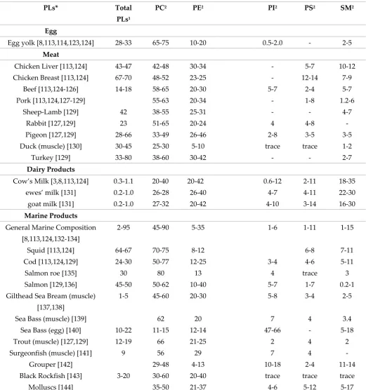
review, table 2 presents the phospholipid composition of a number of animal and marine species, 423
however it is clear from the literature that the study of the phospholipid composition of many 424
animal and marine food sources has been neglected as published research tends to focus solely on 425
the fatty acid composition and not the phospholipid species composition. 426
2.1 Meat Phospholipids 427
Red and white meat contribute several important nutrients to the diet, including vitamins (B12 428
in particular), essential amino acids, iron, selenium, zinc, folic acids and fats. The phospholipid 429
content of white meat from chicken and turkey is not well established in the literature. A study by 430
Ferioli and Caboni [145] indicates that as with red meat, PC is the dominant species of phospholipid 431
in raw chicken, followed by PE, SM, PI and PS. Similar finding were found for turkey meat (Table 2) 432
[146]. For the purpose of this review, red meat is discussed in terms of their phospholipid content 433
Table 2: Typical composition of the phospholipid content in various foods of animal and marine 435
origin. 436
PLs* Total PLs1
PC2 PE2 PI2 PS2 SM2
Egg
Egg yolk [8,113,114,123,124] 28-33 65-75 10-20 0.5-2.0 - 2-5
Meat
Chicken Liver [113,124] 43-47 42-48 30-34 - 5-7 10-12
Chicken Breast [113,124] 67-70 48-52 23-25 - 12-14 7-9
Beef [113,124-126] 14-18 58-65 20-30 5-7 2-4 5-7
Pork [113,124,127-129] 55-63 20-34 - 1-8 1.2-6
Sheep-Lamb [129] 42 38-55 25-31 - - 4-7
Rabbit [127,129] 23 51-65 20-24 4 4-8 -
Pigeon [127,129] 28-66 33-49 26-46 2-8 3-5 3-5
Duck (muscle) [130] 30-45 25-30 5-10 trace trace 1-2
Turkey [129] 33-80 38-60 30-42 - - 2-7
Dairy Products
Cow’s Milk [3,8,113,124] 0.3-1.1 20-40 20-42 0.6-12 2-11 18-35
ewes’ milk [131] 0.2-1.0 26-28 26-40 4-7 4-11 22-30
goat milk [131] 0.2-1.0 27-32 20-42 4-10 3-14 16-30
Marine Products
General Marine Composition [8,113,124,132-134]
2-95 45-90 5-35 1-6 1-11 1-15
Squid [113,124] 64-67 70-75 8-12 6-8 7-11
Cod [113,124,129] 24-30 50-77 12-25 3-4 4-6 5-11
Salmon roe [135] 30 80 13 4 trace 3
Salmon [129,136] 45-50 50-62 10-40 5-7 1-7 0.2-1
Gilthead Sea Bream (muscle)
[137,138]
1-5 45-60 20-30 5-8 3-4 2-5
Sea Bass (muscle) [139] 62 20 7 4 3.4
Sea Bass (egg) [140] 10-22 11-15 12-14 47-66 - 5-18
Trout (muscle) [127,129] 12-19 66 21-25 2 4 2
Surgeonfish (muscle) [141] 9 56 29 7 4 -
Grouper [142] 29-48 4-13 10-18 2-4 11-14
Black Rockfish [143] 3-20 30-60 20-40 trace trace trace
Molluscs [144] 35-50 21-37 4-6 5-12 5-17
*Various Foods are given on the left column with the relevant references. The table contains the 437
typical composition of the referred PLs, which may differ depending on its source and the analytical 438
method employed. 439
1 Mean values expressed as % of total lipid composition. 440
2 Expressed as % of total phospholipids 441
Abbreviations: PLs = phospholipids, PC = phosphatidylcholine = PE phosphatidylethanolamine PI = 442
phosphatidylinositol PS = phosphatidylserine SM = sphingomyelin. 443
The associated health benefits of red meat are controversial, and although contested there are 445
clear indications that excess consumption of red meat and particularly processed meats may be 446
associated with some forms of cancer and the development of CVDs [147,148]. Red meats (beef, veal, 447
pork, lamb and mutton) are a rich source of phospholipids [125,149]; however, their compositions 448
and structures are not well updated in the literature. The phospholipid content of beef, lamb and 449
pork from mechanically deboned meat is reported to be 13.2%, 3.3% and 3.6% respectively of the 450
total lipid content of the meat. In deboned beef, PC represents 56% of the total phospholipid content, 451
followed by PE at 17%. Hamburgers or ground beef is consumed globally. In hamburgers PC 452
(53.4-57.2 % phospholipid content) is the most abundant followed by PE (24%) and lesser quantities 453
of PI (5.4-6.6%), SM (5.3-6.4%), and CL (5.0-5.7%), and PS (1.9-3.7%) [126,150]. The phospholipids 454
present in pork meat are found in similar quantities, where PC (58-63%) and PE (28-34%) are the 455
most abundant followed by lesser quantities of PS and SM [128]. The total PUFA content of meat is 456
generally low. Notably, the PUFA composition of PC in hamburgers is 29.8%, however the PUFA 457
content of PE in hamburgers is 54.3%. The PUFA content in hamburgers is swelled by the enormous 458
amount of arachidonic acid present (39.0%) [150]. This is of note as arachidonic acid is a ω-6 PUFA 459
and is considered to possess pro-inflammatory properties and thus may contribute to CVD 460
development. However the abundant presence of PC in beef, which as highlighted by Lordan and 461
Zabetakis [23] may be cardioprotective in nature, and may offset the inflammatory effects of the high 462
arachidonic acid content of the meat. The arachidonic acid content also relates to the ω-6/ω-3 PUFA 463
ratio, where a 1:1 ratio is considered ideal for a healthy lifestyle, however due to modern food 464
production, the ratio is closer to 15:1 or even 17:1. This imbalance in the ω-6/ω-3 PUFA ratio is 465
associated with the pathogenesis of several systemic inflammatory diseases such as obesity and 466
CVDs [51,151]. 467
There is also considerable concern that red meat consumption elevates levels of choline and 468
L-carnitine. Phosphatidylcholine is broken down to choline, which is transformed by the intestinal 469
microbiota to trimethylamine (TMA), which along with L-carnitine is metabolised to trimethylamine 470
N-oxide (TMAO) [152]. It is thought that excess dietary phosphatidylcholine increases the levels of 471
TMAO resulting in a pro-inflammatory and prothrombotic state leading to insulin resistance, type II 472
diabetes, and cardiovascular disease [153,154]. However, research indicates that dietary choline may 473
not be to blame, and that the presence of specific gut bacteria promotes the conversion of choline 474
into TMAO [155,156]. Research has shown that dietary choline from phosphatidylcholine 475
derivatives in dairy and marine sources possess anti-thrombotic properties, contrary to the effects of 476
TMAO [80,95]. Further research is required to study the structures and composition of 477
phospholipids of animal meat origin, in order to discern their biological effects upon consumption. 478
In addition, a variety of meats consumed as part of the western diet, contain substantial 479
amounts of ether-linked PLs, such as alkylacyl-sn-glycero-3-phosphocholine, choline and 480
ethanolamine plasmalogens [157]. Interestingly, meat TAGs contain greater proportions of SFA than 481
PLs, however ether-linked PLs generally contain more unsaturated FA than the usual and more 482
abundant diacyl PLs. Such dietary ether-linked phospholipids could influence the lipid composition 483
of host tissues to the extent that biological responses produced by ether lipid mediators would be 484
affected. For example, the ingestion ether-linked PLs may provide precursors for the production of 485
either PAF or agonists of PAF (PAF-like molecules) [157].
486 487
2.2 Milk and Dairy Phospholipids 488
The lipid profile of bovine milk is a complex mixture and can be distinguished by the fact that it 489
is the most natural source of short-chain fatty acids (C4 - C8, 4-13wt % total FA), which are generally 490
esterified on the sn-3 position of the triglyceride [158]. The non-polar (neutral) lipids (triglycerides or 491
TG; 96-97 % of milk lipids), the polar lipids (glycerophospholipids, sphingolipids, 492
glycosphingolipids, glycolipids; 0.2 – 2 % of milk lipids) and cholesterol create an oil in water 493
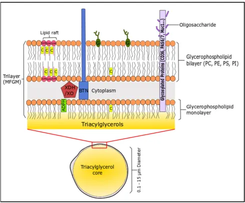
emulsion to form milk. These lipids assemble into spherical milk fat globules of triacylglycerides (0.1 494
phospholipids and sphingolipids, suspended in an aqueous liquid phase, which is derived from 496
mammary endothelial cells [159]. This unique structure is the milk fat globule membrane (MFGM), 497
which consists of lipid (40%), proteins (60%) and cholesterol [160]. The membrane consists of 498
phospholipids (mainly located on the outer leaflet) and cholesterol, which stabilises the TG-rich milk 499
fat globule against coalescence and protects the core from lypolytic degradation and oxidation 500
(Figure 2). Milk is a rich source of SFA, even though cow’s generally follow an unsaturated diet that 501
includes PUFA, due to their presence in forage crops and seeds. The high levels of SFA is due to 502
biohydrogenation of PUFA in the rumen of cattle [158]. 503
The phospholipids present in bovine ovine and caprine milks are quantitatively minor 504
constituents of milk lipids, however they possess beneficial techno-functional properties and are 505
involved in various physiological processes and nutritionally valuable. Other sources of 506
phospholipids in dairy products include MFGM fragments and lipoprotein particles, which are 507
believed to be remnants of the mammary secretory cell membranes. PLs like the MFGM originate 508
from the apical plasma membrane of the mammary gland secretory cell [2,3,159-163]. 509
Although phospholipids only account for 0.32 – 1.0 % of the total lipids of milk, they possess 510
strong biological activity. The PL content of raw bovine milk is reported between 9.4 and 35.5 511
mg/100g. The phospholipid composition consists of PE (19.8-42.0%), PC (19.2-37.3%), PS (1.9-10.5%), 512
and PI (0.6-11.8). The reported sphingolipid composition of raw milk consists of glucosylceramide 513
(GluCer: 2.1-5.0%), lactosylceramide (LacCer: 2.8-6.7%) and SM (18.0-34.1%) [3]. The phospholipid 514
content of small ruminants, such as ovine and caprine animals differs to that of bovine animals. 515
Zancada, Pérez-Díez, Sánchez-Juanes, Alonso, García-Pardo and Hueso [131] reports the presence of 516
27.6 mg/100g in caprine milk, and 29.8 mg/100g in ovine milk. The phospholipid composition of 517
ewes’ milk consists of PE (26.1-40.0%), PC (26.4-27.2%), PS (4.96-10.7%), PI (4.16-6.40%) and SM 518
(22.6-29.7%), whereas in goat milk it has been reported as PE (19.9-41.4%), PC (27.2-31.9%), PS 519
(3.2-14.0%), PI (4.00-9.37%) and SM (16.1-29.2%) [131]. It is well documented that the composition, 520
structure, and properties of the fatty acids in milk are affected by several factors such as the breed, 521
season, milking frequency, stage of lactation, nutritional status, and environmental conditions 522
[164-166]. It has also been reported that these factors as affect the phospholipid content of milk 523
[162,167-172]. As aforementioned, milk fat is characterised by short- and medium-chain fatty acids 524
(C4 – C14). These fatty acids are generally absent in the PL fraction of milk. PE tends to be highly 525
unsaturated followed by PI and PC, whereas PC tends to be saturated compared to other 526
glycerophospholipids. The three most abundant phospholipids present in milk are PE, PC and SM. 527
Sphingolipids are nutritionally beneficial PLs with a characteristic structure containing a sphingoid 528
base, which is a long-chain (12 – 22 carbon atoms) aliphatic amine, containing 2-3 hydroxyl groups. 529
Sphingosine (d18:1) is the most prevalent sphingoid base in milk, that contains 18 carbon atoms, two 530
hydroxyl groups, and one double bond. The fatty-acid pattern of SM is very uncommon with 531
approximately 97% of the fatty acids were saturated, including C16:0, C18:0, C18:1n9, C22:0, C24:0 532
and C23:0. The latter accounts for over 17% of the fatty acid content of SM [3,173,174]. 533
The peculiar fatty acid composition of SM allows the molecule to form in the cellular 534
membranes and rigid domains with cholesterol, called lipid rafts, which are involved in different 535
cellular processes [160]. The major sphingolipids in dairy products are GluCer, LacCer, and SM. 536
Gangliosides are also present in dairy product in low concentrations (0.14-1.10 mg/100mL) [175,176]. 537
Lysophospholipids and PA are generally not present in dairy samples and occur due to the 538
enzymatic activity of phospholipases [3]. Their origin is still unclear, but it is thought that could be 539
formed as a consequence of hydrolysis occurring during milk processing or poor sample storage [2] 540
In terms of health benefits, milk polar lipids are now known to have several nutritional benefits. 541
Sphingolipids and their metabolites including ceramide, sphingosine, and sphingosine phosphate 542
have been found to be highly bioactive, having important effects on cell regulation and are linked to 543
many inflammatory diseases [2,23,177-179]. Sphingolipids exhibit the ability to mediate intestinal 544
inflammation and may prevent colon-related diseases including cancer [180-184]. Research has also 545
indicated that the chemotherapeutic effects of dietary sphingolipids may also extend to other cancers 546
induce white adipose tissue hypertrophy and inflammation but increased colonic goblet cells. These 548
effects were attributed to the anti-inflammatory effects of the milk polar lipids, and in particular 549
sphingomyelin derivatives [187]. Dairy SM has also been shown to reduce cholesterol and FA 550
absorption through modulation of the cholesterol micellular solubility. The greater inhibitory effect 551
of milk SM on lipid absorption appears to be associated with its greater saturation and longer 552
chain-length of its fatty acyl group, which may allow for stronger hydrophobic interactions 553
[188-190]. 554
Recent research on dairy polar lipids has shown that PC derivatives in cheese [93] and yogurts 555
[94,95] have strong anti-thrombotic and anti-inflammatory activities. In yogurts, cardioprotective PC 556
derivatives have been isolated from yogurt polar lipids using TLC [95]. Sphingolipids, PE, and CL 557
also tend to be highly bioactive in these dairy products [95]. Research is has also shown that 558
fermented dairy products have greater antithrombotic and anti-inflammatory properties than 559
unfermented milk [94,191]. Therefore current research is focusing on the structural characterisation 560
of these phospholipids and how they are biosynthesised during dairy product manufacture. 561
Research suggests that these phospholipids may possess cardioprotective properties similar to 562
phospholipids of marine sources, thus further research is warranted to assess the putative benefits of 563
these lipids upon their consumption [23,191]. Overall, dairy product consumption seems to be 564
associated with positive cardiovascular and metabolic health contrary to general perception 565
[23,118,119,191-199], this may be due to the anti-inflammatory of dairy PLs [23]. 566
Figure 2: Illustration of the milk fat globule membrane. The sizes in this schematic are not 567
proteinaceous coat connecting the monolayer to the outer phospholipid bilayer. 569
Adipophilin (ADPH) is located in the inner layer polar lipid layer, xanthine 570
dehydrogenase/oxidase (XDH/XO) is located between both layers. PE, PS and PI are 571
generally concentrated on the inner surface of the membrane, whereas PC, SM, glycolipids 572
(G), cerebrosides and gangliosides are mainly located in the external membrane. SM and 573
cholesterol (C) can form rigid domains in the cellular membrane known as lipid rafts. 574
Glycoproteins are distributed over the external membrane surface, these include 575
butyrophilin (BTN), Mucin 1 (MUC1), PAS 6/7 and CD36. 576
2.3 Egg Phospholipids 577
Eggs are a valuable source of a wide variety of essential nutrients and bioactive compounds that 578
can impact human health including high quality protein, fat-soluble vitamins, B vitamins, minerals, 579
and choline, while providing relatively less saturated fat per gram compared to other animal protein 580
sources. The egg yolk is one of the richest sources of dietary phospholipids. On average, one large 581
egg contains 1.3 g of PLs by weight, which represents approximately the 28%–30% of the total lipids 582
of egg, while the remaining 66% are TAGs and 5% cholesterol. These lipids are almost exclusively 583
found in the yolk, where PC is the predominate PL species accounting for approximately 72% of the 584
total egg PLs. Other PLs are present in lesser quantities including PE (~20%), lyso-PC (3%), PI (2%), 585
and SM (3%). The majority of egg PLs contain long-chain saturated and monounsaturated FAs, 586
while the variety of the distribution of FAs can be somewhat reflective of the hen’s diet, age, and 587
environmental conditions [8,114,123].Because of their wide spectrum of activities, egg PLs have also 588
been extensively used as pharmaceutical excipients for pharmaceutical formulations in oral, dermal, 589
and parenteral products including liposomes [200]. 590
Egg PLs are important contributors to the overall dietary PLs intake in the western diet. Egg 591
PLs contribute 10% - 40% (or 0.8 g) of daily consumed PLs in typical westernised diet typical of the 592
USA [114]. Egg PLs are highly bioavailable; PC is usually absorbed at approximately 90% efficiency. 593
Consumption of eggs results in greater increases circulating HDL levels and enriches the HDL with 594
PLs from egg. This is because dietary PLs are preferentially incorporated into plasma-HDL, where 595
the beneficial effects are likely attributable to the consumption of egg PLs. It was also observed that 596
the incorporation of TAGs in HDL was decreased [114,123,201]. In contrast to egg PLs, the 597
absorption of egg-derived cholesterol is affected by the food matrix composition and can be altered 598
by interactions with dietary PLs, potentially altering the mobilisation of cholesterol from micelles in 599
the intestine [114].Egg PLs reduce cholesterol and FAs absorption by possibly interfering with lipid 600
mobilisation from mixed micelles. Although biliary PC is a critical emulsifier of dietary lipids and 601
aids in their digestion and absorption in the GI tract, excess luminal PC appears to inhibit lipid 602
absorption. Even though egg SM makes up only about 2% of total PLs in egg yolk, SM and other 603
sphingolipids have also been shown to dose-dependently reduce the absorption of cholesterol, 604
TAGs and FAs [114,188,202,203]. The influence of egg PLs on cholesterol absorption appears to be 605
dependent on the FA saturation, (i.e. egg PC or saturated egg-PC inhibited the absorption of 606
cholesterol into the lymphatic system greater than the more unsaturated soy PC) [114].The reduced 607
absorption of cholesterol, TAGs and FAsby dietary egg PLsappears to influence hepatic lipid levels 608
and metabolism, while egg SM was also found to reduce the expression of hepatic nuclear receptors 609
(such as peroxisome proliferator-activated receptor-α, PPAR-α), resulting in reduction of hepatic 610
expression of genes involved in cholesterol biosynthesis and FA metabolism [114,204]. 611
Egg PC and SM appear to regulate lipid absorption, hepatic lipid metabolism, and 612
inflammation. In clinical studies, egg PL intake is associated with beneficial changes in serum 613
biomarkers related to HDL function. In addition to the effects on lipid metabolism, dietary intake of 614
egg PL may also reduce inflammation [114,201,205]. Consuming eggs for 12 weeks resulted in a 615
reduction in plasma C-reactive protein (CRP) and an increase in adiponectin in overweight men; 616
changes which were not observed with yolk-free egg substitute [205]. Egg consumption has also led 617
to improvements in circulating plasma inflammatory markers in adults with metabolic syndrome 618
suffering from CVDs, since they modulate components of cell membranes, contribute to a decrease 620
of cholesterol level and blood pressure [206].In addition, hen egg yolk PLs were found to inhibit in 621
vitro PAF-induced platelet aggregation, using cage-free hen egg yolk PLs. Thus hen egg yolk was 622
found to contain natural PAF-inhibitors that reinforce their nutritional value in terms of protection 623
against CVDs [92]. 624
The majority of research investigating inflammatory properties of PLs has focused on PC.For 625
example, an in vitro study with hepatic cancer cell lines showed a dose dependent growth restraint 626
when cancer cells were cultured in the presence of egg yolk PC (99% pure PC from egg yolk) and 627
menaquinone-4 (vitamin K2). Furthermore, PC alone also showed a statistically significant reduction 628
of cancer cells via death ligands (i.e.TNF-α), thereby promoting apoptosis by the activation of 629
caspase-8 and -3, resulting in PAP (poly(A)-polymerase) inhibition [8,207]. However, as 630
aforementioned, gut bacteria may be responsible for these observations. 631
However, even though Egg-derived PLs have anti-inflammatory properties, they also seem to 632
exhibit pro-inflammatory properties, via both direct and indirect mechanisms. For example, despite 633
the evidence to suggest that phosphatidylcholine is anti-inflammatory, egg intake has also been 634
shown to dose-dependently increase post-prandial trimethylamine-N-oxide (TMAO) concentrations 635
in plasma, and thus egg PLs have recently been implicated in the promotion of inflammation and 636
atherosclerosis due to this increased formation of TMAO [123]. 637
In addition, the existence of small amounts of the pro-inflammatory mediator PAF and its 638
hydroxyl-PAF analogue (that mimics PAF-activities) was also found in the PCs fraction extracted by 639
hen's egg yolk [38]. A predictable finding, since PAF and its analogues are playing crucial roles in 640
fertilisation [208].Thus, even though the existence of small amounts of PAF and its analogues in 641
hen’s egg-yolk seem to participate mainly in the reproductive system of hen, over consumption of 642
eggs (much more than the dietary recommendations) may increase pro-inflammatory PL uptake 643
(such as PAF and its analogues). 644
Given that numerous epidemiological studies have failed to conclusively find an association 645
between egg intake and atherosclerosis, additional long-term studies are required to determine 646
whether egg-induced TMAO production or pro-inflammatory PL uptake has detrimental effects on 647
inflammation and disease risk. It is also crucial to discern whether the natural existence of 648
PAF-inhibitors in egg-yolk and the perceived benefits of egg PLs intake on CVD risk markers will be 649
able to outweigh any risk derived by potential TMAO formation and PAF uptake by 650
over-consumption of eggs. 651
3. Marine Origin: 652
3.1 Sources of Marine Phospholipids 653
Marine lipids can be derived from several fish, shellfish and algae, but also by Antarctic 654
crustacean krill (Euphausia superba) and from marine industry by-products such as fish roe (fully ripe 655
internal egg masses in the ovaries of fish) [43,209,210]. Fish contains between 1%–1.5% PLs, while the 656
amount of PLs in oil extracted from krill is typically around 40% of its total lipids [211,212]. PC 657
derivatives are the predominant phospholipids present in salmon, tuna, rainbow trout, and 658
mackerel; the second most abundant phospholipid is PE, PI, PS, lyso-PC, and sphingomyelin are 659
also present in minor quantities [213]. Fish roe from herring, salmon, pollock, and flying fish contain 660
between 38%–75% of their lipids in the form of PLs with PC being the predominant lipid class. 661
Notably, the main PLs class of marine-derived PLs is PC, predominantly binding with unsaturated 662
ω-3 PUFAs, with the most prevalent being EPA and DHA, but also stearidonic acid and 663
docosapentaenoic acid (DPA). Marine organisms are enriched in these PUFAs by the aquatic food 664
chain since the main source of ω-3 PUFAs are algae that can synthetize them de novo. Humans can 665
only poorly synthesize ω-3 PUFAs from their precursor α-linolenic acid (ALA; 18:3ω-3) and thus the 666
dietary intake of EPA and DHA is essential as they are extensively associated with optimal human 667
health and protection against disease [43]. 668
670
3.2 Oxidation of Marine Phospholipids – Pro-Inflammatory Mediators 671
ω-3 PUFAs such as DHA are highly susceptible to oxidation and the formation of toxic oxidized 672
products such as aldehydes and hydroperoxides [214,215]. Furthermore, oxidized PLs with PAF-like 673
structures that can mimic PAF-activities can be produced by the oxidation of marine PLs [216]. High 674
amounts of oxidized products in the body over a prolonged period can cause oxidative stress, which 675
can induce an inflammatory response [214]. PUFAs, EPA and DHA, need to be protected, and some 676
different strategies have been used to avoid oxidation. Since the most stable ω-3 fatty acids are in the 677
form of PLs, incorporation of ω-3 PUFAs into the PL structure increases their oxidative stability, 678
suggesting that these molecules may be a more beneficial form of PLs [217]. DHA incorporated into 679
PLs has been found to be more resistant to oxidation than both TAGs and ethyl ester bound DHA 680
[218]. 681
Marine PL products have revealed surprisingly high stability against oxidation [43].There is 682
speculation as to whether this is due to the natural content of antioxidants (e.g., astaxanthin) 683
co-extracted with other lipids and PLs from the biomass or if this is a function of the PLs themselves. 684
Research suggests that both assumptions may be correct, due to the fact that other non-marine PLs, 685
even when highly purified and devoid of antioxidants, are usually quite resistant to oxidation [58]. 686
However, the oxidative stability of marine PLs is influenced by the quality, source, chemical 687
composition of marine PLs and the degree of non-enzymatic browning reactions within marine PLs. 688
In general, the non-enzymatic browning reactions in marine PLs are influenced by the marine PLs 689
manufacturing processes. In addition, the use of marine PLs for food fortification is a challenge due 690
to the complex nature of the degradation products that are formed during the handling and storage 691
of marine PLs. Therefore, stabilisation of marine PLs in food systems with the addition of natural 692
antioxidants should be further investigated [219]. For example, the combination of tocopherol, 693
ascorbic acid, and lecithin has a higher protective effect on PUFAs than tocopherol, ascorbic acid, or 694
lecithin individually [220]. To achieve the maximum protective effect, PUFAs such as DHA should 695
be incorporated into PC or PE (one DHA molecule per lipid molecule) and both tocopherol and 696
ascorbic acid should be added during food fortification or the manufacture of nutraceuticals or 697
supplements [221]. Marine PLs products containing ω-3 PUFAs within their structure seem to 698
provide resistance to oxidation PUFAs on their own. 699
3.3 Bioavailability and Biofunctionality of Marine Phospholipids 700
Consumption of ω-3 PUFAs, particularly the long-chain FAs EPA and DHA, has been reported 701
to have beneficial physiological effects, including the reduction in the incidences of cardiovascular 702
disease, cancer, diabetes, arthritis, and central nervous system disorders such as schizophrenia, 703
depression, Alzheimer’s disease [222,223]. Dietary ω-3 PUFAs have also exhibited beneficial effects 704
in respect to essential FA deficiency in infancy (retinal and brain development), autoimmune 705
disorders, Crohn’s disease, and cancers of breast, colon, and prostate [58]. The beneficial health 706
effects of PUFAs has mostly been attributed to their anti-inflammatory and antithrombotic 707
properties by their ability to decrease both the formation and tissue incorporation of ARA. This then 708
prevents the overproduction of ARA-derived eicosanoids and reduces the release of inflammatory 709
acute-phase proteins. By being precursors to lipid mediators (eicosanoids/docosanoids) or as ligands 710
for transcription factors, these ω-3 PUFAs affect cell and tissue physiology and response to external 711
signals. In addition, EPA and DHA can influence cell membrane fluidity, permeability or membrane 712
protein-mediated responses. By these means, EPA and DHA have been proposed to support 713
cardiovascular health as well as cognitive, visual, immune, and reproductive system functions [43]. 714
There are also indications that they confer health benefits regarding tumorigenesis, 715
hypertriglyceridemia, atherosclerosis, mental illness, dementia, bone health, and attention-deficit 716
hyperactivity disorder (ADHD) [43]. Therefore, the development of products containing ω-3 EPA 717