RESEARCH ARTICLE
Smooth muscle cell-specific knockout of neuropilin-1 impairs postnatal lung
development and pathological vascular smooth muscle cell accumulation
Marwa Mahmoud, Ian M. Evans, Vedanta Mehta, Caroline Pellet-Many, Ketevan Paliashvili, and Ian Zachary
Centre for Cardiovascular Biology and Medicine, BHF Laboratories, Division of Medicine, University College London, London, United Kingdom
Submitted 16 October 2018; accepted in final form 7 January 2019
Mahmoud M, Evans IM, Mehta V, Pellet-Many C, Paliashvili K, Zachary I.Smooth muscle cell-specific knockout of neuropilin-1 impairs postnatal lung development and pathological vascular smooth muscle cell accumulation. Am J Physiol Cell Physiol 316: C424 – C433, 2019. First published January 16, 2019; doi:10.1152/ajpcell. 00405.2018.—Neuropilin 1 (NRP1) is important for neuronal and cardiovascular development due to its role in conveying class 3 semaphorin and vascular endothelial growth factor signaling, respec-tively. NRP1 is expressed in smooth muscle cells (SMCs) and medi-ates their migration and proliferation in cell culture and is implicated in pathological SMC remodeling in vivo. To address the importance ofNrp1for SMC function during development, we generated condi-tional inducibleNrp1SMC-specific knockout mice. Induction of early postnatal SMC-specificNrp1knockout led to pulmonary hemorrhage associated with defects in alveogenesis and revealed a specific re-quirement forNrp1in myofibroblast recruitment to the alveolar septae and PDGF-AA-induced migration in vitro. Furthermore, SMC-spe-cificNrp1knockout inhibited PDGF-BB-stimulated SMC outgrowth ex vivo in aortic ring assays and reduced pathological arterial neoin-tima formation in vivo. In contrast, we observed little significant effect of SMC-specific Nrp1knockout on neonatal retinal vascular-ization. Our results point to a requirement ofNrp1in vascular smooth muscle and myofibroblast function in vivo, which may have relevance for postnatal lung development and for pathologies characterized by excessive SMC and/or myofibroblast proliferation.
alveolar myofibroblast; neointima; neuropilin-1; SMC
INTRODUCTION
Neuropilin-1 (NRP1) is a transmembrane glycoprotein re-ceptor essential for both vascular and neuronal development due to its role in mediating vascular endothelial growth factor (VEGF) and class 3 semaphorin signaling, respectively (14, 16, 25, 36).Nrp1-null mice are embryonic lethal between embry-onic day (E) 10.5 and E14.5, dependent on genetic background, and display a spectrum of cardiovascular and neuronal defects (21, 23).
Several studies have reported NRP1 expression in smooth muscle cells (SMCs) and revealed an important role for NRP1 in vascular SMC migration (13, 27, 30, 35). NRP1 expression has also been reported in SMCs in vivo in large vessels (20) and targeted knockdown of NRP1 in vivo using small hairpin
inhibitory RNA-inhibited neointima formation induced by rat carotid artery endovascular injury (32). Mice with constitutive SMC-specific loss of Nrp1 were viable, displaying no overt early postnatal phenotype but exhibiting a late-onset defect in gastrointestinal motility due to loss of visceral SMC contrac-tility (46). The lack of an overt phenotype in constitutively SMC-specific Nrp1-null mice could be indicative of genetic redundancy, as reported for other constitutive knockout mice (5, 17, 42). NRP1 shares a similar domain structure and 44% amino acid sequence homology with neuropilin-2 (NRP2), which is also expressed in vascular smooth muscle cells (10, 30 –32). Mice lackingNrp1andNrp2die earlier in embryo-genesis (E8.5) and display more severe defects in angiogen-esis compared with single knockouts (39). Moreover, Nrp2 compensates for loss of semaphorin binding to NRP1 in Nrp1Sema/Semamice (15). These data suggest a partial genetic
redundancy betweenNrp1andNrp2, which could explain the lack of an overt phenotype in constitutive SMC-specificNrp1 -null mice.
To circumvent genetic redundancy effects due to possible compensatory upregulation ofNrp2expression, we generated a conditional and inducible smMHC-CreERT2transgenic line to
allow controlled Nrp1 knockout in SMCs when crossed to Nrp1floxed mice, by administration of tamoxifen during devel-opment. Our results revealed a specific requirement forNrp1in myogenic cell lineages during pulmonary microvascular expan-sion and alveogenesis.Nrp1was also necessary for pathological neointima formation in vivo, for PDGF-BB-induced SMC out-growth ex vivo in aortic rings, and for PDGF-AA-induced pul-monary myofibroblast migration in cultured cells.
MATERIALS AND METHODS
Generation and characterization of mice.All procedures involving mice were conducted under a UK Home Office license and the approval of the University College London local ethics committee in accordance with the Animal Care and Ethics Guidelines and the 1986 United Kingdom Home Office Animals (Scientific Procedures) Act.
Inducible SMC-specific Nrp1 knockout mice were generated by crossingNrp1floxed
; R26Rmice tosmMHC-CreERT2
transgenic mice, on a C57BL/6 background. Genotypes of progeny were identified by PCR using the primers listed in Table 1. To induce knockout in neonates, mice were given two subcutaneous 0.5 mg tamoxifen injections at P1–2 and P3– 4. For adults (⬎8 wk), 5 consecutive 1-mg tamoxifen injections were administered intraperitoneally over 5 days. Histology.Tissue was fixed in HistoChoice tissue fixative (Sigma) or zinc fixative (BD PharMingen) overnight and then processed to paraffin and cut into 10-m sections, except for the retinas, which Address for reprint requests and other correspondence: I. Zachary, Centre
for Cardiovascular Biology and Medicine, BHF Laboratories, Division of Medicine, University College London, 5 University Street, London WC1E 6JJ UK (e-mail: i.zachary@ucl.ac.uk).
were fixed in 4% paraformaldehyde and stained as whole-mounts. Samples were subjected to hematoxylin-eosin histological staining, immunohistochemistry, or immunofluorescence using the antibodies and conditions listed in Table 2. X-Gal staining was performed using the LacZ Detection Kit by InvivoGen (no. rep-lz-t), according to the manufacturer’s instructions. Alveolar hemorrhaging was identified by the incidence of hemosiderin-laden macrophages, as previously doc-umented (1).
Transwell migration assay. Mouse pulmonary fibroblasts (no. M3300-57, ScienCell) were cultured in poly-l-lysine-coated dishes and treated with 10 ng/ml transforming growth factor-1 (TGF-1; no. AF-100-21C, Peprotech) for 48 h to induce differentiation to myofibroblasts. Cells were transfected with either scrambled siRNA or two different siRNAs targetingNrp1(siRNA cat. nos. 70800 and s70802, respectively; Thermo Fisher Scientific) using RNAiMAX transfection reagent ( no. 13778075, Thermo Fisher Scientific). Trans-fected cells were seeded at a count of ~2.5 ⫻ 104 cells/Transwell
insert (8.0-m pore size, no. 353097, Falcon) and left to migrate for 20 –24 h in response to either serum-free media or serum-free media with 50 ng/ml PDGF-AA (100-13A, Peprotech). Nonmigrated cells were removed, and Transwell inserts were stained using the REASTAIN Quick-Diff Kit, according to manufacturer’s instruc-tions (no. 102164, Gentaur), and migrated cells were counted under a Leica stereo microscope.
Western blotting. Western blot analysis was performed on trans-fected myofibroblast cell lysates as described previously (11), using the following primary antibodies: neuropilin-1 (no. ab81321, Abcam),
␣-smooth muscle actin (␣-SMA; no. ab7817, Abcam), and-actin (no. A2228, Sigma).
Aortic ring assay.Aortas from 8- to 10-wk-old male mice that had been treated with tamoxifen the week before harvest were dissected and cultured as described previously (3, 9) in the presence of 1M 4-hydroxytamoxifen overnight, after which rings were incubated in fresh medium-containing cytokines. Cell growth and sprout formation were monitored using the IncuCyte Live Cell Analysis System (Essen
BioScience). Assays were stopped 6 days postembedding and sub-jected to double immunofluorescence staining for endothelial-specific isolectin B4 (no. DL-1207, Vector Laboratories) and ␣-SMA (no. F3777, Sigma) to visualize vascular sprouts.
Perivascular cuff model. Surgeries were performed as described (29). Briefly, under general anesthesia (using inhaled isoflurane), the femoral artery is exposed, and a polyethylene cuff (with a sagittal opening to allow placement of the artery) is loosely placed around the artery and secured in place with sutures. Sham surgical controls were handled in the same manner, except no cuff placement was performed. All mice were treated with tamoxifen the week before the surgery. Twenty-one days following the surgery, the mice were euthanized by overdose of CO2, followed by cervical dislocation, and the cuffed
femoral arteries and sham controls were harvested for analysis. Measurements and statistical analyses. Pulmonary hemorrhaging was quantified by blinded measurements of the percentage of hemor-rhaging per field of view from at least three different sections per sample. Statistical significance was calculated using the Kruskal-Wallis H test with post hoc Dunn’s multiple comparison test. Quan-tification of␣-SMA expression and numbers of presumptive septal tips with␣-SMA-expressing myofibroblasts was determined using the ImageJ Color Threshold function or the ImageJ Cell Counter plugin, respectively. Data were collected and analyzed blindly from at least three different sections/fields of view per sample.
Outgrowth areas in aortic ring assays were quantified as described (8), using the ImageJ Color Threshold function (with background subtraction).
Unless specified otherwise, all data are presented as means ⫾ SE, and statistical significance was calculated using the two-tailed unpaired Student’st-test, except for the perivascular cuff intima/ media ratio data, which were analyzed using the one-tailed un-paired t-test, and the myofibroblast Transwell migration data, which were analyzed using two-way ANOVA with Bonferroni post test.
Table 1. Details of genotyping primers
Gene Targeted Genotyping Primer Sequence (5=–3=)
FloxedNrp1allele flNrp1_F1 CAA TGA CAC TGA CCA GGC TTA TCA TC
flNrp1_R1 GAT TTT TAT GGT CCC GCC ACA TTT GTC
RecombinedNrp1allele rNrp1_F2 AGG CCA ATC AAA GTC CTG AAA GAC AGT CCC
rNrp1_R2 TCT GCA GAT CAT GTA TAC TGG TGA CCC ACA
smMHC-CreERT2transgene SMWT1 TGA CCC CAT CTC TTC ACT CC
SMWT2 AAC TCC ACG ACC ACC TCA TC
PhCREAS1 AGT CCC TCA CAT CCT CAG GTT
LacZallele LacZ1 AAA GTC GCT CTG AGT TGT TAT
LacZ2 GGA GCG GGA GAA ATG GAT ATG
LacZ3 GCG AAG AGT TTG TCC TCA ACC
Nrp1, neuropilin 1.
Table 2. Antibodies used for histology
Protein Antibody Details Working Dilution Antigen Retrieval/Special Kit Used ␣-SMA Monoclonal anti-actin,␣-smooth muscle,
clone 1A4. Sigma no. A2547.
1/100 M.O.M. Kit (Vector Labs no. BMK-2202)
NG2 Anti-NG2 chondroitin sulfate proteoglycan antibody. Millipore no. AB5320.
1/200 Citrate buffer (pH 6.0)
Isolectin B4 DyLight 594 Labeled Griffonia Simplicifolia
Lectin I (GSL I) isolectin B4. Vector Labs no. DL-1207.
1/100
␣-SMA Monoclonal anti-actin,␣-smooth muscle-FITC antibody, clone 1A4. Sigma no. F3777.
1/100
NRP1 Neuropilin 1 antibody. GeneTex no. GTX127947. 1/100 Citrate buffer (pH 6.0) Ki67 Ki-67 (D3B5) rabbit mAb. Cell Signaling
Technology no. 12202.
1/200 Citrate buffer (pH 6.0)
RESULTS AND DISCUSSION
SMC-specific knockout of Nrp1 in early postnatal develop-ment causes pulmonary hemorrhaging and impaired alveolar development. SmMHC-CreERT2 mice (44) were bred with
Nrp1floxed mice (14), previously crossed to the R26R-LacZ
reporter strain (37), to generateNrp1SMCiKOmice, allowing for
inducible ablation ofNrp1in SMCs (Fig. 1A). Induction of Cre recombinase activity, monitored by expression of -galactosi-dase, following administration of tamoxifen in adult and neo-natal Nrp1SMCiKO mice, was restricted to SMCs in multiple
tissues, including the heart and lungs (Fig. 1B). Nrp1 allele recombination and protein knockdown in SMC were confirmed by genotyping PCR (Fig. 1C) and immunostaining (Fig. 1D). To determine the role of SMC-specificNrp1in early post-natal development, we conditionally ablated Nrp1expression using tamoxifen from postnatal day (P)1–2 and monitored the effect ofNrp1loss. TheNrp1SMCiKOneonates appeared viable
and healthy compared with their littermate controls; however, internal examination revealed hemorrhaging in the lungs of the Nrp1SMCiKO neonatal pups from P8 (Fig. 2A). Hemorrhaging
was generally localized to the lung periphery and characterized by the presence of hemosiderin-laden macrophages, which are an indication of long-term alveolar hemorrhaging (⬎48 h), and evidence of necrosis of the alveolar walls (Fig. 2B). Hemor-rhaging was most marked and statistically significant compared with control littermates at P14 (Fig. 2C) but had partially resolved by P22. This phenotype was not fully penetrant, with 75% and 66.7% ofNrp1SMCiKOmutants displaying pulmonary
hemorrhaging at P8 and P14, respectively (Fig. 2D). Timing of the knockout was critical, as a later induction with tamoxifen at P3– 4 led to a much milder hemorrhaging phenotype (red data points in Fig. 2C).
To determine the underlying cause of the pulmonary hem-orrhaging, we examined SMC and myofibroblast coverage of the lung by immunohistochemical staining for␣-SMA expres-sion. Whereas SMCs are present in larger blood vessels and muscularized airways, myofibroblasts are specialized cells present on the alveolar septa in the developing postnatal lung and are identified by their expression of ␣-SMA and their location at septal tips (7, 45). This revealed a loss of
Neonate
Heart Lung
Cre+ Control Cre - Control
Heart Lung Lung
Lung
Adult
100µm
100µm
200µm
400µm
200µm
400µm
Nrp1
+/+
Nrp1
SMCiKO
Lumen
Lumen
100µm
100µm
A C
B D
Fig. 1. Characterization ofNrp1smooth muscle cell (SMC)-specific inducible knockout mice.A: floxedNrp1mice have exon-2 flanked by two loxP sites, which undergo Cre-mediated recombination exclusively in SMCs when crossed to thesmMHC-CreERT2transgenic mice and given tamoxifen. Location of the floxed
Nrp1and recombinedNrp1PCR primers is depicted by F1 and R1 and F2 and R2, respectively.B: X-gal staining showing Cre recombinase activity inCre⫹
and Cre⫺mice. C: genotyping PCR used to identify transgenic mice. The recombinedNrp1allele is only detected following treatment of the mice with tamoxifen.D: immunostaining for NRP1 in the aortae ofNrp1SMCiKOandNrp1⫹/⫹mice. NRP1, neuropilin 1; SEA, PDZ-binding domain motif; EC, extracellular
alveolar myofibroblasts in the secondary alveolar septae in theNrp1SMCiKOmutants (Fig. 2E, red arrows), suggesting a
defect in the recruitment of alveolar myofibroblasts to the secondary alveolar septae of neonatal mice following Nrp1 depletion. Quantification of alveolar myofibroblast recruit-ment, based on their expression of␣-SMA and their location at septal tips (22, 45), showed a significant reduction in myofibroblasts associated with septae in Nrp1SMCiKO mice
(Fig. 2F). In addition, a significant reduction in overall coverage of␣-SMA-positive cells occurred in the alveoli of the Nrp1SMCiKO mutants (Fig. 2, Gand H). The number of
alveolar septae was not significantly different between the Nrp1SMCiKO mutants and controls when normalized to total
tissue area, although a trend towards a reduction was ob-served (Fig. 2I). The expression of Cre recombinase in areas corresponding to the location of alveolar myofibroblasts was
confirmed by X-Gal staining (Fig. 3A), which demonstrated expression ofsmMHC-CreERT2and recombination of theNrp1
allele in these cells. Recruitment of myofibroblasts to septal tips is important for terminal airway branching and future alveolarization (6, 7), and concomitant with loss of alveolar myofibroblasts in the septae of theNrp1SMCiKOmutants (Figs.
2,EandFand 3C), we observed evidence of impaired alveolar development at P14, as indicated by fewer and larger alveoli in theNrp1SMCiKOmutants versus littermate controls (Fig. 2,
A and G). No significant changes in expression of the pericyte-specific marker neural/glial antigen 2 (NG2) were observed in the lungs of P14Nrp1SMCiKOneonates (Fig. 3B),
suggesting that inducible Nrp1 knockout did not impact pericyte recruitment to the vasculature, consistent with the reported lack of expression ofsmMHC in pericytes (41, 43). The reduction in ␣-SMA expression was also limited to the
A
B
E
C
Red blood cells inside the alveolar
lumens Hemosideren-laden macrophages Necrosis of alveolar walls 400µm P8 P14 P22
Nrp1+/+ Nrp1SMCiKO Nrp1+/+ Nrp1SMCiKO
100µm 100µm 100µm bronchiole artery
F
3mm 400µm 3mm 400µm 1mm 1mm 1mm 1mm 200µm 200µmG
H
Pulmonary Haemorrhaging (P8) +/+ Nrp1 (P8) SMCiKO Nrp1 (P14) +/+ Nrp1 (P14) SMCiKO Nrp1 0 1 2 3 4 5 6 7 8 9 1011 *
P<0.02 P e rcen ta g e H aem o rr h ag in g / Fi e ld of V ie w -SMA Nrp1 SMCiKO Nrp1 +/+
D
400µm Nrp1+/+ -SMA ImageJ Colour Threshold Nrp1SMCiKO p<0.01 *Nrp1+/+ Nrp1SMCiKO
100 75 50 25 0 Percentage α
-SMA positive Septa
Nrp1+/+ Nrp1SMCiKO Nrp1+/+ Nrp1SMCiKO
* (p<0.05)
I
ns (p<0.13) 10.0 7.5 5.0 2.5 0.0 1.00 0.75 0.50 0.25 0.00 Percentage α -SMA staining
/ total tissue area
Tota
l Septa Nor
m
a
lis
e
d to
Total Tissue Area / Field of View
Fig. 2. Pulmonary phenotype inNrp1SMCiKOmice.A: hematoxylin-eosin staining ofNrp1⫹/⫹andNrp1SMCiKOlung sections. Arrows indicate pulmonary hemorrhaging, which is detected from P8, increases to P14, and is reduced by P22.B: high-magnification image indicating hemorrhaging and alveolar wall necrosis inNrp1SMCiKOlung.
C: quantification of hemorrhaging at P14. *P⬍0.02 vs.Nrp1⫹/⫹. Red symbols indicate mice treated with tamoxifen at P3; all other mice were treated at P1.D: incidence
of pulmonary hemorrhaging in theNrp1SMCiKOneonates.E: immunostaining of lung sections for␣-SMA. Black arrows indicate␣-SMA-positive septal tips inNrp1⫹/⫹
alveoli, and red arrows indicate␣-SMA-negative septal tips inNrp1SMCiKOalveoli.F: analyses of the percentage of␣-SMA-positive secondary alveolar septal tips at P14; *P⬍0.01 vs.Nrp1⫹/⫹.G:␣-SMA staining was quantified using the ImageJ Color Threshold function; representative images showing detection of␣-SMA staining
alveolar septae and capillaries, whereas ␣-SMA staining of larger arteries and arterioles did not appear to be affected (Fig. 3, B and C). Given the important role of myofibroblasts in alveolarization during the rapid early postnatal growth and remodeling of the lung, we hypothesize that the observed hemorrhaging resulting from SMC-specific Nrp1 knockout may be due to defective alveolarization consequent upon im-paired myofibroblast recruitment, which in turn results in capillary loss and a reduced capacity of the developing lung to accommodate increased blood flow. Additionally, an impair-ment of capillary stability due to reduced smooth muscle cell recruitment to capillaries and small vessels may also contribute to the hemorrhaging phenotype inNrp1SMCiKOneonates.
Fur-ther work is required to establish more precisely the causal links between defective myofibroblast recruitment and hemor-rhaging in the lungs ofNrp1SMCiKOmice.
PDGF stimulation of pulmonary myofibroblast migration is inhibited following Nrp1 knockout. Platelet-derived growth factor subunit A (PDGF-A) signaling is required for
pulmo-nary myofibroblast differentiation and migration, as evi-denced by the absence of myofibroblasts from the primary alveolar septa in PDGF-A-null mice, resulting in severe alveolarization defects (7, 22, 26). To determine the effect ofNrp1loss on PDGF-induced myofibroblast migration, we performed in vitro Transwell migration assays. Pulmonary fibroblasts isolated from isolated from postnatal day 2 C57BL/6 mouse lung were differentiated into myofibro-blasts in vitro based on cell plating density and treatment with TGF-1, as previously described (28), and PDGF-AA-induced myofibroblast migration was then measured after transfection with either scrambled siRNA or two different Nrp1-specific siRNAs. The results showed a significant reduction in the migration of NRP1-depleted myofibroblasts in response to PDGF-AA (Fig. 4A). The efficiency of Nrp1 protein knockout by siRNA was confirmed by Western blotting (Fig. 4B). Nrp1 siRNA knockout in myofibroblasts caused no observable effects on viability (results not shown) Fig. 3. Pulmonary phenotype in theNrp1SMCiKOmice.A: X-gal expression in P8 lungs showing Cre recombinase expression in cells with locations corresponding to presumptive alveolar myofibroblasts (arrows). B: ␣-SMA and NG2 staining in the lungs of Nrp1SMCiKO mice and littermate controls. Arrows indicate
or ␣-SMA expression levels compared with control myofi-broblasts transfected with scrambled siRNA (Fig. 4B).
PDGF stimulation of SMC migration is inhibited following Nrp1 knockout.NRP1 is implicated in the regulation of PDGF signaling required for migration and proliferation in mesen-chymal stem cells (4) and vascular SMCs (30, 32). The effect of Nrp1 loss on PDGF-induced myogenic cell proliferation, migration, and recruitment to neovascular sprouts was deter-mined ex vivo in aortic ring assays. A marked reduction in SMC outgrowth in response to PDGF-BB was observed in the Nrp1SMCiKO aortic rings versus wild-type controls following
treatment with tamoxifen (Fig. 4,C andD). Although SMCs appeared to be recruited normally to the developing vascular sprouts in this model (Fig. 4E), the sprouts in theNrp1SMCiKO
aortic rings exhibited reduced branching compared with the wild-type aortic rings. Since prevention of VEGF binding to NRP1 blocks endothelial vascular sprouting in aortic rings (12), we also examined the effect of SMC-specific Nrp1 knockout on the VEGF response. In contrast to the impaired response to PDGF-BB, VEGF-induced aortic ring vessel
sprouting was not reduced inNrp1SMCiKOaortic rings,
indicat-ing that the effect ofNrp1ablation was specific for SMCs and did not impact indirectly on endothelial outgrowth.
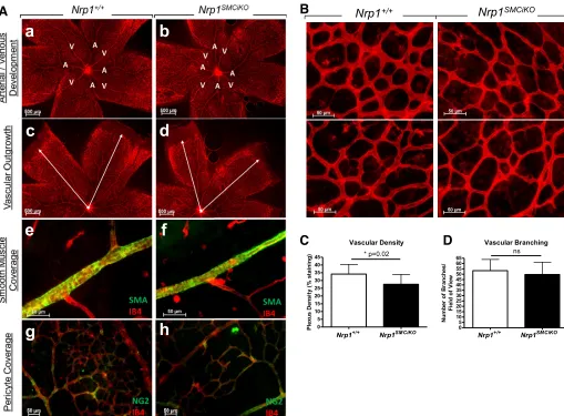
Retinal vascular development is largely unaffected by SMC-specific loss of Nrp1.The impact of SMC-specific loss ofNrp1 in developmental angiogenesis was further investigated in the neonatal mouse retina, in which the vasculature develops radially from the central optic nerve from P0 and matures into a hierarchical network of arteries, veins, and interconnecting capillaries (38). Following ablation of Nrp1 expression in Nrp1SMCiKOneonates from P1, we could not detect any delay
in vascularization, defects in arterial/venous differentiation, or an effect on SMC/pericyte coverage of the retinal vessels in the Nrp1SMCiKOneonates versus their littermate controls (Fig. 5A).
However, a small, statistically significant reduction in vascular density due to smaller capillary diameters was detected in Nrp1SMCiKOretinas, whereas vascular branching appeared
un-affected (Fig. 5, B–D). Since Nrp1 loss in our model is restricted to SMCs and myofibroblasts, whereas pericytes are unaffected, it is possible that the mild retinal phenotype
ob-A
B
C
Aortic Ring Assay Outgrowth Area
+/+
Nrp1 SM
CiKO
Nrp1
+/+
Nrp1 SM
CiKO
Nrp1
+/+
Nrp1 SM
CiKO
Nrp 1 0
5 10 15 20 25 30
**
ns
+50ng/ml PDGFBB
control
P<0.005
+30ng/ml VEGF-A
ns
P<0.0001
***
P<0.005 **
%
Ou
tg
ro
wt
h
A
re
a
D
E
Fig. 4. Nrp1 is required for PDGF-induced migration of myofibroblasts and SMCs.A: pulmonary myofibroblast Transwell migration assays in response to PDGF-AA compared with serum-free (SF; siScrambled,n⫽3; siNrp1 #1,n⫽3; siNrp1 #2,n⫽3). *P⬍0.05 vs. siScr SF and #P⬍0.01 vs. siScr PDGF-AA.
served results from a restricted role of myofibroblast-mediated vascular remodeling in retinal vascularization.
Pathological neointima formation is attenuated following SMC-Nrp1 knockout. To determine the role of SMC-Nrp1 in pathological SMC accumulation giving rise to neointima for-mation and vascular remodeling, we utilized the mouse perivascular cuff model of vessel injury, in which neointima formation is induced by placement of a nonocclusive cuff around the femoral artery, without removal of the endothelial cell layer (29). We detected a significant reduction in neoin-tima formation in theNrp1SMCiKOmutants versus controls at 21
days following cuff placement and tamoxifen treatment to reduceNrp1expression in arterial SMC (Fig. 6, AandC–F). Cell proliferation in the media in Nrp1SMCiKO mice was
de-creased versus controls, as measured by ki67 staining (Fig. 6,G–J), and the reduction in proliferation was statistically significant (Fig. 6B), indicating that reduced SMC prolifer-ation at least partly explains reduced neointima formprolifer-ation in Nrp1SMCiKO mice (24). It is likely that medial proliferation
of the SMCs is followed by the migration of these cells through the internal elastic lamina into the intima. Studies have also implicated adventitial myofibroblasts in the de-velopment of neointima (33, 34). These results are consis-tent with inhibition of SMC/myofibroblast migration and pro-liferation following genetic ablation of Nrp1 expression in these cells in vivo.
In conclusion, this study identifies a novel requirement for Nrp1 in SMCs and myofibroblasts during alveolar develop-ment in vivo. A role for Nrp1 in lung development is also supported by an earlier finding that constitutive loss of sema-phorin-NRP1 signaling due to knock-in of mutantNrp1unable to bind Sema3 ligands led to acute respiratory distress and high neonatal mortality, which was associated with loss of alveolar myofibroblasts at sites of presumptive septal tips (18), as we found in Nrp1SMCiKO mice. However, Joza et al. did not
identify the cell types responsible for the phenotypes observed. Since manifestation of the phenotype waned later in postnatal life and later induction of knockout resulted in a milder
a
b
c
d
e
f
g
h
Nrp1SMCiKO
Nrp1+/+
B
Nrp1+/+ Nrp1SMCiKO
0 5 10 15 20 25 30 35 40 45
Vascular Density
* p=0.02
P
lexu
s D
e
n
s
it
y (
%
st
ai
n
in
g
)
Vascular Branching
Nrp1+/+ Nrp1SMCiKO
0 5 10 15 20 25 30 35 40 45 50 55 60 65
ns
N
u
m
b
er
o
f B
ran
ch
es/
Fi
e
ld of
V
ie
w
A
C
D
Fig. 5. SMC-specific Nrp1 loss results in a mild reduction in retinal vascular density, but retinal vascular development is largely unaffected.A: retinal vascular development showing arterial/venous differentiation (aandb), vascular outgrowth (candd), SMC coverage (eandf), and pericyte coverage (gandh) in the
Nrp1SMCiKOmutants (b,d,fandh) vs. controls (a,c,e, andg) at P8.B: representative images from P8 wild-type andNrp1SMCiKOretinas stained with endothelial isolectin B4.C: quantification of vascular density at P8. *P⫽0.02 vs.Nrp1⫹/⫹.D: quantification of vascular branching at P8;P⫽ns vs.Nrp1⫹/⫹.n⫽3
phenotype, our findings indicate a time-specific requirement forNrp1-expressing SMCs in the early stages of postnatal lung development, whereas Nrp1 loss in SMCs at later stages of postnatal lung development may not be required or could be compensated by NRP2 and/or other mechanisms. This conclu-sion is consistent with the report that postnatal deletion ofNrp1 at P5, using the tamoxifen-inducibleEsr1-Cretransgene, only caused a mild, transient alveolar and vascular phenotype, indicating that expression ofNrp1after P5 is not essential for alveolar development or vascular function (19). Given that PDGF-A is essential for alveolar (septal) myofibroblast devel-opment and alveogenesis (7), the impairment of Nrp1-deficient pulmonary myofibroblast migration in response to PDGF-AA in vitro and the impairment of PDGF-BB-induced SMC out-growth inNrp1SMCiKO aortic rings suggest that Nrp1 may be
important for PDGF signaling in both SMCs and myofibroblast migration and recruitment to septae during alveolar maturation. Our findings may have relevance for neonatal respiratory disorders such as bronchopulmonary dysplasia (BPD). Ab-sence of alveolar myofibroblasts has been implicated in the pathology of BPD (2), and levels of VEGF and VEGFR1/R2 are decreased in BPD (8), whereas downregulation of Nrp1, Vegfr1, and Vegfr2was reported in a baboon model of BPD (40). Conversely, though alveolar myofibroblasts are abundant
during alveolarization, they are absent in adult lungs except in fibrotic lung diseases such as interstitial fibrosis where they are implicated in disease pathogenesis (45). NRP1 may therefore be a therapeutic target in fibrotic diseases of the lung and other pathologies in which excessive SMC/myofibroblast prolifera-tion plays a role.
Our findings demonstrate that SMC expression of Nrp1is largely dispensable for early postnatal vascular development in the retina and, since these mice appear normal and viable, is therefore seemingly not required for SMC maturation in arte-riogenesis more generally. The apparent restriction of early postnatal defects inNrp1SMCiKOto the lung may be due to the
important role of NRP1 in myofibroblast recruitment during postnatal alveolar development, as revealed in this study; in contrast, myofibroblasts may be less essential for postnatal retinal vascularization or in development and expansion of other vascular beds.
Our results also demonstrate a requirement for Nrp1 in pathological SMC/myofibroblast proliferation and neointima formation in vivo in a mouse perivascular cuff model, in agreement with our previous findings demonstrating inhibition of neointima formation due to targeted shRNA-mediated knockdown ofNrp1andNrp2in the rat balloon carotid artery injury model (32). Taken together, these data support a role for
A
100µm 100µm
200µm 200µm
Nrp1+/+
C
D
E F
G
H
H&E
NRP1
Ki-67
Nrp1SMCiKO
Nrp1+/+ Nrp1SM CiKO 0.0
0.1 0.2 0.3 0.4 0.5
*
P<0.05
In
ti
m
a
/M
e
d
ia
R
a
ti
o
Ki-67
,
-SMA
&
DAPI
Nrp1+/+ Nrp1SM CiKO
0 5 10
15 P<0.05
*
K
i-6
7 P
o
si
ti
ve C
e
ll
s/
Ar
te
ry
Cr
o
s
s
S
e
c
ti
o
n
B
I
J
Fig. 6. Neointima formation is reduced following SMC-Nrp1knockout.A: intima/media ratios of cuffed femoral arteries from tamoxifen treated maleNrp1⫹/⫹
(n⫽9) andNrp1SMCiKO(n⫽10) mice.B: quantification ofK
i-67-positive proliferating cells at 21 days postsurgery. *P⬍0.05 vs. tamoxifen-treatedNrp1⫹/⫹
(n⫽5Nrp1⫹/⫹, andn⫽5Nrp1SMCiKO).C–J: representative sections of cuffed arteries were H&E-stained (CandD) or immunostained for NRP1 (EandF),
Ki-67 (GandH), or costained forKi-67 and␣-SMA (IandJ). Dotted lines indicate the internal elastic lamina, which separates the intima from the media, and
the external elastic lamina, which separates the media from the adventitia. Arrows inGandHandIandJdenote Ki67-positive SMCs. Boxed regions inIand
Nrp1in pathological neointimal SMC remodeling in response to vascular injury, findings that may be relevant for vascu-loproliferative diseases such as atherosclerosis and arterial stenosis following angioplasty and transplantation.
ACKNOWLEDGMENTS
We thank the staff at the University College London Biological Services Unit for mouse husbandry, maintenance, and welfare.
GRANTS
This work was supported by British Heart Foundation program Grant RG/06/003.
DISCLOSURES
No conflicts of interest, financial or otherwise, are declared by the authors.
AUTHOR CONTRIBUTIONS
I.Z. conceived and designed research; M.M., I.M.E., V.M., C.P.-M., and K.P. performed experiments; M.M. and I.Z. analyzed data; M.M. and I.Z. interpreted results of experiments; M.M. and I.M.E. prepared figures; M.M. and I.Z. drafted manuscript; I.M.E. and I.Z. edited and revised manuscript; I.Z. approved final version of manuscript.
REFERENCES
1. Afessa B, Tefferi A, Litzow MR, Krowka MJ, Wylam ME, Peters SG.
Diffuse alveolar hemorrhage in hematopoietic stem cell transplant recip-ients.Am J Respir Crit Care Med166: 641–645, 2002. doi:10.1164/rccm.
200112-141CC.
2. Alejandre-Alcázar MA, Kwapiszewska G, Reiss I, Amarie OV, Marsh LM, Sevilla-Pérez J, Wygrecka M, Eul B, Köbrich S, Hesse M, Schermuly RT, Seeger W, Eickelberg O, Morty RE.Hyperoxia mod-ulates TGF-/BMP signaling in a mouse model of bronchopulmonary dysplasia.Am J Physiol Lung Cell Mol Physiol292: L537–L549, 2007.
doi:10.1152/ajplung.00050.2006.
3. Baker M, Robinson SD, Lechertier T, Barber PR, Tavora B, D’Amico G, Jones DT, Vojnovic B, Hodivala-Dilke K.Use of the mouse aortic ring assay to study angiogenesis.Nat Protoc 7: 89 –104, 2012. doi:10.
1038/nprot.2011.435.
4. Ball SG, Bayley C, Shuttleworth CA, Kielty CM.Neuropilin-1 regulates platelet-derived growth factor receptor signalling in mesenchymal stem cells.Biochem J427: 29 –40, 2010. doi:10.1042/BJ20091512.
5. Barbaric I, Miller G, Dear TN.Appearances can be deceiving: pheno-types of knockout mice. Brief Funct Genomics Proteomics6: 91–103, 2007. doi:10.1093/bfgp/elm008.
6. Benjamin JT, Smith RJ, Halloran BA, Day TJ, Kelly DR, Prince LS.
FGF-10 is decreased in bronchopulmonary dysplasia and suppressed by Toll-like receptor activation.Am J Physiol Lung Cell Mol Physiol292: L550 –L558, 2007. doi:10.1152/ajplung.00329.2006.
7. Boström H, Willetts K, Pekny M, Levéen P, Lindahl P, Hedstrand H, Pekna M, Hellström M, Gebre-Medhin S, Schalling M, Nilsson M, Kurland S, Törnell J, Heath JK, Betsholtz C.PDGF-A signaling is a critical event in lung alveolar myofibroblast development and alveogen-esis.Cell85: 863–873, 1996. doi:10.1016/S0092-8674(00)81270-2. 8. Bourbon J, Boucherat O, Chailley-Heu B, Delacourt C.Control
mech-anisms of lung alveolar development and their disorders in bronchopul-monary dysplasia.Pediatr Res57: 38R–46R, 2005. doi:10.1203/01.PDR.
0000159630.35883.BE.
9. Chau CH, Figg WD. Whole or partial vessel outgrowth assays. In:
Angiogenesis Assays: A Critical Appraisal of Current Techniques, edited by Staton CA, Lewis C, Bicknell R. Chichester, UK: Wiley, 2006.
doi:10.1002/9780470029350.ch6.
10. Chen H, Chédotal A, He Z, Goodman CS, Tessier-Lavigne M. Neu-ropilin-2, a novel member of the neuropilin family, is a high affinity receptor for the semaphorins Sema E and Sema IV but not Sema III.
Neuron19: 547–559, 1997. doi:10.1016/S0896-6273(00)80371-2. 11. Evans IM, Yamaji M, Britton G, Pellet-Many C, Lockie C, Zachary
IC, Frankel P.Neuropilin-1 signaling through p130Cas tyrosine phos-phorylation is essential for growth factor-dependent migration of glioma and endothelial cells.Mol Cell Biol31: 1174 –1185, 2011. doi:10.1128/
MCB.00903-10.
12. Fantin A, Herzog B, Mahmoud M, Yamaji M, Plein A, Denti L, Ruhrberg C, Zachary I.Neuropilin 1 (NRP1) hypomorphism combined with defective VEGF-A binding reveals novel roles for NRP1 in devel-opmental and pathological angiogenesis. Development 141: 556 –562, 2014. doi:10.1242/dev.103028.
13. Frankel P, Pellet-Many C, Lehtolainen P, D’Abaco GM, Tickner ML, Cheng L, Zachary IC. Chondroitin sulphate-modified neuropilin 1 is expressed in human tumour cells and modulates 3D invasion in the U87MG human glioblastoma cell line through a p130Cas-mediated path-way.EMBO Rep9: 983–989, 2008. doi:10.1038/embor.2008.151. 14. Geretti E, Shimizu A, Klagsbrun M. Neuropilin structure governs
VEGF and semaphorin binding and regulates angiogenesis.Angiogenesis
11: 31–39, 2008. doi:10.1007/s10456-008-9097-1.
15. Gu C, Rodriguez ER, Reimert DV, Shu T, Fritzsch B, Richards LJ, Kolodkin AL, Ginty DD.Neuropilin-1 conveys semaphorin and VEGF signaling during neural and cardiovascular development. Dev Cell 5: 45–57, 2003. doi:10.1016/S1534-5807(03)00169-2.
16. He Z, Tessier-Lavigne M. Neuropilin is a receptor for the axonal chemorepellent Semaphorin III. Cell 90: 739 –751, 1997. doi:10.1016/
S0092-8674(00)80534-6.
17. Jaisser F.Inducible gene expression and gene modification in transgenic mice.J Am Soc Nephrol11,Suppl16: S95–S100, 2000.
18. Joza S, Wang J, Fox E, Hillman V, Ackerley C, Post M. Loss of semaphorin-neuropilin-1 signaling causes dysmorphic vascularization reminiscent of alveolar capillary dysplasia.Am J Pathol181: 2003–2017, 2012. doi:10.1016/j.ajpath.2012.08.037.
19. Joza S, Wang J, Tseu I, Ackerley C, Post M.Fetal, but not postnatal, deletion of semaphorin-neuropilin-1 signaling affects murine alveolar development.Am J Respir Cell Mol Biol49: 627–636, 2013. doi:10.1165/
rcmb.2012-0407OC.
20. Jubb AM, Strickland LA, Liu SD, Mak J, Schmidt M, Koeppen H.
Neuropilin-1 expression in cancer and development.J Pathol226: 50 –60, 2012. doi:10.1002/path.2989.
21. Kawasaki T, Kitsukawa T, Bekku Y, Matsuda Y, Sanbo M, Yagi T, Fujisawa H.A requirement for neuropilin-1 in embryonic vessel forma-tion.Development126: 4895–4902, 1999.
22. Kim N, Vu TH.Parabronchial smooth muscle cells and alveolar myofi-broblasts in lung development.Birth Defects Res C Embryo Today78: 80 –89, 2006. doi:10.1002/bdrc.20062.
23. Kitsukawa T, Shimizu M, Sanbo M, Hirata T, Taniguchi M, Bekku Y, Yagi T, Fujisawa H.Neuropilin-semaphorin III/D-mediated chemorepul-sive signals play a crucial role in peripheral nerve projection in mice.
Neuron19: 995–1005, 1997. doi:10.1016/S0896-6273(00)80392-X. 24. Kockx MM, De Meyer GR, Andries LJ, Bult H, Jacob WA, Herman
AG. The endothelium during cuff-induced neointima formation in the rabbit carotid artery.Arterioscler Thromb13: 1874 –1884, 1993. doi:10.
1161/01.ATV.13.12.1874.
25. Kolodkin AL, Levengood DV, Rowe EG, Tai YT, Giger RJ, Ginty DD.
Neuropilin is a semaphorin III receptor.Cell90: 753–762, 1997. doi:10.
1016/S0092-8674(00)80535-8.
26. Lindahl P, Karlsson L, Hellström M, Gebre-Medhin S, Willetts K, Heath JK, Betsholtz C.Alveogenesis failure in PDGF-A-deficient mice is coupled to lack of distal spreading of alveolar smooth muscle cell progenitors during lung development. Development 124: 3943–3953, 1997.
27. Liu W, Parikh AA, Stoeltzing O, Fan F, McCarty MF, Wey J, Hicklin DJ, Ellis LM.Upregulation of neuropilin-1 by basic fibroblast growth factor enhances vascular smooth muscle cell migration in response to VEGF.Cytokine32: 206 –212, 2005. doi:10.1016/j.cyto.2005.09.009. 28. Masur SK, Dewal HS, Dinh TT, Erenburg I, Petridou S.
Myofibro-blasts differentiate from fibroMyofibro-blasts when plated at low density.Proc Natl Acad Sci USA93: 4219 –4223, 1996. doi:10.1073/pnas.93.9.4219. 29. Moroi M, Zhang L, Yasuda T, Virmani R, Gold HK, Fishman MC,
Huang PL.Interaction of genetic deficiency of endothelial nitric oxide, gender, and pregnancy in vascular response to injury in mice.J Clin Invest
101: 1225–1232, 1998. doi:10.1172/JCI1293.
30. Pellet-Many C, Frankel P, Evans IM, Herzog B, Jünemann-Ramírez M, Zachary IC. Neuropilin-1 mediates PDGF stimulation of vascular smooth muscle cell migration and signalling via p130Cas.Biochem J435: 609 –618, 2011. doi:10.1042/BJ20100580.
31. Pellet-Many C, Frankel P, Jia H, Zachary I. Neuropilins: structure, function and role in disease.Biochem J411: 211–226, 2008. doi:10.1042/
32. Pellet-Many C, Mehta V, Fields L, Mahmoud M, Lowe V, Evans I, Ruivo J, Zachary I.Neuropilins 1 and 2 mediate neointimal hyperplasia and re-endothelialization following arterial injury. Cardiovasc Res108: 288 –298, 2015. doi:10.1093/cvr/cvv229.
33. Sartore S, Chiavegato A, Faggin E, Franch R, Puato M, Ausoni S, Pauletto P.Contribution of adventitial fibroblasts to neointima formation and vascular remodeling: from innocent bystander to active participant.
Circ Res89: 1111–1121, 2001. doi:10.1161/hh2401.100844.
34. Shi Y, O’Brien JE, Fard A, Mannion JD, Wang D, Zalewski A.
Adventitial myofibroblasts contribute to neointimal formation in injured porcine coronary arteries.Circulation94: 1655–1664, 1996. doi:10.1161/
01.CIR.94.7.1655.
35. Shintani Y, Takashima S, Asano Y, Kato H, Liao Y, Yamazaki S, Tsukamoto O, Seguchi O, Yamamoto H, Fukushima T, Sugahara K, Kitakaze M, Hori M.Glycosaminoglycan modification of neuropilin-1 modulates VEGFR2 signaling.EMBO J 25: 3045–3055, 2006. doi:10.
1038/sj.emboj.7601188.
36. Soker S, Takashima S, Miao HQ, Neufeld G, Klagsbrun M. Neuropi-lin-1 is expressed by endothelial and tumor cells as an isoform-specific receptor for vascular endothelial growth factor.Cell92: 735–745, 1998.
doi:10.1016/S0092-8674(00)81402-6.
37. Soriano P.Generalized lacZ expression with the ROSA26 Cre reporter strain.Nat Genet21: 70 –71, 1999. doi:10.1038/5007.
38. Stahl A, Connor KM, Sapieha P, Chen J, Dennison RJ, Krah NM, Seaward MR, Willett KL, Aderman CM, Guerin KI, Hua J, Löfqvist C, Hellström A, Smith LE.The mouse retina as an angiogenesis model.
Invest Ophthalmol Vis Sci 51: 2813–2826, 2010. doi:
10.1167/iovs.10-5176.
39. Takashima S, Kitakaze M, Asakura M, Asanuma H, Sanada S, Tashiro F, Niwa H, Miyazaki J, Hirota S, Kitamura Y, Kitsukawa T, Fujisawa H, Klagsbrun M, Hori M.Targeting of both mouse
neuropi-lin-1 and neuropilin-2 genes severely impairs developmental yolk sac and embryonic angiogenesis.Proc Natl Acad Sci USA99: 3657–3662, 2002.
doi:10.1073/pnas.022017899.
40. Tambunting F, Beharry KD, Waltzman J, Modanlou HD.Impaired lung vascular endothelial growth factor in extremely premature baboons developing bronchopulmonary dysplasia/chronic lung disease.J Investig Med53: 253–263, 2005. doi:10.2310/6650.2005.53508.
41. Volz KS, Jacobs AH, Chen HI, Poduri A, McKay AS, Riordan DP, Kofler N, Kitajewski J, Weissman I, Red-Horse K. Pericytes are progenitors for coronary artery smooth muscle.eLife4: e10036, 2015.
doi:10.7554/eLife.10036.
42. Wang Y, Schnegelsberg PN, Dausman J, Jaenisch R.Functional re-dundancy of the muscle-specific transcription factors Myf5 and myogenin.
Nature379: 823–825, 1996. doi:10.1038/379823a0.
43. Wanjare M, Kusuma S, Gerecht S.Perivascular cells in blood vessel regeneration.Biotechnol J8: 434 –447, 2013. doi:10.1002/biot.201200199. 44. Wirth A, Benyó Z, Lukasova M, Leutgeb B, Wettschureck N, Gorbey
S, Orsy P, Horváth B, Maser-Gluth C, Greiner E, Lemmer B, Schütz G, Gutkind JS, Offermanns S.G12-G13-LARG-mediated signaling in vascular smooth muscle is required for salt-induced hypertension. Nat Med14: 64 –68, 2008 [Erratum inNat Med14: 222, 2008]. doi:10.1038/
nm1666.
45. Yamada M, Kurihara H, Kinoshita K, Sakai T.Temporal expression of alpha-smooth muscle actin and drebrin in septal interstitial cells during alveolar maturation.J Histochem Cytochem53: 735–744, 2005. doi:10.
1369/jhc.4A6483.2005.
46. Yamaji M, Mahmoud M, Evans IM, Zachary IC. Neuropilin 1 is essential for gastrointestinal smooth muscle contractility and motility in aged mice. PLoS One 10: e0115563, 2015. doi:10.1371/journal.pone.