Review
Insulin resistance in diabetes: the promise of using
induced pluripotent stem cell technology
Ahmed K. Elsayed1, Selvaraj Vimalraj2, Manjula Nandakumar1, Essam M. Abdelalim1,3*
1 Diabetes Research Center, Qatar Biomedical Research Institute (QBRI), Hamad Bin Khalifa
University (HBKU), Qatar Foundation (QF), PO Box 34110, Doha, Qatar.
2 Centre for Biotechnology, Anna University, Chennai, 600025, Tamil Nadu, India.
3 College of Health and Life Sciences, Hamad Bin Khalifa University (HBKU), Qatar Foundation, Education City,
Doha, Qatar.
* Correspondence: e-mail: emohamed@hbku.edu.qa; Tel.: +974-44546432 / Fax: +974-44541770
Abstract: In this review, we discuss the insulin resistance (IR) and its development in the insulin target tissues that leads to diabetes. Also, we highlight the use of induced pluripotent stem cells (iPSCs) to understand the mechanisms underlying the development of IR. IR is associated with several metabolic disorders, including type 2 diabetes (T2D). The development of IR in insulin target tissues involves genetic and acquired factors. Persons at genetic risk for T2D tend to develop IR several years before glucose intolerance. Although there are currently several mouse models for both IR and T2D that had provided a lot of information about the disease, these models cannot recapitulate all the aspects of this complex disease as seen in each individual. Patient-specific iPSCs can overcome the hurdles faced with the classical mouse models for studying IR. iPSC technology can generate cells genetically identical to IR individuals, which can help in distinguishing between genetic and acquired defects in insulin sensitivity. Combining the technologies of the genome editing and iPSCs may provide important information about the inherited factors underlying the development of different forms of IR. Further studies are required to fill the gaps in understanding the pathogenesis of IR and diabetes.
Keywords: Type 2 diabetes. insulin target tissues. iPSCs. genetic factors. disease modeling
Introduction
Insulin resistance (IR) is a hallmark of type 2 diabetes (T2D) and other related metabolic disorders. Several hereditary and environmental factors are known to be involved in the development of IR in individuals at risk for T2D. Persons at genetic risk for T2D tend to develop IR several years before glucose intolerance [1,2]. Diabetes is associated with several complications, such as diabetic ketoacidosis, nonketotic hyperosmolar coma or death, heart disease, stroke, kidney failure, foot ulcers etc. Glucose metabolism is regulated by a feedback loop between islet β-cell and insulin -target tissues. Under IR condition, the β-cells control normal glucose tolerance by increasing the level of insulin secretion [3,4].
Although, several genetic and environmental factors are known to be involved in the development of IR, the molecular and cellular mechanisms underlying IR development and its progression to T2D remain not completely understood. This is due to lack of the appropriate human models to study the pathophysiology of different forms of IR. Induced pluripotent stem cell (iPSC) technology offers new tools to study the genetic factors involved in the development of human diseases. Therefore, iPSCs provide a source to be used as human model to study the IR in insulin target tissues and pancreatic β-cell dysfunction [8]. iPSCs can generate β-cells genetically identical to insulin resistant individuals, which can help in distinguishing between genetic and acquired defects in insulin sensitivity. In the current review, we mainly focus on the IR associated with diabetes and the mechanism involved. Also, we discuss the use of iPSC technology to understand and treat these disorders and explain the challenges and limitations of using the human iPSC-based models.
INSULIN SIGNALING AND ITS PHYSIOLOGICAL ROLE
Insulin is an anabolic polypeptide hormone synthetized in pancreatic β-cells as a pre-proinsulin and stored in its secretory vesicles as an immature proinsulin, which secreted into portal vein as mature insulin and c-peptide [9,10]. This hormone plays a critical role in a wide range of cells and tissues in the body through its main action in regulating the cellular energy supply and metabolic processes of the macronutrients (carbohydrate, protein, and lipid) in addition to its growth promoting action through its mitogenic effect. These basal requirements for every individual tissue indicate the great necessity of insulin as any impairment in insulin can influence on the functionality of most organs and affecting the normal physiology of the whole body.
Figure 1. Schematic illustration of insulin signaling pathways. Insulin binding activates the IR, which enables the recruitment of IRS isoforms and subsequent activation of the PI3K. The downstream event of PI3K enhances glucose uptake by translocation of glucose transporter proteins over cell membrane, enhances glycogen, lipid and protein synthesis and regulates lipolysis and gluconeogenesis. Alternative pathway for GLUT-4 translocation by insulin stimulation. Insulin binding activates the IR, which enables F binding to CAP Phosphorylates Cbl and recruit CrkII/C3G complex. This complex converts GDP into GTP on TC10. The stimulated GTP containing TC10 involved in GLUT-4 translocation by actin remodeling on GLUT-4. Insulin receptor (INSR), Insulin receptor substrate (IRS), phosphatidylinositol 3-kinase (PI3K), phosphatidylinositol 4,5 bisphosphate (PIP2), phosphatidylinositol 3,4,5 trisphosphate (PIP3), phosphatidylinositidedependent protein kinase 1 (PDK1), atypical protein kinase C (aPKC), glycogen synthase kinase 3 (GSK3), forkhead box O (FoxO), mTOR complex (mTORC), Akt substrate 160kDa (AS160), Flotillin (F), Cbl associated protein (CAP). CRK proto-oncogene, adaptor protein (CRK), transcript variant II (CrkII). Guanine nucleotide exchange factor (C3G), guanosine triphosphate (GTP), guanosine diphosphate (GDP), small GTP binding protein TC10 (TC10), Glucose transporter type 4 (GLUT-4).
INSULIN RESISTANCE AND ITS CONSEQUENCES
IR is the condition in which the cells respond inappropriately to the circulating insulin. In other terms it is the impaired sensitivity to insulin-mediated actions [16]. It is known that insulin controls the energy production to be mainly through glucose oxidation and inhibiting the other sources such as lipolysis, protein catabolism, glycogenolysis and gluconeogenesis. However, in case of IR, these processes directed to be activated as an alternative source of energy to glucose. These catabolic processes are accompanied by accumulation of toxic metabolic byproducts and inflammatory factors, which have harmful effects on the insulin-target tissues. The muscular glycogen and protein synthesis is impaired with decrease in glucose uptake leading to sarcopenia [15].
Under IR conditions, the lipolysis is enhanced leading to the release of triglyceride, free fatty acids (FFAs) and inflammatory cytokines (e.g. IL-6, TNFα, Leptin) into the circulation [17]. The metabolic toxic derivatives of FFAs and the inflammatory cytokines affect the functionality of most other tissues either directly through its lipo-toxic and lipo-apoptotic effects or indirectly through impairment in the insulin signaling pathways [18-20]. The liver responds to IR and the demand of other cells to glucose by stimulation of the glycogenolysis process and so more glucose output [21,22]. The released FFAs from fat (due to IR) transported to liver causes nonalcoholic fatty liver disease (NAFLD) (Steatohepatitis), which is subsequently followed by liver cirrhosis [23]. This impairment in the liver function leads to decrease in the insulin clearance with hyperinsulinemia. IR induces the impaired mitochondrial oxidative metabolism and the endocrine disorders like polycystic ovary syndrome (PCOS), adrenal disorders and thyroid function abnormalities [24,25]. It has been discussed that the insulin sensitive brain areas are hypothalamus, prefrontal cortex, hippocampus and fusiform gyrus. So the IR in brain cause mild cognitive impairment (MCI) and dementia and Alzheimer's disease [26,27]. IR in gut leads to alteration in microbiota thereby resulting in dysregulated short chain fatty acid (SCFAs) production and dysregulated gut hormone production [18,28].
INSULIN RESISTANCE AND TYPE 2 DIABETES (T2D)
Impairment in insulin sensitivity in skeletal muscle, adipose tissue and liver are mainly responsible for the IR progression in the entire body and are the precursor of T2D and other metabolic disorders. The relation between the IR in these tissues and its link to T2D is discussed below.
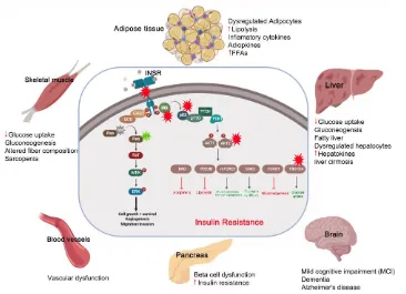
Figure 2. The pathological effect and consequence of insulin resistance (IR). Genetic alterations and mutations in the insulin signaling pathway can lead to IR in the insulin-target tissues. The red stars indicate the reported genetically defective molecules, such as insulin receptor (INS), insulin receptor substrates (IRS), p85, AKT and TBC1D4. In response to insulin resistance, the insulin target tissues (adipose tissue, skeletal muscle, liver, pancreas, brain and blood vessels) show activation of the catabolic processes and accumulation of toxic metabolic byproducts and the inflammatory cytokines, leading to pancreatic β-cell dysfunction and other metabolic disorders.
Skeletal Muscles
the insulin signaling, glucose transport, glycogen synthesis, and mitochondrial activity are main dysfunctional features associated with the IR in the skeletal muscle.
Adipocytes
Adipose tissue is considered the main regulator of insulin action in the body; therefore, defects in insulin signaling in adipocytes cause systemic IR, indicating that impairments in adipose insulin signaling are a common hallmark of IR [39]. Molecular dysfunctions of this process, such as defects in insulin-signaling pathway, involving suppression of lipolysis and glucose transport, leads to the development of IR in adipocyte tissues. [40]. Glucose transporter, GLUT4, is significantly down-regulated in subcutaneous adipose cells from T2D patients and in healthy individuals with a genetic susceptibility for T2D [41]. This reduction in GLUT4 expression is associated with changes in the secretion of adipokines. Also, activation of peroxisome proliferator activated receptors (PPARs), which are responsible for the regulation of adipogenesis as well as lipid metabolism in adipocytes, leads to an improvement in insulin sensitivity through the induction of the expression of several genes that are related to insulin signaling pathway. In addition to its role in adipogenesis, PPAR is involved in regulating lipid, metabolism in mature adipocytes by increasing fatty acid trapping [42]. The importance of insulin signaling in the development of adipocyte IR has been reported in T2D, where previous studies reported that the substrate of insulin receptor (IRS-1) is involved in IR in adipocytes by inhibiting insulin-signaling [43]. Also, in T2D patients, several IRS-1 mutations have been found [44-46] and a reduced IRS-1 protein level has been observed in adipocytes from other T2D patients [47], relatives of T2D patients, and obese individuals [48]. In addition to insulin signaling defects, dysregulation of adipokines and lipolysis process are linked to IR and T2D. The adiponectin gene is considered a candidate susceptibility gene for T2D, because it has been detected on chromosome 3q27, which is linked to T2D and the metabolic disorders [49-51].
Liver cells
The insulin signaling in the hepatic cells is crucial in maintaining normal liver function and regulating glucose homeostasis [52]. The liver activates the glucose uptake and glycogen storage, but it inhibits glycogenolysis and gluconeogenesis. Hepatic IR progression plays a critical role in the pathogenesis of T2D. Loss of INSR signaling in hepatic tissue leads to an increase in the gluconeogenesis and a decrease in the lipogenesis [52-54]. The IRS-1/2 is involved in the suppression of gluconeogenesis and stimulation of lipogenesis. In the liver, insulin inhibits IRS-2 expression at the transcriptional level and doesn’t influence on IRS-1 expression [53,55]. Hepatic IR characterized by inability of insulin to inhibit the production of glucose in the liver [56]. This impairment in the liver function leads to decrease in the insulin clearance with hyperinsulinemia. The released FFAs from fat tissues due to IR transports to liver causing nonalcoholic fatty liver disease (NAFLD) (Steatohepatitis), which followed by liver cirrhosis [23]. Liver-derived proteins termed as hepatokines are released into the circulation which causes defective insulin signaling [57]. The hepatokine fetuin-A was identified as an endogenous ligand for TLR4 through which saturated fatty acids induce proinflammatory cascade and regulate insulin action [58]. Other than fetuin-A, hepatokines Fibroblast growth factor 21 (FGF21) and Selenoprotein P are major drivers of IR [59,60].
GENETIC FACTORS INVOLVED IN INSULIN RESISTANCE DEVELOPMENT
The IR arises due to counteracting insulin action or any defect affecting the insulin signaling pathway starting from its receptor (INSR) followed by a series of downstream branching pathways which involve complex cross talks and feedback effect on each other. This complexity widens the variety of IR etiologies from environmental and genetic perspectives. Several factors and effectors are involved in IR including; diet habit, obesity, lifestyle, physical activity, pharmacological agents, drugs, stress, cytokines, hormones and any factor antagonist the insulin action.
direct effect on the cellular insulin signaling pathway or indirectly through affecting other targets with subsequent secondary effect on the insulin pathway. The disruption and/or mutations in the genes encoding proteins involved in insulin signal transduction impairs insulin action and reduces rate of glucose uptake leading to IR.
Mutations in INSR and Insulin Signaling Genes
There are several mutations in the INSR, which have been classified into several classes: class1 (impaired receptor biosynthesis); class II (receptors with impaired processing and trafficking to plasma membrane); class III (decreased affinity to insulin binding); class IV (receptors with defective phosphorylation and kinase activity); and class V (receptor recycling defects and receptor degradation in lysosome) [61]. There are two types of INSR mutations, autosomal recessive and mild type mutations. The autosomal recessive condition is more severe as it is caused by mutation in both alleles of INSR gene and is usually presented in the infantile and early childhood stage as in Donohue and Rabson Mendenhall syndromes. These syndromes are characterized by an increase in the plasma insulin level with postprandial hyperglycemia in addition to severe growth retardation with poor prognosis history [62,63]. The mild form of INSR mutation is caused by mutation in one allele or mutation in the intra cellular domain only of the INSR and is commonly presented around puberty period and is usually accompanied by lipoatrophy and hyperandrogenism [64,65].
Homozygous mutation in INSR (INSR-/-) develops diabetes rapidly and causes lethality within the first week after birth in mice [66,67]. Nonsense and deletion mutations have been correlated in INSR and cause IR through decreasing the insulin receptor mRNA level or by deletion of the important domains of INSR [68,69]. Also, INSR mutation might cause the reduction of INSR tyrosine kinase activity [70]. Several mutations and polymorphisms in IRSs, particularly IRS1 and IRS2 have been proved to be involved in IR [71].
Genetic loss-function of the molecule(s) involved in intracellular signal transmitting from phosphorylated INSR impairs insulin signaling leading to development of IR. Several post-receptor genetic defects have been reported. Loss of function and mutation of PI3K showed a severe IR which is common in SHORT syndrome, which is characterized by short stature, joint hyperextensibility and hernias, ocular depression, Rieger anomaly and teething delay [72-74]. A family with mutation in gene encoding for AKT2 inherited with severe IR and diabetes mellitus. R274H mutation disrupted the conformation of the activation and catalytic loops of the enzyme depleting its catalytic activity [75]. In another study, a family with Acantho nigrans was found to possess a heterozygous premature stop mutation in AS160 (TBC1D4), which led to GLUT4 translocation inhibition to the membrane and decrease in the intracellular transportation of glucose and hyperglycemia [76]. TBC1D4 nonsense p. Arg684Ter variant (arginine replaced by a termination codon) was also described with severe insulin resistant case of muscle selective form with, impaired glucose tolerance, T2D and postprandial hyperglycemia and IR [77-81].
Other Mutations Associated with Insulin Resistance
complex syndrome exhibiting severe IR caused by heterozygous mutations in POLD1 encoding DNA polymerase Delta [87].
IR is associated with genetic syndrome linked to lipodystrophy and dyslipidemia. Although, several studies proved that the obesity is a strong predisposing risk factor for IR, the pathogenesis of this process is critically related to adipose tissue defects rather than the obesity degree and adipose tissue capacity. In lipodystrophy, the impaired functionality of the adipose tissue is considered as a sump for flux of free fatty acids into the circulation causing harmful effects, including lipotoxicity and IR. Inherited lipodystrophy is classified according to the degree of adipose tissue loss into; recessive congenital generalized lipodystrophy (CGL) with complete adipose absence and familial partial lipodystrophies (FPLD) with defects in only some parts of adipose tissue [88]. In both types, the clinical cases are associated with fatty liver, hypertriglyceridemia, dyslipidemia and sever IR. CGL is mainly caused by mutation of both allele of either AGPAT2, which is a key factor in the synthesis of triglyceride, or BSCL2 genes, which is important for droplet formation and adipocyte differentiation [89-91]. These two genes mutations represent about 90-95% of the etiologies of CGL. Defects in other genes as CAV1 and PTRF can lead to CGL through perturbation in small plasma membrane investigation (caveolae) and the disturbance in lipid trafficking [92,93]. In some cases, the genetic alteration in phosphate cytidylyltransferase 1 (PCYT1A) also reported in CGL patients [94]. There are seven mutations in genes regulating the adipocyte biological processes involved in FPLD; LMNA, PPARG, PLN1, CIDEC, PIK3R1, and AKT2 with high incidence in LMNA and PPARG [88]. LMNA is essential in supporting the structural cytoskeleton and protein network surrounding the nuclear envelop; therefore, its mutation alter the gene expression as a result of the defect in the nuclear envelop and abnormal binding of the transcription factor and chromatin in adipose tissue causing lipodystrophy [90]. The LMNA mutant patient lose the adipose subcutaneously in leg and trunk region only with cushingoid topography [95]. PPARG is essential nuclear receptor hormone for adipocyte differentiation and for sensing the flux of fatty acids. The recorded heterozygous PPARG mutation sometimes causes sever dyslipidemia with reduction in the subcutaneous fat [96,97]. Miscellaneous mutation in important lipid droplet proteins (perilipin and CIDEC) that regulate the triglyceride mobilization also implicated in the FPLD [98,99]. Loss of fat droplets and femoro-gluteal adipose tissue is accompanied by severe IR in the primary insulin signaling disorders especially SHORT syndrome and PI3K/AKT mutation where the insulin signaling is very important for adipocyte differentiation causing lipodystrophy in these syndromes [72,100,101].
INSULIN RESISTANCE AND Β-CELL DYSFUNCTION
hyperglycemia, FFAs influences on the biosynthesis and expression of the insulin gene, leading to suppression of the adequate insulin secretion in response to glucose (GSIS) [108,117-119]. Increased FFAs leads to intrapancreatic and intra β-cell accumulation of triglyceride and fat droplets, triggering the β-cell dysfunction and death due to an increase in the inflammation process [120,121]. Inflammation and the proinflammatory cytokines are increasingly being recognized as an important contributor to β-cell dysfunction [122]. IR associated inflammatory cytokines such as IL6, TNFα, IFNγ, NF-κB and others cause the dysfunctionality and death of β-cells via damage in the mitochondria, cellular proteins, lipids and nucleic acids and ER stress [123-126]. The inflammatory cytokines and the recruited immune cells in the inflamed dysregulated pancreatic islet trigger the β-cell dysfunction [127,128]. Proinflammatory cytokines mediate reactive oxygen species (ROS) and reactive nitrogen species (RNS) production and reduce the ATP production and eventually lead to β-cell dysfunction [129].
The β-cell dysfunction and inadequate β-cell mass expansion can be due to the defect in the insulin signaling pathway in the pancreatic β-cell itself. Improper glucose sensing has been noticed in mouse β-cells, which lack the INSR or IGF-1R. Additionally, loss of INSR led to β-cell mass reduction and early onset diabetes [130-132]. Another study showed similar diabetic phenotypes in mice with PDK1 deficiency in β-cells [133]. The impairment of cell cycle progression is hypothesized to be engaged in the β-cell mass reduction and dysfunction. It has been found that the cell cycle inhibitor, p27Kip1, is accumulated in the nucleus of the β-cells of hyperglycemic IRS2-deficient mice and the deletion of p27Kip1 gene ameliorates β-cell proliferation and the hyperglycemia, reflecting the role of cell cycle inhibition in the β-cell function [106,134].
ANIMAL MODELS OF INSULIN RESISTANCE AND THEIR LIMITATIONS
Studies on this model demonstrated the contribution of hepatic tumor factor, NF-kB/RelA, TNF and interleukin4 in the development of IR [147-149]. Zucker fatty (fa/fa) rat exhibit hyperinsulinemia, glucose intolerance and hyperlipidemia due to deficiency in the leptin receptor and autosomal recessive fa gene [150]. Defect in leptin transportation through the blood brain barrier represented in New Zealand mice strain (NZO), which is a hepatic IR model [151]. The spontaneous deletion mutation in CD36 fatty acid transporter gene developed the OLETF rat as a model to study the IR syndrome in hypertensive, hyper insulinemic glucose intolerant animal [152] as it resemble the clinical and pathological features of human non-insulin-dependent diabetes mellitus (NIDDM). KK/Ay mouse model with defects in the yellow obese gene represents a condition of severe hyperinsulinaemia, hyperglycaemia, glucose intolerance and obesity due to defective insulin receptor and post-receptor signaling [153]. There are other models of the spontaneously developed IR strains [154]. Although the above-mentioned animal models provided a lot of information about the mechanisms of IR, they did not answer all the questions related to the development of IR in human tissues and relationship between IR and β-cell dysfunction.
in the oxidative stress [165]. All the derived INSR-Mut hiPSCs showed reduced proliferation and defected INSR phosphorylation and defects in its downstream signaling pathway such as, AKT, GSK3, ERK1 and ERK2 [158]. Differentiation of INSR-Mut hiPSCs towards skeletal myotubes exhibits defect in insulin signaling, glucose uptake, glycogen accumulation and altered insulin signaling gene expression [166], indicating the genetic defects in the skeletal myotubes. Another iPSC model for IR, in which iPSCs have been generated from the fibroblast of insulin resistant patient with congenital generalized lipodystrophy (CGL), an autosomal recessive disease due to BSCL2 mutation [90]. These patient-specific iPSCs have been used as an in vitro model to study the physiopathology of lipid accumulation and lipodystrophy and its relation to IR. Adipocytes derived from these BSCL2-Mut iPSCs showed reduced lipid droplet formation and dispersed cytoplasmic distribution of adipose differentiation related protein (ADRP) [167]. Recent studies showed the ability to generate adipocytes from hiPSCs. It has been reported that hiPSC-derived adipocytes, transplanted into mice, are able to sustain their functional characteristics for several weeks [168,169], suggesting that these cells can be also used therapeutically to improve metabolic disorder conditions in patients. Therefore, differentiation of patient-specific hiPSCs into white adipocytes can offer a large number of functional adipocytes for transplantation as a possible way to treat adipocytes-associated disorders as well as studying IR. Since the liver is an important insulin target tissue, the iPSCs were used to interpret the etiology of steatosis due to the NAFLD, which is accompanied by IR and hyperlipidemia. The iPSCs generated from patients with liver steatosis showed a decrease in the AKT/mTOR signaling molecule and the phenotypes of IR are observed in both liver and skin fibroblasts of the patients. Additionally, it showed that the transcription factor, sterol regulatory element binding transcription factor 1 (SREBF1) and its downstream targets like LIPIN1 (LPIN) and low density lipoprotein receptor are involved in glycerolipid and fatty acid biosynthesis [170]. Recently, we generated the first iPSCs from patients with psoriasis and insulin resistance. Our study showed that iPSC-derived keratinocytes carry genetic defects associated with insulin resistance [171], indicating that the IR in patients with psoriasis is due to genetic defects in signaling pathways regulating insulin sensitivity in the epidermal keratinocytes. Further studies are required to fill the gaps in understanding the pathogenesis of IR and diabetes. There are several genetic factors involved in the development of IR; therefore, patient-specific iPSCs can be used to study the most common form of IR leading to T2D. Also, hiPSCs/hESCs can be genetically edited using genome editing tools, such as Crisper/CAS9. The combination of these technologies may provide more details about the inherited factors underlying the development of different forms of IR.
LIMITATIONS OF IPSCS AS AN IN VITRO MODEL
reports highlighting the use of genome editing tool for generation of iPSC-based disease models [175,177].
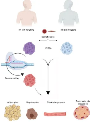
Figure 3. Using hiPSCs to study insulin resistance (IR). A schematic diagram showing the possibility of using iPSCs to study IR with special focus on the genetic factors. Somatic cells are reprogrammed into pluripotency to generate iPSCs carrying the genetic signatures of insulin sensitive (IS) and IR individuals. The generated iPSCs can be differentiated in vitro into the main insulin-target cells, including hepatocyte, adipocyte, and skeletal myotubes
as well as insulin producing β-cells. Genome editing tools can be used to correct specific mutations in the generated
iPSCs to establish the isogenic iPSC control. Studying those cells can help in understanding the signaling pathways involved in the development of IR and T2D. Also, these iPSC-based models can be used for drug screening.
CONCLUSIONS AND FUTURE PERSPECTIVES
disease specific cells are brought forward to avail a successful point to characterize specific gene. However, combining genome editing tool and iPSCs have certain limitations. Most of the metabolic diseases are not caused by a single gene dysfunction, but a group of complex genes are involved in regulation of metabolic diseases. Development of iPSC differentiation protocols is very important to generate a homogenous population of the target cell. One of the limitations of the genome editing tools is the off-target effect. Therefore, improving the gene-editing process is critical to avoid the off-target effects.
Competing interests
The authors declare that they have no conflict of interest.
Ethical approval
This article does not contain any studies with human or animal subjects performed by any of the authors.
Funding
This work was funded by grants from Qatar National Research Fund (QNRF) (Grant No. NPRP9-283-3-056 & NPRP10-1221-160041).
REFERENCES
1.
Martin, B.C.; Warram, J.H.; Rosner, B.; Rich, S.S.; Soeldner, J.S.; Krolewski, A.S.
Familial clustering of insulin sensitivity.
Diabetes 1992
,
41
, 850-854.
2.
Shulman, R.G. Nuclear magnetic resonance studies of glucose metabolism in
non-insulin-dependent diabetes mellitus subjects.
Mol Med 1996
,
2
, 533-540.
3.
Ling, C.; Rönn, T. Epigenetics in human obesity and type 2 diabetes.
Cell metabolism
2019
.
4.
Ling, C.; Rönn, T. Epigenetic markers to further understand insulin resistance.
Diabetologia 2016
,
59
, 2295-2297.
5.
Elgzyri, T.; Parikh, H.; Zhou, Y.; Dekker Nitert, M.; Ronn, T.; Segerstrom, A.B.;
Ling, C.; Franks, P.W.; Wollmer, P.; Eriksson, K.F., et al. First-degree relatives of
type 2 diabetic patients have reduced expression of genes involved in fatty acid
metabolism in skeletal muscle.
J Clin Endocrinol Metab 2012
,
97
, E1332-1337,
doi:10.1210/jc.2011-3037.
6.
Patti, M.E.; Butte, A.J.; Crunkhorn, S.; Cusi, K.; Berria, R.; Kashyap, S.; Miyazaki,
Y.; Kohane, I.; Costello, M.; Saccone, R., et al. Coordinated reduction of genes of
oxidative metabolism in humans with insulin resistance and diabetes: Potential role of
PGC1 and NRF1.
Proc Natl Acad Sci U S A 2003
,
100
, 8466-8471,
doi:10.1073/pnas.1032913100.
7.
Mootha, V.K.; Lindgren, C.M.; Eriksson, K.F.; Subramanian, A.; Sihag, S.; Lehar, J.;
Puigserver, P.; Carlsson, E.; Ridderstrale, M.; Laurila, E., et al.
PGC-1alpha-responsive genes involved in oxidative phosphorylation are coordinately
downregulated in human diabetes.
Nat Genet 2003
,
34
, 267-273, doi:10.1038/ng1180.
8.
Al-Khawaga, S.; Memon, B.; Butler, A.E.; Taheri, S.; Abou-Samra, A.B.; Abdelalim,
E.M. Pathways governing development of stem cell-derived pancreatic beta cells:
lessons from embryogenesis.
Biol Rev Camb Philos Soc 2018
,
93
, 364-389,
doi:10.1111/brv.12349.
mechanisms, granule pools, and biphasic insulin secretion.
2002
,
10.2337/diabetes.51.2007.s83, doi:10.2337/diabetes.51.2007.s83.
10.
Dodson, G.; Don, S. The role of assembly in insulin's biosynthesis.
Current Opinion
in Structural Biology 1998
, 10.1016/s0959-440x(98)80037-7,
doi:10.1016/s0959-440x(98)80037-7.
11.
Burks, D.J.; F., W.M. IRS proteins and beta-cell function.
Diabetes 2001
,
10.2337/diabetes.50.2007.s140, doi:10.2337/diabetes.50.2007.s140.
12.
Saltiel, A.R.; Kahn, C.R. Insulin signalling and the regulation of glucose and lipid
metabolism. 2001; 10.1038/414799a.
13.
Smith, U. Impaired ('diabetic') insulin signaling and action occur in fat cells long
before glucose intolerance - Is insulin resistance initiated in the adipose tissue? 2002;
10.1038/sj.ijo.0802028.
14.
Khan, A.; Pessin, J. Insulin regulation of glucose uptake: a complex interplay of
intracellular signalling pathways.
Diabetologia 2002
,
45
, 1475-1483.
15.
Hunter, S.J.; Garvey, W.T. Insulin action and insulin resistance: Diseases involving
defects in insulin receptors, signal transduction, and the glucose transport effector
system. 1998; 10.1016/s0002-9343(98)00300-3.
16.
Wilcox, G. Insulin and Insulin Resistance Gisela.
2005
,
26
, 19--39.
17.
Hotamisligil, G.S. Inflammatory pathways and insulin action.
International Journal of
Obesity 2003
, 10.1038/sj.ijo.0802502, doi:10.1038/sj.ijo.0802502.
18.
Artunc, F.; Erwin, S.; Cora, W.; Andreas, F.; Norbert, S.; Ulrich, H.H. The impact of
insulin resistance on the kidney and vasculature. 2016; 10.1038/nrneph.2016.145.
19.
Gauthier, M.S.; B., R.N. Adipose tissue inflammation and insulin resistance: All
obese humans are not created equal. 2010; 10.1042/bj20101062.
20.
Krauss, R.M.; W., S.P. Metabolic abnormalities: Triglyceride and low-density
lipoprotein. 2004; 10.1016/j.ecl.2004.03.016.
21.
Devaraj, S.; Rosenson, R.S.; Jialal, I. Metabolic syndrome: An appraisal of the
pro-inflammatory and procoagulant status. 2004; 10.1016/j.ecl.2004.03.008.
22.
Krauss, R.M.; Siri, P.W. Metabolic abnormalities: Triglyceride and low-density
lipoprotein. 2004; 10.1016/j.ecl.2004.03.016.
23.
Reaven, G. The metabolic syndrome or the insulin resistance syndrome? Different
names, different concepts, and different goals. 2004; 10.1016/j.ecl.2004.03.002.
24.
Morrison, S.A.; Goss, A.M.; Azziz, R.; Raju, D.A.; Gower, B.A. Peri-muscular
adipose tissue may play a unique role in determining insulin sensitivity/resistance in
women with polycystic ovary syndrome.
Human Reproduction 2017
,
10.1093/humrep/dew279, doi:10.1093/humrep/dew279.
25.
Peppa, M.; Chrysi, K.; Panagiotis, N.; A., R.S. Skeletal muscle insulin resistance in
endocrine disease. 2010; 10.1155/2010/527850.
26.
M. de la Monte, S. Brain Insulin Resistance and Deficiency as Therapeutic Targets in
Alzheimers Disease.
Current Alzheimer Research 2012
,
10.2174/156720512799015037, doi:10.2174/156720512799015037.
27.
Heni, M.; Kullmann, S.; Preissl, H.; Fritsche, A.; Häring, H.U. Impaired insulin action
in the human brain: Causes and metabolic consequences. 2015;
10.1038/nrendo.2015.173.
28.
Burcelin, R. Gut microbiota and immune crosstalk in metabolic disease. 2016;
10.1016/j.molmet.2016.05.016.
30.
Warram, J.H.; Martin, B.C.; Krolewski, A.S.; Soeldner, J.S.; Kahn, C.R. Slow
glucose removal rate and hyperinsulinemia precede the development of type II
diabetes in the offspring of diabetic parents.
Ann Intern Med 1990
,
113
, 909-915.
31.
Thiebaud, D.; Jacot, E.; DeFronzo, R.A.; Maeder, E.; Jequier, E.; Felber, J.P. The
effect of graded doses of insulin on total glucose uptake, glucose oxidation, and
glucose storage in man.
Diabetes 1982
,
31
, 957-963.
32.
Petersen, K.F.; Dufour, S.; Befroy, D.; Garcia, R.; Shulman, G.I. Impaired
mitochondrial activity in the insulin-resistant offspring of patients with type 2
diabetes.
N Engl J Med 2004
,
350
, 664-671, doi:10.1056/NEJMoa031314.
33.
Kelley, D.E.; He, J.; Menshikova, E.V.; Ritov, V.B. Dysfunction of mitochondria in
human skeletal muscle in type 2 diabetes.
Diabetes 2002
,
51
, 2944-2950.
34.
Krook, A.; Bjornholm, M.; Galuska, D.; Jiang, X.J.; Fahlman, R.; Myers, M.G., Jr.;
Wallberg-Henriksson, H.; Zierath, J.R. Characterization of signal transduction and
glucose transport in skeletal muscle from type 2 diabetic patients.
Diabetes 2000
,
49
,
284-292.
35.
Morino, K.; Petersen, K.F.; Dufour, S.; Befroy, D.; Frattini, J.; Shatzkes, N.; Neschen,
S.; White, M.F.; Bilz, S.; Sono, S., et al. Reduced mitochondrial density and increased
IRS-1 serine phosphorylation in muscle of insulin-resistant offspring of type 2
diabetic parents.
J Clin Invest 2005
,
115
, 3587-3593, doi:10.1172/JCI25151.
36.
Beck-Nielsen, H. Mechanisms of insulin resistance in non-oxidative glucose
metabolism: the role of glycogen synthase.
J Basic Clin Physiol Pharmacol 1998
,
9
,
255-279.
37.
Damsbo, P.; Vaag, A.; Hother-Nielsen, O.; Beck-Nielsen, H. Reduced glycogen
synthase activity in skeletal muscle from obese patients with and without type 2
(non-insulin-dependent) diabetes mellitus.
Diabetologia 1991
,
34
, 239-245.
38.
Iovino, S.; Burkart, A.M.; Warren, L.; Patti, M.E.; Kahn, C.R. Myotubes derived from
human-induced pluripotent stem cells mirror in vivo insulin resistance.
Proc Natl
Acad Sci U S A 2016
,
113
, 1889-1894, doi:10.1073/pnas.1525665113.
39.
Adams-Huet, B.; Devaraj, S.; Siegel, D.; Jialal, I. Increased adipose tissue insulin
resistance in metabolic syndrome: relationship to circulating adipokines.
Metab Syndr
Relat Disord 2014
,
12
, 503-507, doi:10.1089/met.2014.0092.
40.
Saltiel, A.R.; Kahn, C.R. Insulin signalling and the regulation of glucose and lipid
metabolism.
Nature 2001
,
414
, 799-806, doi:10.1038/414799a.
41.
Gustafson, B.; Hedjazifar, S.; Gogg, S.; Hammarstedt, A.; Smith, U. Insulin resistance
and impaired adipogenesis.
Trends Endocrinol Metab 2015
,
26
, 193-200,
doi:10.1016/j.tem.2015.01.006.
42.
Tamori, Y.; Masugi, J.; Nishino, N.; Kasuga, M. Role of peroxisome
proliferator-activated receptor-gamma in maintenance of the characteristics of mature 3T3-L1
adipocytes.
Diabetes 2002
,
51
, 2045-2055.
43.
White, M.F. IRS proteins and the common path to diabetes.
Am J Physiol Endocrinol
Metab 2002
,
283
, E413-422, doi:10.1152/ajpendo.00514.2001.
44.
Almind, K.; Inoue, G.; Pedersen, O.; Kahn, C.R. A common amino acid
polymorphism in insulin receptor substrate-1 causes impaired insulin signaling.
Evidence from transfection studies.
J Clin Invest 1996
,
97
, 2569-2575,
doi:10.1172/JCI118705.
46.
Yoshimura, R.; Araki, E.; Ura, S.; Todaka, M.; Tsuruzoe, K.; Furukawa, N.;
Motoshima, H.; Yoshizato, K.; Kaneko, K.; Matsuda, K., et al. Impact of natural
IRS-1 mutations on insulin signals: mutations of IRS-IRS-1 in the PTB domain and near SH2
protein binding sites result in impaired function at different steps of IRS-1 signaling.
Diabetes 1997
,
46
, 929-936.
47.
Rondinone, C.M.; Wang, L.M.; Lonnroth, P.; Wesslau, C.; Pierce, J.H.; Smith, U.
Insulin receptor substrate (IRS) 1 is reduced and IRS-2 is the main docking protein
for phosphatidylinositol 3-kinase in adipocytes from subjects with
non-insulin-dependent diabetes mellitus.
Proc Natl Acad Sci U S A 1997
,
94
, 4171-4175.
48.
Carvalho, E.; Jansson, P.A.; Axelsen, M.; Eriksson, J.W.; Huang, X.; Groop, L.;
Rondinone, C.; Sjostrom, L.; Smith, U. Low cellular IRS 1 gene and protein
expression predict insulin resistance and NIDDM.
FASEB J 1999
,
13
, 2173-2178.
49.
Kissebah, A.H.; Sonnenberg, G.E.; Myklebust, J.; Goldstein, M.; Broman, K.; James,
R.G.; Marks, J.A.; Krakower, G.R.; Jacob, H.J.; Weber, J., et al. Quantitative trait loci
on chromosomes 3 and 17 influence phenotypes of the metabolic syndrome.
Proc
Natl Acad Sci U S A 2000
,
97
, 14478-14483, doi:10.1073/pnas.97.26.14478.
50.
Mori, Y.; Otabe, S.; Dina, C.; Yasuda, K.; Populaire, C.; Lecoeur, C.; Vatin, V.;
Durand, E.; Hara, K.; Okada, T., et al. Genome-wide search for type 2 diabetes in
Japanese affected sib-pairs confirms susceptibility genes on 3q, 15q, and 20q and
identifies two new candidate Loci on 7p and 11p.
Diabetes 2002
,
51
, 1247-1255.
51.
Vasseur, F.; Helbecque, N.; Dina, C.; Lobbens, S.; Delannoy, V.; Gaget, S.; Boutin,
P.; Vaxillaire, M.; Lepretre, F.; Dupont, S., et al. Single-nucleotide polymorphism
haplotypes in the both proximal promoter and exon 3 of the APM1 gene modulate
adipocyte-secreted adiponectin hormone levels and contribute to the genetic risk for
type 2 diabetes in French Caucasians.
Hum Mol Genet 2002
,
11
, 2607-2614.
52.
Michael, M.D.; Kulkarni, R.N.; Postic, C.; Previs, S.F.; Shulman, G.I.; Magnuson,
M.A.; Kahn, C.R. Loss of insulin signaling in hepatocytes leads to severe insulin
resistance and progressive hepatic dysfunction.
Mol Cell 2000
,
6
, 87-97.
53.
Thirone, A.C.; Huang, C.; Klip, A. Tissue-specific roles of IRS proteins in insulin
signaling and glucose transport.
Trends Endocrinol Metab 2006
,
17
, 72-78,
doi:10.1016/j.tem.2006.01.005.
54.
Kubota, N.; Kubota, T.; Itoh, S.; Kumagai, H.; Kozono, H.; Takamoto, I.; Mineyama,
T.; Ogata, H.; Tokuyama, K.; Ohsugi, M., et al. Dynamic functional relay between
insulin receptor substrate 1 and 2 in hepatic insulin signaling during fasting and
feeding.
Cell Metab 2008
,
8
, 49-64, doi:10.1016/j.cmet.2008.05.007.
55.
Hirashima, Y.; Tsuruzoe, K.; Kodama, S.; Igata, M.; Toyonaga, T.; Ueki, K.; Kahn,
C.R.; Araki, E. Insulin down-regulates insulin receptor substrate-2 expression through
the phosphatidylinositol 3-kinase/Akt pathway.
J Endocrinol 2003
,
179
, 253-266.
56.
Farese, R.V., Jr.; Zechner, R.; Newgard, C.B.; Walther, T.C. The problem of
establishing relationships between hepatic steatosis and hepatic insulin resistance.
Cell Metab 2012
,
15
, 570-573, doi:10.1016/j.cmet.2012.03.004.
57.
Stefan, N.; Häring, H.U. The role of hepatokines in metabolism. 2013;
10.1038/nrendo.2012.258.
58.
Pal, D.; Dasgupta, S.; Kundu, R.; Maitra, S.; Das, G.; Mukhopadhyay, S.; Ray, S.;
Majumdar, S.S.; Bhattacharya, S. Fetuin-A acts as an endogenous ligand of TLR4 to
promote lipid-induced insulin resistance.
Nature Medicine 2012
, 10.1038/nm.2851,
doi:10.1038/nm.2851.
of Hepatic Lipid Metabolism in Ketotic States.
Cell Metabolism 2007
,
10.1016/j.cmet.2007.05.002, doi:10.1016/j.cmet.2007.05.002.
60.
Misu, H.; Takamura, T.; Takayama, H.; Hayashi, H.; Matsuzawa-Nagata, N.; Kurita,
S.; Ishikura, K.; Ando, H.; Takeshita, Y.; Ota, T., et al. A liver-derived secretory
protein, selenoprotein P, causes insulin resistance.
Cell Metabolism 2010
,
10.1016/j.cmet.2010.09.015, doi:10.1016/j.cmet.2010.09.015.
61.
Hashiramoto, M.; Osawa, H.; Ando, M.; Murakami, A.; Nishimiya, T.; Nakano, M.;
Nishida, W.; Onuma, H.; Makino, H. A nonsense mutation in the Arg345 of the
insulin receptor gene in a Japanese type A insulin-resistant patient.
Endocr J 2005
,
52
, 499-504.
62.
Kosztolnyi, G. Leprechaunism/Donohue syndrome/insulin receptor gene mutations: A
syndrome delineation story from clinicopathological description to molecular
understanding.
European Journal of Pediatrics 1997
, 10.1007/s004310050594,
doi:10.1007/s004310050594.
63.
Longo, N.; Yuhuan, W.; Marzia, P. Progressive decline in insulin levels in
Rabson-Mendenhall syndrome.
Journal of Clinical Endocrinology and Metabolism 1999
,
10.1210/jc.84.8.2623, doi:10.1210/jc.84.8.2623.
64.
Musso, C.; Elaine, C.; Ann, M.S.; and, S.M.C. Clinical course of genetic diseases of
the insulin receptor (type A and Rabson-Mendenhall syndromes): A 30-year
prospective.
Medicine 2004
, 10.1097/01.md.0000133625.73570.54,
doi:10.1097/01.md.0000133625.73570.54.
65.
Taylor, S.I.; T., K.; H., K.; D., A.; A., C.; C., M. Mutations in insulin-receptor gene in
insulin-resistant patients.
1990
, 10.2337/diacare.13.3.257,
doi:10.2337/diacare.13.3.257.
66.
Accili, D.; Drago, J.; Lee, E.J.; Johnson, M.D.; Cool, M.H.; Salvatore, P.; Asico,
L.D.; José, P.A.; Taylor, S.I.; Westphal, H. Early neonatal death in mice homozygous
for a null allele of the insulin receptor gene.
Nature genetics 1996
,
12
, 106.
67.
Joshi, R.L.; Lamothe, B.; Cordonnier, N.; Mesbah, K.; Monthioux, E.; Jami, J.;
Bucchini, D. Targeted disruption of the insulin receptor gene in the mouse results in
neonatal lethality.
The EMBO journal 1996
,
15
, 1542-1547.
68.
Kadowaki, T.; Bevins, C.L.; Cama, A.; Ojamaa, K.; Marcus-Samuels, B.; Kadowaki,
H.; Beitz, L.; McKeon, C.; Taylor, S.I. Two mutant alleles of the insulin receptor
gene in a patient with extreme insulin resistance.
Science 1988
,
240
, 787-790.
69.
Yoshimasa, Y.; Seino, S.; Whittaker, J.; Kakehi, T.; Kosaki, A.; Kuzuya, H.; Imura,
H.; Bell, G.I.; Steiner, D.F. Insulin-resistant diabetes due to a point mutation that
prevents insulin proreceptor processing.
Science 1988
,
240
, 784-787.
70.
Odawara, M.; Kadowaki, T.; Yamamoto, R.; Shibasaki, Y.; Tobe, K.; Accili, D.;
Bevins, C.; Mikami, Y.; Matsuura, N.; Akanuma, Y. Human diabetes associated with
a mutation in the tyrosine kinase domain of the insulin receptor.
Science 1989
,
245
,
66-68.
71.
Sigal, R.; Doria, A.; Warram, J.; Krolewski, A. Codon 972 polymorphism in the
insulin receptor substrate-1 gene, obesity, and risk of noninsulin-dependent diabetes
mellitus.
The Journal of Clinical Endocrinology & Metabolism 1996
,
81
, 1657-1659.
72.
Chudasama, K.K.; Jonathon, W.; Stefan, J.; Tor, C.; Rainer, K.; Ingfrid, H.; Bente, J.;
Rang, W.J.; Dagfinn, A.; V., S.J., et al. SHORT syndrome with partial lipodystrophy
due to impaired phosphatidylinositol 3 kinase signaling.
American Journal of Human
Genetics 2013
, 10.1016/j.ajhg.2013.05.023, doi:10.1016/j.ajhg.2013.05.023.
syndrome.
American Journal of Human Genetics 2013
, 10.1016/j.ajhg.2013.06.005,
doi:10.1016/j.ajhg.2013.06.005.
74.
Thauvin-Robinet, C.; Auclair, M.; Duplomb, L.; Caron-Debarle, M.; Avila, M.;
St-Onge, J.; Le Merrer, M.; Le Luyer, B.; Héron, D.; Mathieu-Dramard, M., et al.
PIK3R1 mutations cause syndromic insulin resistance with lipoatrophy.
American
Journal of Human Genetics 2013
, 10.1016/j.ajhg.2013.05.019,
doi:10.1016/j.ajhg.2013.05.019.
75.
George, S.; Rochford, J.J.; Wolfrum, C.; Gray, S.L.; Schinner, S.; Wilson, J.C.; Soos,
M.A.; Murgatroyd, P.R.; Williams, R.M.; Acerini, C.L., et al. A family with severe
insulin resistance and diabetes due to a mutation in AKT2.
Science 2004
,
304
,
1325-1328, doi:10.1126/science.1096706.
76.
Dash, S.; Sano, H.; Rochford, J.J.; Semple, R.K.; Yeo, G.; Hyden, C.S.; Soos, M.A.;
Clark, J.; Rodin, A.; Langenberg, C., et al. A truncation mutation in TBC1D4 in a
family with acanthosis nigricans and postprandial hyperinsulinemia.
Proc Natl Acad
Sci U S A 2009
,
106
, 9350-9355, doi:10.1073/pnas.0900909106.
77.
Dash, S.; Hiroyuki, S.; J., R.J.; K., S.R.; Giles, Y.; S., H.C.S.; A., S.M.; James, C.;
Andrew, R.; Claudia, L., et al. A truncation mutation in TBC1D4 in a family with
acanthosis nigricans and postprandial hyperinsulinemia.
Proceedings of the National
Academy of Sciences of the United States of America 2009
,
10.1073/pnas.0900909106, doi:10.1073/pnas.0900909106.
78.
JØRgensen, M.E.; Tvermosegaard, M.; RØNn, P.F.; Bjerregaard, P.; Dahl-Petersen,
I.K.; Larsen, C.V.; Pedersen, M.L.; Albrechtsen, A.; Moltke, I.D.A.; Grarup, N., et al.
Do Greenlandic Carriers of the TBC1D4 p.Arg684Ter Variant Have Increased Risk
of Cardiovascular Disease?
Diabetes 2018
, 10.2337/db18-439-p,
doi:10.2337/db18-439-p.
79.
Moltke, I.; Grarup, N.; Jørgensen, M.E.; Bjerregaard, P.; Treebak, J.T.; Fumagalli,
M.; Korneliussen, T.S.; Andersen, M.A.; Nielsen, T.S.; Krarup, N.T., et al. A
common Greenlandic TBC1D4 variant confers muscle insulin resistance and type 2
diabetes.
Nature 2014
, 10.1038/nature13425, doi:10.1038/nature13425.
80.
Jrgensen, M.E.; Maria, T.; F., R.N.N.P.; Peter, B.; K., D.-P.I.; V., L.C.; L., P.M.;
Anders, A.; A., M.I.D.; Niels, G., et al. Do Greenlandic Carriers of the TBC1D4
p.Arg684Ter Variant Have Increased Risk of Cardiovascular Disease?
Diabetes 2018
,
10.2337/db18-439-p, doi:10.2337/db18-439-p.
81.
Moltke, I.; Niels, G.; E., J.M.; Peter, B.; T., T.J.; Matteo, F.; S., K.T.; A., A.M.; S.,
N.T.; T., K.N., et al. A common Greenlandic TBC1D4 variant confers muscle insulin
resistance and type 2 diabetes.
Nature 2014
, 10.1038/nature13425,
doi:10.1038/nature13425.
82.
Minton, J.A.; Owen, K.R.; Ricketts, C.J.; Crabtree, N.; Shaikh, G.; Ehtisham, S.;
Porter, J.R.; Carey, C.; Hodge, D.; Paisey, R., et al. Syndromic obesity and diabetes:
changes in body composition with age and mutation analysis of ALMS1 in 12 United
Kingdom kindreds with Alstrom syndrome.
J Clin Endocrinol Metab 2006
,
91
,
3110-3116, doi:10.1210/jc.2005-2633.
83.
Savage, D.B.; Zhai, L.; Ravikumar, B.; Choi, C.S.; Snaar, J.E.; McGuire, A.C.; Wou,
S.E.; Medina-Gomez, G.; Kim, S.; Bock, C.B., et al. A prevalent variant in PPP1R3A
impairs glycogen synthesis and reduces muscle glycogen content in humans and mice.
PLoS Med 2008
,
5
, e27, doi:10.1371/journal.pmed.0050027.
84.
Savage, D.B.; Agostini, M.; Barroso, I.; Gurnell, M.; Luan, J.; Meirhaeghe, A.;
Harding, A.H.; Ihrke, G.; Rajanayagam, O.; Soos, M.A., et al. Digenic inheritance of
severe insulin resistance in a human pedigree.
Nat Genet 2002
,
31
, 379-384,
85.
Yamada, K.; Ikegami, H.; Yoneda, H.; Miki, T.; Ogihara, T. All patients with
Werner's syndrome are insulin resistant, but only those who also have impaired
insulin secretion develop overt diabetes.
Diabetes Care 1999
,
22
, 2094-2095.
86.
Payne, F.; Colnaghi, R.; Rocha, N.; Seth, A.; Harris, J.; Carpenter, G.; Bottomley,
W.E.; Wheeler, E.; Wong, S.; Saudek, V., et al. Hypomorphism in human NSMCE2
linked to primordial dwarfism and insulin resistance.
J Clin Invest 2014
,
124
,
4028-4038, doi:10.1172/JCI73264.
87.
Weedon, M.N.; Ellard, S.; Prindle, M.J.; Caswell, R.; Lango Allen, H.; Oram, R.;
Godbole, K.; Yajnik, C.S.; Sbraccia, P.; Novelli, G., et al. An in-frame deletion at the
polymerase active site of POLD1 causes a multisystem disorder with lipodystrophy.
Nat Genet 2013
,
45
, 947-950, doi:10.1038/ng.2670.
88.
Semple, R.K. How does insulin resistance arise, and how does it cause disease?
Human genetic lessons. 2016; 10.1530/eje-15-1131.
89.
Agarwal, A.K.; Elif, A.; Salome, d.A.; Nurullah, A.; I., T.S.; M., B.A.; I., B.R.;
Abhimanyu, G. AGPAT2 is mutated in congenital generalized lipodystrophy linked to
chromosome 9q34.
Nature Genetics 2002
, 10.1038/ng880, doi:10.1038/ng880.
90.
Garg, A. Acquired and Inherited Lipodystrophies. 2004; 10.1056/NEJMra025261.
91.
Magré, J.; Delépine, M.; Khallouf, E.; Gedde-Dahl, T.; Van Maldergem, L.; Sobel,
E.; Papp, J.; Meier, M.; Mégarbané, A.; Bachy, A., et al. Identification of the gene
altered in Berardinelli-Seip congenital lipodystrophy on chromosome 11q13.
Nature
Genetics 2001
, 10.1038/ng585, doi:10.1038/ng585.
92.
Hayashi, Y.K.; Chie, M.; Megumu, O.; Kanako, G.; Kayo, T.; Satomi, M.; Eun, P.Y.;
Ikuya, N.; Naomi, H.-F.; Kazuhiro, H., et al. Human PTRF mutations cause secondary
deficiency of caveolins resulting in muscular dystrophy with generalized
lipodystrophy.
Journal of Clinical Investigation 2009
, 10.1172/jci38660,
doi:10.1172/jci38660.
93.
Kim, C.A.; Marc, D.; and, B.E. Association of a homozygous nonsense caveolin-1
mutation with berardinelli-seip congenital lipodystrophy.
Journal of Clinical
Endocrinology and Metabolism 2008
, 10.1210/jc.2007-1328,
doi:10.1210/jc.2007-1328.
94.
Payne, F.; Koini, L.; Amandine, G.; J., B.R.; Nora, K.; Ann, R.; Yali, X.; Alison, S.;
Elaine, C.; Claire, A., et al. Mutations disrupting the Kennedy phosphatidylcholine
pathway in humans with congenital lipodystrophy and fatty liver disease.
Proceedings
of the National Academy of Sciences of the United States of America 2014
,
10.1073/pnas.1408523111, doi:10.1073/pnas.1408523111.
95.
Shackleton, S.; J., L.D.; J., J.S.N.; Richard, E.; F., N.M.; M., S.B.; Hartmut, S.;
Georg, B.; Sudesh, K.; N., D.P., et al. LMNA, encoding lamin A/C, is mutated in
partial lipodystrophy.
Nature Genetics 2000
, 10.1038/72807, doi:10.1038/72807.
96.
Semple, R.K.; Chatterjee, V.K.K.; O'Rahilly, S. PPAR gamma and human metabolic
disease.
Journal of Clinical Investigation 2006
.
97.
Ali, A.T.; Hochfeld, W.E.; Myburgh, R.; Pepper, M.S. Adipocyte and adipogenesis.
European journal of cell biology 2013
,
92
, 229-236.
98.
Gandotra, S.a. Perilipin deficiency and autosomal dominant partial lipodystrophy.
New England Journal of Medicine 2011
, 10.1056/NEJMoa1007487,
doi:10.1056/NEJMoa1007487.
99.
Kozusko, K.; Satish, P.; B., S.D. Human congenital perilipin deficiency and insulin
resistance.
2013
, 10.1159/000342511, doi:10.1159/000342511.
American Journal of Human Genetics 2013
, 10.1016/j.ajhg.2013.05.019,
doi:10.1016/j.ajhg.2013.05.019.
101.
George, S.; J., R.J.; Christian, W.; L., G.S.; Sven, S.; C., W.J.; A., S.M.; R., M.P.; M.,
W.R.; L., A.C., et al. A family with severe insulin resistance and diabetes due to a
mutation in AKT2.
Science 2004
, 10.1126/science.1096706,
doi:10.1126/science.1096706.
102.
Ashcroft, F.M.; Rorsman, P. Diabetes mellitus and the β cell: The last ten years. 2012;
10.1016/j.cell.2012.02.010.
103.
Ferrannini, E. The Stunned β Cell: A Brief History.
Cell metabolism 2010
.
104.
Talchai, C.; Xuan, S.; Lin, H.V.; Sussel, L.; Accili, D. Pancreatic β cell
dedifferentiation as a mechanism of diabetic β cell failure.
Cell 2012
,
10.1016/j.cell.2012.07.029, doi:10.1016/j.cell.2012.07.029.
105.
Prentki, M.; Nolan, C.J. Islet β cell failure in type 2 diabetes. 2006; 10.1172/jci29103.
106.
Kasuga, M. Insulin resistance and pancreatic β cell failure. 2006; 10.1172/jci29189.
107.
Hasnain, S.Z.; Borg, D.J.; Harcourt, B.E.; Tong, H.; Sheng, Y.H.; Ng, C.P.; Das, I.;
Wang, R.; Chen, A.C.H.; Loudovaris, T., et al. Glycemic control in diabetes is
restored by therapeutic manipulation of cytokines that regulate beta cell stress.
Nature
Medicine 2014
, 10.1038/nm.3705, doi:10.1038/nm.3705.
108.
Poitout, V.; Robertson, R.P. Glucolipotoxicity: Fuel excess and β-cell dysfunction.
2008; 10.1210/er.2007-0023.
109.
Khaldi, M.Z.; Guiot, Y.; Gilon, P.; Henquin, J.C.; Jonas, J.C. Increased glucose
sensitivity of both triggering and amplifying pathways of insulin secretion in rat islets
cultured for 1 wk in high glucose.
American Journal of Physiology - Endocrinology
and Metabolism 2004
, 10.1152/ajpendo.00426.2003,
doi:10.1152/ajpendo.00426.2003.
110.
Patanè, G.; Anello, M.; Piro, S.; Vigneri, R.; Purrello, F.; Rabuazzo, A.M. Role of
ATP production and uncoupling protein-2 in the insulin secretory defect induced by
chronic exposure to high glucose or free fatty acids and effects of peroxisome
proliferator-activated receptor-γ inhibition.
Diabetes 2002
,
10.2337/diabetes.51.9.2749, doi:10.2337/diabetes.51.9.2749.
111.
Federici, M.; Hribal, M.; Perego, L.; Ranalli, M.; Caradonna, Z.; Perego, C.; Usellini,
L.; Nano, R.; Bonini, P.; Bertuzzi, F., et al. High glucose causes apoptosis in cultured
human pancreatic islets of Langerhans: A potential role for regulation of specific Bcl
family genes toward an apoptotic cell death program.
Diabetes 2001
,
10.2337/diabetes.50.6.1290, doi:10.2337/diabetes.50.6.1290.
112.
Van Raalte, D.H.; Diamant, M. Glucolipotoxicity and beta cells in type 2 diabetes
mellitus: Target for durable therapy?
Diabetes Research and Clinical Practice 2011
,
10.1016/s0168-8227(11)70012-2, doi:10.1016/s0168-8227(11)70012-2.
113.
Cunha, D.A.; Hekerman, P.; Ladrière, L.; Bazarra-Castro, A.; Ortis, F.; Wakeham,
M.C.; Moore, F.; Rasschaert, J.; Cardozo, A.K.; Bellomo, E. Initiation and execution
of lipotoxic ER stress in pancreatic β-cells.
Journal of cell science 2008
,
121
,
2308-2318.
114.
Cnop, M.; Ladrière, L.; Igoillo‐Esteve, M.; Moura, R.F.; Cunha, D. Causes and cures
for endoplasmic reticulum stress in lipotoxic β‐cell dysfunction.
Diabetes, obesity and
metabolism 2010
,
12
, 76-82.
116.
Boslem, E.; MacIntosh, G.; Preston, A.M.; Bartley, C.; Busch, A.K.; Fuller, M.;
Laybutt, D.R.; Meikle, P.J.; Biden, T.J. A lipidomic screen of palmitate-treated MIN6
β-cells links sphingolipid metabolites with endoplasmic reticulum (ER) stress and
impaired protein trafficking.
Biochemical Journal 2011
,
435
, 267-276.
117.
Kelpe, C.L.; Moore, P.C.; Parazzoli, S.D.; Wicksteed, B.; Rhodes, C.J.; Poitout, V.
Palmitate inhibition of insulin gene expression is mediated at the transcriptional level
via ceramide synthesis.
Journal of Biological Chemistry 2003
,
10.1074/jbc.M302548200, doi:10.1074/jbc.M302548200.
118.
Solinas, G.; Naugler, W.; Galimi, F.; Lee, M.S.; Karin, M. Saturated fatty acids
inhibit induction of insulin gene transcription by JNK-mediated phosphorylation of
insulin-receptor substrates.
Proceedings of the National Academy of Sciences of the
United States of America 2006
, 10.1073/pnas.0607626103,
doi:10.1073/pnas.0607626103.
119.
Yaney, G.; Corkey, B. Fatty acid metabolism and insulin secretion in pancreatic beta
cells.
Diabetologia 2003
,
46
, 1297-1312.
120.
Gastaldelli, A. Role of beta-cell dysfunction, ectopic fat accumulation and insulin
resistance in the pathogenesis of type 2 diabetes mellitus.
Diabetes Research and
Clinical Practice 2011
, 10.1016/s0168-8227(11)70015-8,
doi:10.1016/s0168-8227(11)70015-8.
121.
Lim, E.L.; Hollingsworth, K.G.; Aribisala, B.S.; Chen, M.J.; Mathers, J.C.; Taylor, R.
Reversal of type 2 diabetes: Normalisation of beta cell function in association with
decreased pancreas and liver triacylglycerol.
Diabetologia 2011
,
10.1007/s00125-011-2204-7, doi:10.1007/s00125-011-2204-7.
122.
Donath, M.Y.; Dalmas, É.; Sauter, N.S.; Böni-Schnetzler, M. Inflammation in obesity
and diabetes: Islet dysfunction and therapeutic opportunity. 2013;
10.1016/j.cmet.2013.05.001.
123.
Choi, H.J.; Hwang, S.; Lee, S.H.; Lee, Y.R.; Shin, J.; Park, K.S.; Cho, Y.M.
Genome-wide identification of palmitate-regulated immediate early genes and target genes in
pancreatic beta-cells reveals a central role of NF-κB.
Molecular Biology Reports
2012
, 10.1007/s11033-012-1503-5, doi:10.1007/s11033-012-1503-5.
124.
Cnop, M.; Welsh, N.; Jonas, J.C.; Jörns, A.; Lenzen, S.; Eizirik, D.L. Mechanisms of
pancreatic β-cell death in type 1 and type 2 diabetes: Many differences, few
similarities. 2005; 10.2337/diabetes.54.suppl_2.S97.
125.
Eizirik, D.L.; Cnop, M. ER stress in pancreatic ?? cells: The thin red line between
adaptation and failure. 2010; 10.1126/scisignal.3110pe7.
126.
Gurgul-Convey, E.; Mehmeti, I.; Lortz, S.; Lenzen, S. Cytokine toxicity in
insulin-producing cells is mediated by nitro-oxidative stress-induced hydroxyl radical
formation in mitochondria.
Journal of Molecular Medicine 2011
,
10.1007/s00109-011-0747-1, doi:10.1007/s00109-011-0747-1.
127.
Donath, M.Y.; E., S.S. Type 2 diabetes as an inflammatory disease. 2011;
10.1038/nri2925.
128.
Lin, X.; Qin, Y.; Jia, J.; Lin, T.; Lin, X.; Chen, L.; Zeng, H.; Han, Y.; Wu, L.; Huang,
S. MiR-155 enhances insulin sensitivity by coordinated regulation of multiple genes
in mice.
PLoS genetics 2016
,
12
, e1006308.
129.
Robertson, R.; Huarong, Z.; Tao, Z.; S., H.J. Chronic oxidative stress as a mechanism
for glucose toxicity of the beta cell in type 2 diabetes.
Cell Biochemistry and
Biophysics 2007
, 10.1007/s12013-007-0026-5, doi:10.1007/s12013-007-0026-5.
an insulin secretory defect similar to that in type 2 diabetes.
Cell 1999
,
10.1016/s0092-8674(00)80546-2, doi:10.1016/s0092-8674(00)80546-2.
131.
Kulkarni, R.N.; Holzenberger, M.; Shih, D.Q.; Ozcan, U.; Stoffel, M.; Magnuson,
M.A.; Kahn, C.R. β-cell-specific deletion of the Igf1 receptor leads to
hyperinsulinemia and glucose intolerance but does not alter β-cell mass.
Nature
Genetics 2002
, 10.1038/ng872, doi:10.1038/ng872.
132.
Xuan, S.; Kitamura, T.; Nakae, J.; Politi, K.; Kido, Y.; Fisher, P.; Morroni, M.; Cinti,
S.; White, M.; Herrera, P., et al. Defective insulin secretion in pancreatic β cells
lacking type 1 IGF receptor.
Journal of Clinical Investigation 2002
,
10.1172/jci15276, doi:10.1172/jci15276.
133.
Hashimoto, N.; Kido, Y.; Uchida, T.; Asahara, S.I.; Shigeyama, Y.; Matsuda, T.;
Takeda, A.; Tsuchihashi, D.; Nishizawa, A.; Ogawa, W., et al. Ablation of PDK1 in
pancreatic β cells induces diabetes as a result of loss of β cell mass.
Nature Genetics
2006
, 10.1038/ng1774, doi:10.1038/ng1774.
134.
Uchida, T.; Nakamura, T.; Hashimoto, N.; Matsuda, T.; Kotani, K.; Sakaue, H.; Kido,
Y.; Hayashi, Y.; Nakayama, K.I.; White, M.F., et al. Deletion of Cdkn1b ameliorates
hyperglycemia by maintaining compensatory hyperinsulinemia in diabetic mice.
Nature Medicine 2005
, 10.1038/nm1187, doi:10.1038/nm1187.
135.
Sah, S.P.; Singh, B.; Choudhary, S.; Kumar, A. Animal models of insulin resistance:
A review. 2016; 10.1016/j.pharep.2016.07.010.
136.
Islam, M.S.; du Loots, T. Experimental rodent models of type 2 diabetes: a review.
Methods and findings in experimental and clinical pharmacology 2009
,
31
, 249-261.
137.
Qi, J.; Yang, B.; Ren, C.; Fu, J.; Zhang, J. Swimming exercise alleviated insulin
resistance by regulating tripartite motif family protein 72 expression and AKT signal
pathway in Sprague-Dawley rats fed with high-fat diet.
Journal of diabetes research
2016
,
2016
.
138.
Sone, H.; Suzuki, H.; Takahashi, A.; Yamada, N. Disease model: Hyperinsulinemia
and insulin resistance. Part A - Targeted disruption of insulin signaling or glucose
transport. 2001; 10.1016/s1471-4914(01)02041-x.
139.
Cho, H.; Mu, J.; Kim, J.K.; Thorvaldsen, J.L.; Chu, Q.; Crenshaw, E.B.; Kaestner,
K.H.; Bartolomei, M.S.; Shulman, G.I.; Birnbaum, M.J. Insulin resistance and a
diabetes mellitus-like syndrome in mice lacking the protein kinase Akt2 (PKBβ).
Science 2001
, 10.1126/science.292.5522.1728, doi:10.1126/science.292.5522.1728.
140.
Guillam, M.T.; Hümmler, E.; Schaerer, E.; Wu, J.Y.; Birnbaum, M.J.; Beermann, F.;
Schmidt, A.; Dériaz, N.; Thorens, B. Early diabetes and abnormal postnatal pancreatic
islet development in mice lacking Glut-2.
Nature Genetics 1997
,
10.1038/ng1197-327, doi:10.1038/ng1197-327.
141.
Duan, W.; Guo, Z.; Jiang, H.; Ware, M.; Mattson, M.P. Reversal of behavioral and
metabolic abnormalities, and insulin resistance syndrome, by dietary restriction in
mice deficient in brain-derived neurotrophic factor. 2003.
142.
Yang, C.; Coker, K.J.; Kim, J.K.; Mora, S.; Thurmond, D.C.; Davis, A.C.; Yang, B.;
Williamson, R.A.; Shulman, G.I.; Pessin, J.E. Syntaxin 4 heterozygous knockout mice
develop muscle insulin resistance.
Journal of Clinical Investigation 2001
,
10.1172/jci12274, doi:10.1172/jci12274.
143.
Shimomura, I.; Hammer, R.E.; Richardson, J.A.; Ikemoto, S.; Bashmakov, Y.;
Goldstein, J.L.; Brown, M.S. Insulin resistance and diabetes mellitus in transgenic
mice expressing nuclear SREBP-1c in adipose tissue: Model for congenital
144.
Shimano, H.; Horton, J.D.; Shimomura, I.; Hammer, R.E.; Brown, M.S.; Goldstein,
J.L. Isoform 1c of sterol regulatory element binding protein is less active than isoform
1a in livers of transgenic mice and in cultured cells.
Journal of Clinical Investigation
1997
, 10.1172/jci119248, doi:10.1172/jci119248.
145.
Kim, J.K.; Gavrilova, O.; Chen, Y.; Reitman, M.L.; Shulman, G.I. Mechanism of
insulin resistance in A-ZIP/F-1 fatless mice.
Journal of Biological Chemistry 2000
,
10.1074/jbc.275.12.8456, doi:10.1074/jbc.275.12.8456.
146.
Shi, H.; V., K.M.; Karen, I.; Iphigenia, T.; Huali, Y.; S., F.J. TLR4 links innate
immunity and fatty acid-induced insulin resistance.
Journal of Clinical Investigation
2006
, 10.1172/jci28898, doi:10.1172/jci28898.
147.
Tartaglia, L.A.; Dembski, M.; Weng, X.; Deng, N.; Culpepper, J.; Devos, R.;
Richards, G.J.; Campfield, L.A.; Clark, F.T.; Deeds, J., et al. Identification and
expression cloning of a leptin receptor, OB-R.
Cell 1995
,
10.1016/0092-8674(95)90151-5, doi:10.1016/0092-8674(95)90151-5.
148.
Zhang, Y.; Proenca, R.; Maffei, M.; Barone, M.; Leopold, L.; Friedman, J.M.
Positional cloning of the mouse obese gene and its human homologue.
Nature 1994
,
10.1038/372425a0, doi:10.1038/372425a0.
149.
Yekollu, S.K.; Thomas, R.; O'Sullivan, B. Targeting curcusomes to inflammatory
dendritic cells inhibits NF-κB and improves insulin resistance in obese mice.
Diabetes
2011
, 10.2337/db11-0275, doi:10.2337/db11-0275.
150.
Oana, F.; Takeda, H.; Hayakawa, K.; Matsuzawa, A.; Akahane, S.; Isaji, M.;
Akahane, M. Physiological difference between obese (fa/fa) Zucker rats and lean
Zucker rats concerning adiponectin.
Metabolism: Clinical and Experimental 2005
,
10.1016/j.metabol.2005.02.016, doi:10.1016/j.metabol.2005.02.016.
151.
King, A.J.F. The use of animal models in diabetes research. 2012;
10.1111/j.1476-5381.2012.01911.x.
152.
Pravenec, M.; Landa, V.; Zidek, V.; Musilova, A.; Kren, V.; Kazdova, L.; Aitman,
T.J.; Glazier, A.M.; Ibrahimi, A.; Abumrad, N.A., et al. Transgenic rescue of
defective Cd36 ameliorates insulin resistance in spontaneously hypertensive rats.
Nature Genetics 2001
, 10.1038/84777, doi:10.1038/84777.
153.
Srinivasan, K.; Ramarao, P. Animal models in type 2 diabetes research: An overview
K. 2012.
154.
King, A.J. The use of animal models in diabetes research.
Br J Pharmacol 2012
,
166
,
877-894, doi:10.1111/j.1476-5381.2012.01911.x.
155.
Méndez-Hernández, P.; Dosamantes-Carrasco, L.D.; Siani, C.; Pierlot, R.;
Martínez-Gómez, M.; Rivera-Paredez, B.; Cervantes-Popoca, L.; Rojas-Lima, E.;
Salazar-Martínez, E.; Flores, Y.N., et al. Mealtime habits and risk of developing the metabolic
syndrome or insulin resistance among Mexican adults.
British Journal of Nutrition
2016
, 10.1017/s0007114516003329, doi:10.1017/s0007114516003329.
156.
Wu, C.; Gang, X.; A., T.S.Y.; J., F.W.; Ting, L.C. Transcriptional profiles of type 2
diabetes in human skeletal muscle reveal insulin resistance, metabolic defects,
apoptosis, and molecular signatures of immune activation in response to infections.
Biochemical and Biophysical Research Communications 2017
,
10.1016/j.bbrc.2016.11.055, doi:10.1016/j.bbrc.2016.11.055.
157.
Onat, D.; David, B.; C., C.P.; Marie, S.A. Human vascular endothelial cells: A model
system for studying vascular inflammation in diabetes and atherosclerosis. 2011;
10.1007/s11892-011-0182-2.
expression and proliferation in human iPS cells.
Diabetes 2014
,
63
, 4130-4142,
doi:10.2337/db14-0109.
159.
Burkart, A.M.; Tan, K.; Warren, L.; Iovino, S.; Hughes, K.J.; Kahn, C.R.; Patti, M.E.
Insulin Resistance in Human iPS Cells Reduces Mitochondrial Size and Function.
Sci
Rep 2016
,
6
, 22788, doi:10.1038/srep22788.
160.
Balhara, B.; Burkart, A.; Topcu, V.; Lee, Y.K.; Cowan, C.; Kahn, C.R.; Patti, M.E.
Severe insulin resistance alters metabolism in mesenchymal progenitor cells.
Endocrinology 2015
,
156
, 2039-2048, doi:10.1210/en.2014-1403.
161.
Wang, L.; Schulz, T.C.; Sherrer, E.S.; Dauphin, D.S.; Shin, S.; Nelson, A.M.; Ware,
C.B.; Zhan, M.; Song, C.Z.; Chen, X., et al. Self-renewal of human embryonic stem
cells requires insulin-like growth factor-1 receptor and ERBB2 receptor signaling.
Blood 2007
,
110
, 4111-4119, doi:10.1182/blood-2007-03-082586.
162.
Armstrong, L.; Hughes, O.; Yung, S.; Hyslop, L.; Stewart, R.; Wappler, I.; Peters, H.;
Walter, T.; Stojkovic, P.; Evans, J., et al. The role of PI3K/AKT, MAPK/ERK and
NFkappabeta signalling in the maintenance of human embryonic stem cell
pluripotency and viability highlighted by transcriptional profiling and functional
analysis.
Hum Mol Genet 2006
,
15
, 1894-1913, doi:10.1093/hmg/ddl112.
163.
Anello, M.; Lupi, R.; Spampinato, D.; Piro, S.; Masini, M.; Boggi, U.; Del Prato, S.;
Rabuazzo, A.M.; Purrello, F.; Marchetti, P. Functional and morphological alterations
of mitochondria in pancreatic beta cells from type 2 diabetic patients.
Diabetologia
2005
,
48
, 282-289, doi:10.1007/s00125-004-1627-9.
164.
Pravenec, M.; Hyakukoku, M.; Houstek, J.; Zidek, V.; Landa, V.; Mlejnek, P.;
Miksik, I.; Dudova-Mothejzikova, K.; Pecina, P.; Vrbacky, M., et al. Direct linkage of
mitochondrial genome variation to risk factors for type 2 diabetes in conplastic
strains.
Genome Res 2007
,
17
, 1319-1326, doi:10.1101/gr.6548207.
165.
Balhara, B.; Alison, B.; Vehap, T.; Kyoung, L.Y.; Chad, C.; Ronald, K.C.; Elizabeth,
P.M. Severe insulin resistance alters metabolism in mesenchymal progenitor cells.
Endocrinology 2015
,
156
, 2039--2048, doi:10.1210/en.2014-1403.
166.
Iovino, S.; M., B.A.; Laura, W.; Elizabeth, P.M.; Ronald, K.C. Myotubes derived
from human-induced pluripotent stem cells mirror in vivo insulin resistance.
Proceedings of the National Academy of Sciences of the United States of America
2016
, 10.1073/pnas.1525665113, doi:10.1073/pnas.1525665113.
167.
Mori, E.; Junji, F.; Michio, N.; Kazuhiro, N.; Masaki, M.; Masakatsu, S.; Daisuke, T.;