http://www.ijcmph.com pISSN 2394-6032 | eISSN 2394-6040
Original Research Article
Breast papillary lesions: a retrospective analysis from oncology set up
of Pakistan
Waqas Ahmad
1, Palwasha Gul
1*, Shahper Aqeel
2, Eisha Tahir
3INTRODUCTION
Papillary lesions of the breast are a heterogeneous group of breast lesions that are difficult to diagnose as benign or malignant. These lesions have different morphologic features that carry variable prognostic implications for affected patients.1 They develop as tufts of epithelium with a fibro vascular core that arborize into branching papillae and protrude into the duct lumen. The benign variety includes intra-ductal papilloma of the breast,
papilloma with foci of atypia and juvenile papillomatosis of the breast. Papilloma with carcinoma in situ, papillary carcinoma of the breast, intra-ductal adenocarcinoma with papillary growth pattern are the malignant types of papillary lesions.2 The papillary lesions in the breast can show variable appearances both clinical and radiologically. Clinically, it may present with nipple discharge with a palpable mass. On mammogram patients might have multiple bilateral lesions of different sizes and micro calcifications may be present or absent. ABSTRACT
Background: Papillary lesions of the breast are a heterogeneous group of breast lesions that are difficult to diagnose as benign or malignant. These lesions have varied morphologic features that carry differing prognostic implications for affected patients. Accurate diagnosis is required to ensure that effective treatment is achieved. Papillary lesions can have increased risk of carcinoma and recurrence, in these patients even for lesions yielding a benign concordant diagnosis of papilloma at percutaneous biopsy, surgical excision may be warranted. Malignant lesions are usually surgically treated. Depending on stage-adjuvant chemotherapy and/or radiation therapy is given.
Methods: A retrospective study was conducted at Shaukat Khanum Memorial hospital and Research Centre Lahore Pakistan. We reviewed the electronic records of diagnostic and registered patients from January 2007 till December 2017 in women imaging section, in age range of 25 to 75 years. Total 150 diagnosed patients with benign or malignant breast papillary lesions were selected and their conventional breast imaging (mammography and ultrasound) and histopathology was retrospectively analyzed on SPSS.
Results: Patients were predominantly asymptomatic or on follow-up to an abnormal mammogram. Of the 150 cases most of the patients had intra-ductal papilloma followed by invasive papillary carcinoma and intra cystic papillary carcinoma. Few patients had intra-ductal papillomatosis and invasive micro papillary carcinoma.
Conclusions: Conventional breast imaging remains the first main stay and quite sensitive in detecting breast papillary lesions leading to early detection and management.
Keywords: Breast, Papillary lesions, Carcinoma
1
Department of Diagnostic Radiology, Shaukat Khanum Memorial Cancer Hospital and Research Center Lahore, Punjab, Pakistan
2Department of Radiology, Bahria Hospital, Lahore, Punjab, Pakistan 3
Department of Radiology, Shalamar Hospital, Lahore, Punjab, Pakistan
Received: 12 October 2019 Revised: 26 November 2019 Accepted: 04 December 2019
*Correspondence: Dr. Palwasha Gul,
E-mail: [email protected]
Copyright: © the author(s), publisher and licensee Medip Academy. This is an open-access article distributed under the terms of the Creative Commons Attribution Non-Commercial License, which permits unrestricted non-commercial use, distribution, and reproduction in any medium, provided the original work is properly cited.
Sonographically, the lesion present as a complex intra cystic lesion or homogenous solid lesion. The imaging appearance alone cannot precisely predict the benign or malignant nature of the lesion.3,4 Tissue diagnosis through biopsy or surgical removal is required in all cases to ensure proper and effective management and need for follow up depending upon the case.
METHODS
We reviewed electronic records of diagnostic and registered patients from January 2007 till December 2017 in women imaging section at our tertiary care cancer hospital. Most of the patients presented with nipple discharge (serous or bloody), few had lesions picked up on screening and a couple of patients with diagnosed breast cancer had incidental ancillary findings of ductal lesions on same side or contralateral breast. Total 150 diagnosed patients (age range of 25-75 years) with benign or malignant breast papillary lesions were selected and their conventional breast imaging (mammography and ultrasound) and histopathology was retrospectively analyzed on SPSS. Mammographic Images were obtained in two standard planes (mediolateral oblique and cranio-
caudal). Coned magnified views were also obtained in some patients. Dedicated film-screen equipment was used. The mammographic findings were reported according to the American College of Radiology, Breast Imaging Reporting and Data System. All sonographic examinations were performed with a 10-MHz linear array transducer in different planes. All examinations were performed by breast imaging fellows and consultants. All grey scale and color Doppler images of the lesions and axillary lymph nodes identified were also saved.
RESULTS
Patients were predominantly asymptomatic or on follow-up to an abnormal mammogram. Of the 150 cases, 68 (45.3%) patients had intra-ductal papilloma, 41 (27.3%) had invasive papillary carcinoma, 17 (11.3%) had intra-ductal micro papillary, 10 (6%) came out to be intra cystic papillary carcinoma and 6 (4%) had invasive micro papillary carcinoma. Few patients, 8 (5.3%) had intra-ductal papillomatosis and invasive micro papillary carcinoma (Table 1). Imaging findings of benign and malignant papillary lesions were also analyzed (Table 2).
Table 1: Patient distribution on the basis of histopathology.
Histopathology No. of patients (n=150)
Intra-ductal papilloma 68
Invasive papillary carcinoma 41
Intra-ductal micro papillary 17
Intra-cystic papillary carcinoma 10
Intraductal papillomatosis and invasive micropapillary carcinoma 8 Invasive micropapillary carcinoma 6
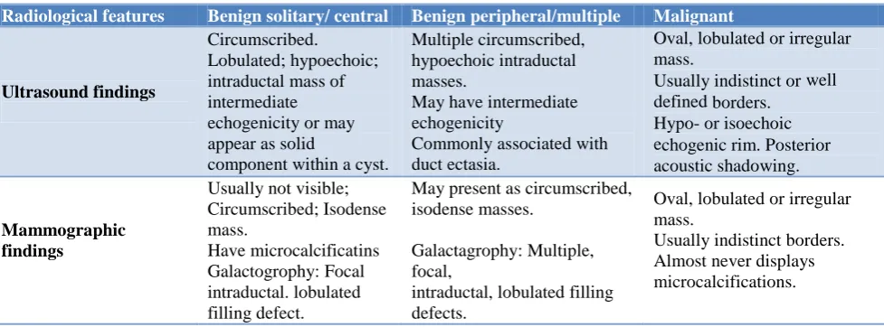
Table 2: Imaging features of various papillary lesions.
Radiological features Benign solitary/ central Benign peripheral/multiple Malignant
Ultrasound findings
Circumscribed. Lobulated; hypoechoic; intraductal mass of intermediate echogenicity or may appear as solid
component within a cyst.
Multiple circumscribed, hypoechoic intraductal masses.
May have intermediate echogenicity
Commonly associated with duct ectasia.
Oval, lobulated or irregular mass.
Usually indistinct or well defined borders.
Hypo- or isoechoic echogenic rim. Posterior acoustic shadowing.
Mammographic findings
Usually not visible; Circumscribed; Isodense mass. Have microcalcificatins Galactogrophy: Focal intraductal. lobulated filling defect.
May present as circumscribed, isodense masses.
Galactagrophy: Multiple, focal,
intraductal, lobulated filling defects.
Oval, lobulated or irregular mass.
Usually indistinct borders. Almost never displays microcalcifications.
DISCUSSION
The differential diagnosis of papillary lesions includes papilloma, papillomatosis, sclerosing papilloma, atypical papilloma and papillary carcinoma.5 Papillary breast lesions constitute wide spectrum of pathologies ranging
carcinoma from benign papillary lesions is essential. Papillary carcinoma accounts for fewer than 2% of all breast malignancies.7 Other benign and malignant pathologies can also mimic papillary lesions on imaging and tissue diagnosis is essential. Imaging plays an important role in lesion identification, assessment of extent, tissue sampling, and follow-up.6
Solitary breast papillomata are potentially malignant and are associated with high risk of breast malignancies.8 On imaging smaller lesions may be occult on mammography. Larger lesions are seen as rounded or ovoid, well-circumscribed retro-areolar mass. Multiple papillomas are usually peripheral in location and can be bilateral. Calcifications are uncommon and include both coarse dense calcifications and micro calcifications.
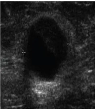
Figure 1 (A): Cystic lesion with mural pedunculated mass.
Figure 1 (B): Complex solid and cystic nodule.
Figure 1 (C): Intraductal echogenic lesion with vascular flow.
Figure 1 (D): Dilated duct (white arrow head) with lesion having internal calcifications (white arrow).
In our study malignant lesions showed no evidence of micro calcifications which means that not all malignant lesions will present with micro calcifications. Mammographic findings of papillary carcinomas include round or oval, circumscribed solitary or clustered masses, which may be associated with micro calcifications. Spiculations are fairly uncommon. On US, the lesion may be seen as an intra-ductal mass with or without ductal dilatation, a complex solid cystic mass, or single or multiple solid nodules. These lesions are usually vascular and have a tendency to bleed spontaneously, resulting in intra cystic fluid-debris levels.1,11 However, papillary lesions can be mammographically or sonographically occult if small. The overall sensitivity for detection of papillary lesions on mammography is low.5 A nonparallel orientation, echogenic halo, posterior acoustic enhancement, and associated micro calcification are reported to be more frequent in malignant lesions.12
Calcifications are uncommon in papillomas.5 However, presence of micro calcifications are common in malignant papillary lesions. On mammography analysis of characteristics of micro calcifications is slightly more sensitive than analysis of the characteristics of the mass in differentiating benign and malignant lesions.5,13 The benign papillary lesions show calcifications or micro calcifications only in 25% of the cases.11,14 Papillary carcinomas, on the contrary, usually show up in mammography as an ill-defined mass with stromal distortion. The presence pleomorphic micro calcifications have also been described in this type of lesion.11,15,16
Intra-ductal content such as blood products, inspissated secretions in ductal ectasia, and neoplastic cells in DCIS can mimic a papillary lesion. A papillary lesion appearing as a well-defined solid nodule may be indistinguishable from a fibro adenoma. Additionally non-papillary and papillary carcinomas can have similar appearances.6
Imaging is important for lesion identification and local staging, guiding tissue diagnosis, and follow-up. Mammography and US are the main imaging modalities for identification. However, small papillomas or papillary DCIS, may be occult or have nonspecific segmental ductal dilatation.6 The best test for detecting image papillary lesions is the breast US. According to Boin et al 100% of patients showed an altered US.17 Various studies report an increased sensitivity of US to detect breast papillary lesions compared to mammography.14,15
Tissue sampling of papillary lesions can utilize different options; however, US-guided percutaneous core biopsy is used most frequently because it allows for real-time visualization, a feature that is essential for sampling the solid component of the lesion. Nevertheless, definitive histopathological categorization and results of biopsy samples may be limited due to tissue fragmentation and under sampling. These findings have resulted in surgical excision being recommended even for imaging concordant, percutaneous, biopsy-proven benign
papillomas.6 Other authors have found a low risk of malignancy for benign papillomas diagnosed by core-needle biopsy and recommend mammographic follow-up for benign and excision for atypical papillomas.6,18
CONCLUSION
Papillary lesions are an uncommon group of breast diseases that present with diverse imaging and pathological findings and pose diagnostic and management challenges. Conventional breast imaging such as sonography and mammography remains the first main stay and quite sensitive in detecting breast papillary lesions leading to early detection and management. The radiologist plays an important role in the diagnosis and management of these lesions. Knowledge of the types and imaging spectra of various papillary lesions and the role of imaging in their evaluation are thus essential.
Funding: No funding sources Conflict of interest: None declared
Ethical approval: The study was approved by the Institutional Ethics Committee
REFERENCES
1. Muttarak M, Lerttumnongtum P, Chaiwun B, Peh WC. Spectrum of papillary lesions of the breast: clinical, imaging, and pathologic correlation. Am J Roentgenol. 2008;191(3):700-7.
2. Mercado CL, Hamele-Bena D, Oken SM, Singer CI, Cangiarella J. Papillary lesions of the breast at percutaneous core-needle biopsy. Radiology. 2006;238(3):801-8.
3. Tavassoli FA. Papillary lesions. In: Tavassoli FA, ed. Pathology of the breast. Norwalk, CT: Appleton and Lange; 1992: 193-227.
4. Rosen EL, Bentley RC, Baker JA, Soo MS. Imaging-guided core-needle biopsy of papillary lesions of the breast. AJR. 2002;179:1185-92. 5. Lam WW, Chu WC, Tang AP, Tse G, Ma TK. Role
of radiologic features in the management of papillary lesions of the breast. Am J Roentgenol. 2006;186(5):1322-7.
6. Jagmohan P, Pool FJ, Putti TC, Wong J. Papillary lesions of the breast: imaging findings and diagnostic challenges. Diagnost Intervention Radiol. 2013;19(6):471.
7. Schneider JA. Invasive papillary breast carcinoma: mammographic and sonographic appearance. Radiology. 1989;171(2):377-9.
8. Gutman H, Schachter J, Wasserberg N, Shechtman I, Greiff F. Are solitary breast papillomas entirely benign?. Arch Surg. 2003;138(12):1330-3.
9. Cardenosa G, Eklund GW. Benign papillary neoplasms of the breast: mammographic appearances. Radiology. 1991;181:751–5.
11. Eiada R, Chong J, Kulkarni S, Goldberg F, Muradali D. Papillary lesions of the breast: MRI, ultrasound and mammographic appearances. AJR Am J Roentgenol. 2012;198:264–71.
12. Kim TH, Kang DK, Kim SY, Lee EJ, Jung YS, Yim H. Sonographic differentiation of benign and malignant papillary lesions of the breast. J Ultrasound Med. 2008;27:75–82.
13. Yang WT, Suen M, Metreweli C. Sonographic features of benign papillary neoplasms of the breast: review of 22 patients. J Ultrasound Med. 1997;16(3):161-8.
14. Brookes MJ and Bourke AG. Radiological appearances of papillary breast lesions. Clin Radiol. 2008;63(11):1265–73.
15. McCulloch GL, Stanislavsky A, Radswiki. Radiological features of papillary carcinoma of the breast. Clin Radiol. 1997;52(11):865–8.
16. Kestelman FP, Gomes CF, Fontes FB, Marchiori E. Imaging findings of papillary breast lesions: a pictorial review. Clin Radiol. 2014;69(4):436-41. 17. Boin DP, Baez JJ, Guajardo MP, Benavides DO,
Ortega ME, Valdés DR, et al. Breast papillary lesions: an analysis of 70 cases. Cancer Med Sci. 2014;8:461.
18. Liberman L, Bracero N, Vuolo MA, Dershaw DD, Morris EA, Abramson AF, et al. Percutaneous large-core biopsy of papillary breast lesions. Am J Roentgenol. 1999;172(2):331-7.