www.fm.viamedica.pl
Address for correspondence: Prof. Witold Woźniak, Department of Anatomy, University School of Medical Sciences, ul. Święcickiego 6, 60–195 Poznań, Poland, tel: +48 61 86 99 181 ext. 564, e-mail: [email protected]
The development of the epidural
space in human embryos
Magdalena Patelska-Banaszewska, Witold Woźniak
Department of Anatomy, University School of Medical Sciences, Poznań, Poland
[Received 25 April 2004; Accepted 25 June 2004]
The epidural space is seen in embryos at stage 17 (41 days) on the periphery of the primary meninx. During stage 18 (44 days) the dura mater proper appears and the epidural space is located between this meninx and the perichondrium and contains blood vessels. During the last week of the embryonic period (stages 20–23) the epidural space is evident around the circumference of the spinal cord. On the posterior surface it is found between the dura mater and the me-soderm of the dorsal body wall.
Key words: human neuroembryology, primary meninx, epidural space
INTRODUCTION
The epidural space lies between the spinal dura mater and the periosteum of the vertebral canal. This periosteum is formed by the outer endosteal layer of the dura mater. The epidural space contains loose connective tissue, venous plexuses and adipose tis-sue, which is particularly evident in the lumbar re-gion [8]. There is some evidence that it is only a po-tential space [2].
Most papers devoted to the development of the meningeal spaces deal with such spaces in the skull [1, 3, 4, 6, 7].
The development of the epidural space in the vertebral canal was observed by Sensenig [9] in em-bryos at 30 mm C-R length. Lüdinghausen and Dzial-las [5] found the space in foetuses of 180–280 mm C-R length.
The aim of the present study is to describe the formation of the epidural space in staged human embryos.
MATERIAL AND METHODS
The study was performed in 47 human embryos of 12 to 30 mm C-R length, from stage 17 to stage 23 (Table 1). All the embryos were sectioned in the
horizontal, frontal, and sagittal planes and stained according to various methods (chiefly Mallory, hae-matoxylin and eosin and with silver salts). In some embryos graphic reconstructions were prepared at each of the stages investigated.
RESULTS
The primordium of the epidural space appears in embryos at the end of the 6th week (stage 17). In
these embryos a more cellular zone appears on the periphery of the primary meninx, which is observed close to the vertebral bodies covered by the peri-chondrium (Fig. 1). In some places between the per-ichondrium and this zone there are irregular spaces containing blood vessels. These spaces may be con-sidered the early epidural space.
In embryos at the beginning of the 7th week
(sta-ge 18) the condensed cellular layers appear in the most peripheral part of the primary meninx (Fig. 2, 3). These layers are present in the cervical and upper thoracic portions of the vertebral canal.
274
Folia Morphol., 2004, Vol. 63, No. 3
Table 1. C-R length, developmental stage and age of the embryos investigated
Catalogue No. C-R length [mm] Developmental stage Age [days] Plane of section
B-70 12.0 17 41 Horizontal
A-12 12.5 17 41 Horizontal
B-180 13.0 17 41 Sagittal
B-64 13.5 17 41 Frontal
PJK-2 13.5 17 41 Horizontal
PJK-14 13.5 17 41 Sagittal
B-68 14.0 17 41 Horizontal
A-1 14.0 17 41 Horizontal
B-129 14.0 17 41 Sagittal
B-122 14.5 18 44 Frontal
Bł-4 15.0 18 44 Horizontal
B-128 15.0 18 44 Sagittal
B-65 16.0 18 44 Horizontal
B-66 16.5 19 46 Horizontal
Bł-5 17.0 19 46 Horizontal
Z-13b 17.0 19 46 Frontal
Bł-10 17.5 19 46 Horizontal
B-123 17.5 19 46 Sagittal
X-19 17.5 19 46 Sagittal
KA-2 18.0 19 46 Horizontal
B-112 18.0 19 46 Sagittal
A-10 18.0 19 46 Horizontal
PJK-1 19.0 19 46 Sagittal
KA-3 19.0 19 46 Sagittal
PJK-28 19.0 19 46 Horizontal
PJK-13 19.0 19 46 Horizontal
B-99 19.5 20 49 Horizontal
Bł-2 20.0 20 49 Sagittal
Bł-1 20.5 20 49 Horizontal
PJK-27 21.0 20 49 Horizontal
B-126 22.0 21 51 Horizontal
B-170 22.5 21 51 Horizontal
B-127 23.5 21 51 Sagittal
A-4 23.5 21 51 Frontal
PK-61 24.0 21 51 Sagittal
WR-II 25.0 22 53 Horizontal
ZJ-2 26.0 22 53 Sagittal
Z-3 26.5 22 53 Horizontal
B-114 27.0 23 56 Sagittal
WW 28.5 23 56 Frontal
B-177 28.5 23 56 Horizontal
A-4 29.0 23 56 Horizontal
A-2 29.0 23 56 Frontal
A-71 29.0 23 56 Horizontal
A-3 29.0 23 56 Horizontal
Kub-2 29.5 23 56 Horizontal
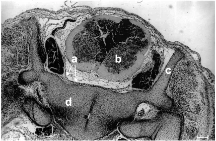
Figure 1. Cross-section of embryo at stage 17. a — spinal
cord, b — primary meninx, c — perichondrium, d — verte-bral body, e — blood vessels, f — primordium of the epidu-ral space. H+E. Scale bar: 100 µm.
Figure 2. Cross-section of embryo at stage 18. a — spinal
cord, b — spinal ganglion, c — primary meninx, d — ex-ternal layer of dura mater, e — epidural space, f — inex-ternal condensed layer forming dura mater proper, g — body of vertebra. H+E. Scale bar: 100 µm.
Fig. 1
276
Folia Morphol., 2004, Vol. 63, No. 3
Figure 4. Cross-section of embryo at stage 19. Nissl staining. a — dura mater, b — spinal cord, c — vertebral arch, d — body
of vertebra. Scale bar: 100 µm.
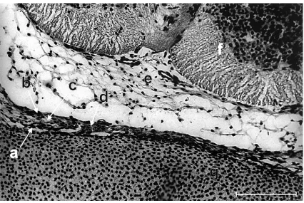
Figure 3. Cross-section of embryo at stage 18. a — primary meninx, b — external layer of dura mater, c — epidural space,
Figure 6. Cross-section of embryo at stage 19. Nissl staining. a — external layer of dura mater, b — dura mater proper, c — epidural
space, d — blood vessels, e — primary meninx, f — spinal cord, g — body of vertebra. Scale bar: 100 µm.
278
Folia Morphol., 2004, Vol. 63, No. 3
Figure 7. Sagittal section of embryo at stage 21. a — spinal cord,
b — primary meninx, c — dura mater, d — epidural space, e — body of vertebra, f — intervertebral disc. H+E. Scale bar: 100 µm.
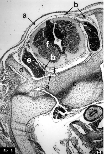
Figure 8. Cross-section of embryo at stage 22. Nissl staining.
a — epidural space, b — dura mater, c — body of vertebra, d — arch of vertebra, e — spinal ganglion, f — spinal cord. Scale bar: 100 µm.
Figure 9. Cross-section of embryo at stage 21. a — spinal cord,
b — primary meninx, c — blood vessels, d — body of vertebra, e — epidural space, f — dura mater. H+E. Scale bar: 100 µm.
Fig. 7 Fig. 8
In embryos at stage 19 the internal condensed layer forming the dura mater proper becomes thin-ner and more fibrous and is present around the whole circumference of the spinal cord (Fig. 4, 5). The epi-dural space is well developed and contains many blood vessels (Fig. 5, 6). It extends throughout the length of the vertebral canal.
During the last week of embryonic development (stages 21–23) the dura mater forms a distinct layer within the vertebral canal (Fig. 7, 8). The epidural space is wide and extends around all the surfaces of the vertebral canal. Within this space blood vessels are present (Fig. 9).
DISCUSSION
Sensenig [9] described the epidural space in em-bryos at the end of the 8th week (stage 23). He wrote:
“In older embryos of this group the epidural cavity is forming by the separation of ventrolateral and lat-eral dura rudiments from the adjacent perichondri-um”. In the present study it was found that the epi-dural space is already marked in embryos at stage 17. During the 8th week this space is well formed
through the walls of the vertebral canal. It contains blood vessels from the very beginning and the num-ber of blood vessels in this space increases with age.
The development of the epidural space proceeds in the rostro-caudal direction and is first observed on the ventral part of the vertebral canal. This was also noted by Lüdinghausen and Dziallas [5].
REFERENCES
1. Cohen H, Davis S (1938) The morphology and perme-ability of the roof of the fourth ventricle in some mam-malian embryos. J Anat, 72: 430–458.
2. Harrison GR, Parkin JG (1985) Resin injection studies of the lumbar extradural space. Br J Anaesth, 57: 333– –336.
3. Hochstetter F (1934) Über die Entwicklung und differ-enzierung der Hüllen der Rückenmarkes beim Men-schen. Morph Jb, 74: 1–104.
4. Klika E (1968) L’ultrastructure des meninges en ontoge-nese de l’homme. Z mikrosk-anat Forsch, 79: 209–222. 5. Lüdinghausen von M, Dziallas P (1972) Zur
Entwick-lung des menschlichen Epiduralraumes. Anat Anz, 130: 571–584.
6. O’Rahilly R, Müller F (1986) The meninges in human development. J Neuropath Exp Neurol, 45: 588–608. 7. Osaka K, Matsumoto S, Yasuda M (1977) The
devel-opment of the cerebro-spinal fluid pathway in human embryos. No Shinkei Geka, 5: 1047–1055.