
Open Access
Research
JNK pathway is involved in the inhibition of inflammatory target
gene expression and NF-kappaB activation by melittin
Hye Ji Park
1, Hwa Jeong Lee
1, Myung Sook Choi
1, Dong Ju Son
1,
Ho Sueb Song
2, Min Jong Song
3, Jeong Min Lee
4, Sang Bae Han
1,
Youngsoo Kim
1and Jin Tae Hong*
1Address: 1College of Pharmacy, Chungbuk National University, 12 Gaesin-dong, Heungduk-gu, Cheongju, Chungbuk 361-763, Korea, 2College
of Oriental Medicine, Kyungwon University, 65 Bukjeong-Dong, Sujeong-gu, Seongnam, Gyeonggi 461-701, Korea, 3Department of Obstetrics
and Gynecology, St. Vincent's Hospital, The Catholic University, Suwon 442-723, Korea and 4Life Science R&D Center, Sinil Pharmaceutical Co,
San 5-1, Bonpyung-Ri, Angsung-Myun, Chungju, Chungbuk,380-862, Korea
Email: Hye Ji Park - [email protected]; Hwa Jeong Lee - [email protected]; Myung Sook Choi - [email protected]; Dong Ju Son - [email protected]; Ho Sueb Song - [email protected]; Min Jong Song - [email protected]; Jeong Min Lee - [email protected]; Sang Bae Han - [email protected]; Youngsoo Kim - [email protected]; Jin Tae Hong* - [email protected]
* Corresponding author
Abstract
Background: Bee venom therapy has been used to treat inflammatory diseases including rheumatoid arthritis in humans and in experimental animals. We previously found that bee venom and melittin (a major component of bee venom) have anti-inflammatory effect by reacting with the sulfhydryl group of p50 of nuclear factor-kappa B (NF-κB) and IκB kinases (IKKs). Since mitogen activated protein (MAP) kinase family is implicated in the NF-κB activation and inflammatory reaction, we further investigated whether activation of MAP kinase may be also involved in the anti-inflammatory effect of melittin and bee venom.
Methods: The anti-inflammatory effects of melittin and bee venom were investigated in cultured Raw 264.7 cells, THP-1 human monocytic cells and Synoviocytes. The activation of NF-κB was investigated by electrophoretic mobility shift assay. Nitric oxide (NO) and prostaglandin E2 (PGE2) were determined either by Enzyme Linked Immuno Sorbent Assay or by biochemical assay. Expression of IκB, p50, p65, inducible nitric oxide synthetase (iNOS), cyclooxygenase-2 (COX-2) as well as phosphorylation of MAP kinase family was determined by Western blot.
Results: Melittin (0.5–5 μg/ml) and bee venom (5 and 10 μg/ml) inhibited lipopolysaccharide (LPS, 1 μg/ml) and sodium nitroprusside (SNP, 200 μM)-induced activation of c-Jun NH2-terminal kinase (JNK) in RAW 264.7 cells in a dose dependent manner. However, JNK inhibitor, anthra [1,9-cd]pyrazole-6 (2H)-one (SP600215, 10–50 μM) dose dependently suppressed the inhibitory effects of melittin and bee venom on NF-κB dependent luciferase and DNA binding activity via suppression of the inhibitory effect of melittin and bee venom on the LPS and SNP-induced translocation of p65 and p50 into nucleus as well as cytosolic release of IκB. Moreover, JNK inhibitor suppressed the inhibitory effects of melittin and bee venom on iNOS and COX-2 expression, and on NO and PGE2 generation.
Conclusion: These data show that melittin and bee venom prevent LPS and SNP-induced NO and PGE2 production via JNK pathway dependent inactivation of NF-κB, and suggest that inactivation of JNK pathways may also contribute to the anti-inflammatory and anti-arthritis effects of melittin and bee venom.
Published: 29 May 2008
Journal of Inflammation 2008, 5:7 doi:10.1186/1476-9255-5-7
Received: 14 March 2007 Accepted: 29 May 2008
This article is available from: http://www.journal-inflammation.com/content/5/1/7
© 2008 Park et al; licensee BioMed Central Ltd.
Background
Bee venom therapy has been used to relieve pain and to treat inflammatory diseases including rheumatoid arthri-tis in humans [1] and in experimental animals [2]. Bee venom contains melittin, a 26 amino acid peptide, which forms an amphipathic helix with a highly charged car-boxyl terminus [3]. We previously found that bee venom and its major component, melittin inhibited
lipopolysac-charide (LPS), tumor necrosis factor-α (TNF-α), and
sodium nitroprusside (SNP)-induced NF-κB activation by
preventing p50 translocation through interaction of
melittin and sulfhydryl residue of p50 and/or IκB kinases
(IKKα and IKKβ), and that these inhibit inflammatory
reaction in the development of rheumatoid arthritis [4,5] through reduction of large amounts of nitric oxide (NO) and prostaglandins (PGs) which are synthesized systemi-cally in animal models of arthritis and in patients with rheumatoid arthritis [6-10].
NF-κB and IKKs have been suggested to play important
roles in the regulation of inflammatory genes, such as, inducible nitric oxide synthetase (iNOS),
cyclooxygenase-2 (COX-cyclooxygenase-2), cytosolic phospholipase A2 (cPLA2), and
tumor necrosis factor-α (TNF-α). Functionally active
NF-κB exists mainly as a heterodimer consisting of subunits
of the Rel family, and this heterodimer is normally sequestered in the cytosol as an inactive complex by
bind-ing to inhibitory κB (IκBs) in unstimulated cells [11]. The
mechanism of NF-κB activation involves the
phosphor-ylation of IκBs via IKK activation [12]. Once IκBs are
phosphorylated, they are targeted for ubiquitination and subsequent degradation by the 26s proteosome [13]. The
resulting free NF-κB is translocated to the nucleus, where
it binds to the κB binding sites in the promoter regions of
target genes, thereby controls their expression [14]. In
sev-eral studies, potent inhibitors of IKKs preventing NF-κB
activity through blockage of IκB release can be useful for
the treatment of inflammatory diseases such as rheuma-toid arthritis (RA) [15-18].
Mitogen activated protein (MAP) kinases are a group of signaling molecules that also appear to play important roles in inflammatory processes. At least three MAP kinase cascades; ERK (extracellular signal-regulated kinase), JNK (c-Jun N-terminal kinase) and p38 are well described, and have been reported to differentially activate depending on the stimuli and cell types [19]. Several studies have dem-onstrated that activation of MAP kinase is significant in the regulation of inflammation via controlling the
activa-tion of NF-κB and IKKs [19-22].
In the present study, we therefore investigated whether
melittin and bee venom inhibit NF-κB via disrupting MAP
kinase signals, and thereby inhibit the inflammatory
response in Raw 264.7 macrophages and in the synovio-cytes of rheumatoid arthritis patients.
Methods
ChemicalsRabbit polyclonal antibodies to cPLA2 (dilution 1:500),
and goat polyclonal antibody to COX-2 (1:500), TNF-α
(1:500), p50 (1:500), p65 (1:500), IκBα (1:500),
phos-pho-IκBα (1:200), IκBβ (1:500) and mouse polyclonal
antibody to iNOS (1:500), IκB kinases (1:500), mouse
monoclonal ERK, JNK and phospho-p38 antibodies (1:500), and rabbit polyclonal ERK, JNK and p38 (1;500), and all of the secondary antibodies used in Western blot analysis were purchased from Santa Cruz Biotechnology (Santa Cruz, CA, USA). T4 polynucleotide kinase was obtained from Promega (Madison, WI). Poly (dI·dC), horseradish peroxidase-labeled donkey anti-rab-bit second antibody, and the ECL detection reagent were obtained from Amersham Pharmacia Biotech (Centennial Ave, NJ, USA). SNP, LPS, Griess reagent, monoclonal
anti-β-actin antibody,
3-(4,5-dimethyl-2-thiazolyl)-2,5-diphe-nyl tetrazolium bromide (MTT) and melittin, a compo-nent of bee venom were purchased from Sigma-Aldrich (St. Louis, MO, USA). U0126 (ERK inhibitor, 1,4-diamino-2,3-dicyano-1,4-bis (2-aminophenylthio)buta-diene) and SP600125 (JNK inhibitor, anthra [1,9-cd]pyra-zole-6 (2H)-one) were purchased from Calbiochem (San Diego, CA, USA). Bee venom was purchased from You-Miel BV Ltd (Hwasoon, Korea). The compositions are fol-lowings: melittin (45–50%), apamin (2.5–3%), MCD peptide (2–3%), PLA2 (12%), Lyso PLA (1%), hyaluroni-dase (2–3%), histidine (1–1.5%), secarpin (0.5%), ter-tiapin (0.1%), procamine (0.1%), amine (2–3%), carbohydrate (4–5%), 6pp lipids (4–5%), and others (19–27%, protease inhibitor, glucosidase, invertase, acid phosphomonoesterase, dopamine, norepinephrine and unknown amino acid).
Cell culture
Raw 264.7, a mouse macrophage-like cell line and THP-1, a human monocytic cell line were obtained from the American Type Culture Collection (Cryosite, Lane Cove NSW, Australia). Dulbecco's modified Eagle medium (DMEM), penicillin, streptomycin, and fetal bovine serum were purchased from Gibco Life Technologies (Rockville, MD, USA). Raw 264.7 cells were grown in DMEM with 10% fetal bovine serum, 100 U/ml
penicil-lin, and 100 μg/ml streptomycin at 37°C in 5% CO2
humidified air. THP-1 cells were grown in RPMI 1640 with L-glutamine and 25 mM HEPES buffer (Gibco Life Technologies, Rockville, MD, USA) supplemented with 10% fetal bovine serum, 100 units/ml penicillin and 100
Synoviocyte culture
Synovial tissues were obtained, with consent, from nine RA patients who were undergoing total knee replacement or arthroscopic synovectomy. All patients satisfied the 1987 revised diagnostic criteria of the American College of Rheumatology [23]. The method of synoviocyte culture was described in elsewhere [4,5].
Determination of Nitric Oxide and Prostaglandin E2
The NO accumulation in the supernatant was assessed by Griess reaction described in elsewhere [4], and the
deter-mination of PGE2 was performed as described in
else-where [4].
DNA binding activity of NF-κB
EMSA was performed according to the manufacturer's rec-ommendations (Promega, Madison, WI) as described in previous study [4,5]. Briefly, nuclear extract was incubated
with κB consensus oligonucleotides end-labeled using T4
polynucleotide kinase and [γ-32P] ATP for 10 min at
37°C. Gel shift reactions were assembled and allowed to incubate at room temperature for 10 min followed by the
addition of 1 μl (50,000–200,000 cpm) of 32P-labeled
oligonucleotide and another 20 min of incubation at room temperature. For the competition assay, 100× or 200× excesses of unlabeled double-stranded
oligonucle-otide of the κB binding site (or 100× irrelevant
oligonu-cleotide of AP-1 or SP-1) were used as specific competitors. Supershift assay was done in the presence of
p50 or p65 subunit of NF-κB (2 μg). Subsequently 1 μl of
gel loading buffer was added to each reaction and loaded onto a 4% nondenaturing gel and electrophoresed until the dye was three-fourths of the way down the gel. The gel was dried at 80°C for 1 hr and exposed to film overnight at 70°C. The relative density of the protein bands was scanned by densitometry using MyImage (SLB, Seoul, Korea), and quantified by Labworks 4.0 software (UVP Inc., Upland, California).
Transfection and assay of Luciferase activity
Raw 264.7 or THP-1 cells were transfected with pNF-κ
B-Luc plasmid (5× NF-κB; Stratagene, CA, USA) using a
mix-ture of plasmid and lipofectAMINE PLUS in OPTI-MEN according to manufacture's specification (Invitrogen, Carlsbad, CA, USA). The control pCMV (Clontech, CA, USA) was co-transfected to monitor the transfection effi-ciency. After 24 hr, the cells were then co-treated with BV (or melittin) and LPS or SNP. Luciferase activity was meas-ured by using the luciferase assay kit (Promega) according to the manufacturer's instructions (WinGlow, Bad Wild-bad, Germany).
Western blot analysis
Cell lysates were prepared as described in the previous
study [12]. Equal amount of lysate proteins (80 μg) were
separated on a SDS/12%-polyacrylamide gel, and then transferred to a nitrocellulose membrane (Hybond ECL, Amersham Pharmacia Biotech Inc., Piscataway, NJ). Blots were blocked for 2 hr at room temperature with 5% (w/v) non-fat dried milk in Tris-buffered saline [10 mM Tris (pH 8.0) and 150 mM NaCl] solution containing 0.05% tween-20. The membrane was incubated for 5 hr at room temperature with specific antibodies. The blot was then incubated with the corresponding conjugated anti-rabbit immunoglobulin G-horseradish peroxidase (Santa Cruz Biotechnology Inc.). Immunoreactive proteins were detected with the ECL western blotting detection system. The relative density of the protein bands was scanned by densitometry using MyImage (SLB, Seoul, Korea), and quantified by Labworks 4.0 software (UVP Inc., Upland, California).
Immunofluorescence staining
Cells were plated in chambered tissue culture slides at a
density of 2 × 10³ cells/well in DMEM. The Raw 264.7
cells were then cultured with serum-free medium contain-ing LPS, BV and SP for 2 hr, and then the intracellular location of p50 was determined by immunofluorescence confocal scanning microscope (magnification, 630×) as described in elsewhere [4]. Twenty-four hours later, the cells were washed once with PBS and fixed with 4% para-formaldehyde for 20 min, membrane-permeabilized by exposure for 2 min to 0.1% Triton ×-100 in phosphate-buffered saline, and placed in blocking serum (5% bovine serum albumin in phosphate-buffered saline) at room temperature for 1 hr. The cells were then exposed to pri-mary polyclonal antibodies for p50 (1:100 dilution) over-night at 4°C, After washes with ice-cold PBS followed by treatment with anti-goat- or anti- rabbit- biotinylated sec-ondary antibodies Alexa Fluor 568 (p50) or Alexa Fluor 633 (DAPI) (Molecular Probes Inc., Eugene, OR, USA), 1:200 dilution, for 4 hr at room temperature. Nuclear stain and mount in antifade medium with DAPI (Vector Laboratory Inc.), immunofluorescence images were acquired using a confocal laser scanning microscope (TCS SP2, Leica Microsystems AG, Wetzlar, Germany) equipped with a 630×oil immersion objective.
Statistical analysis
Data were analyzed using one-way analysis of variance followed by Tukey's test as a post hoc test. Differences were considered significant at p < 0.05.
Results
Melittin inhibited LPS and SNP-induced activation of JNK in RAW 264.7 cells
We previously found that bee venom and its major
com-ponent, melittin inhibits LPS, TNF-α and SNP-induced
inflammatory responses through inactivation of NF-κB
play an important role in the transcriptional regulation of LPS-induced iNOS and COX-2 expression via suppression
of the activation of transcription factor NF-κB. To
investi-gate the involvement of MAP kinase pathway in the
inhib-itory effect by melttin and bee venom on NO and PGE2
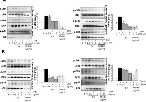
production, the activation of MAP kinase (phosphoryla-tion of ERK, JNK and p38) induced by LPS and SNP was evaluated in both Raw 264.7 cells as well as synoviocytes. The densitometry analysis from individual three different
experiments showed that melittin (0.5–5 μg/ml) and bee
venom (5 and 10 μg/ml) strongly blocked LPS (1 μg/ml)
and SNP (200 μM)-induced activation of JNK in the Raw
264.7 cells (Fig. 1A) as well as synoviocytes (Fig. 1B). We
also found that significant inhibitory effects of melittin
(0.5–5 μg/ml) on the activation of ERK in LPS treated Raw
264.7 cells and synoviocytes, and SNP treated synovio-vytes. Activation of p38 was also significantly reduced in the LPS treated synoviocytes, and SNP treated Raw 264.7 cells and synoviocytes, but the expression ERK and p38 was also reduced, indicating that blocking of the activa-tion of p38 and ERK was not specific (Fig. 1A and 1B). Similar effect of bee venom was also found (Fig. 1). These results suggest that JNK could be the most specific and important signal involved in the melittin and BV-induced
inhibition of NO and PGE2 generation.
Effect of melittin and bee venom on LPS and SNP-induced phosphorylation of MAPKs Figure 1
Effect of melittin and bee venom on LPS and SNP-induced phosphorylation of MAPKs. A, Raw 264.7 macrophages were treated with 5 or 10 μg/ml melittin or 0.5–5 μg/ml bee venom in the presence of LPS (1 μg/ml) or SNP (200 μM) at 37°C for 24 hr. B, Synoviocytes were treated with 5 or 10 μg/ml melittin or 0.5–5 μg/ml bee venom in the presence of 1 μg/ml LPS or 200 μM SNP at 37°C for 24 hr. Equal amounts of total proteins (80 μg/lane) were subjected to 10% SDS ± PAGE, and the expression of p-ERK/ERK, p-JNK/JNK, or p-p38/p38 were detected by western blotting using specific antibodies. Each panel representative of three independent experiments. Quantification of band intensities from three independent experimental results was determined by a densitometry (Imaging System). Data was described as means ± S.E. from three experiments per-formed in triplicate for p-ERK/ERK, p-JNK/JNK, or p-p38/p38. *p < 0.05 indicate statistically significant differences from the LPS or SNP-treated group.
ERK p-ERK p-JNK p-JNK ERK p-ERK
A
p38 p-p38 * P -JNK/JNK Related density
u (% of LP S gr oup) - + + + + + + 0 0 0.5 1 5 5 10
BV Melittin (μg/ml)
LPS (1 μg/ml)
p38 JNK ERK p-ERK p-JNK p-p38 p-JNK/JNK Related density ( % of LP S gr oup) SNP (200 μM) * * * * * - + + + + + + 0 0 0.5 1 5 5 10
BV Melittin (μg/ml) * * * p38 JNK p-p38 P -E RK/E RK Related density ( % of LP S gr oup) - + + + + + + 0 0 0.5 1 5 5 10
BV Melittin (μg/ml)
LPS (1 μg/ml)
p38 JNK p-JNK p-p38 p-RE Rk/E
RK Related den
s ity (% of LPS gr oup) SNP (200 μM) - + + + + + + 0 0 0.5 1 5 5 10
BV Melittin (μg/ml) * * * * * * * * * JNK ERK p-ERK
B
SNP (200 μM) - + + + + + + 0 0 0.5 1 5 5 10BV Melittin (μg/ml) - + + + + + +
0 0 0.5 1 5 5 10
BV Melittin (μg/ml)
LPS (1 μg/ml)
- + + + + + + 0 0 0.5 1 5 5 10
BV Melittin (μg/ml)
LPS (1 μg/ml)
- + + + + + + 0 0 0.5 1 5 5 10
BV Melittin (μg/ml)
JNK inhibitor suppressed the inhibitory effects of melittin
and bee venom on NF-κB dependent Luciferase and DNA
binding activity
To further examine the involvement of JNK pathway in
the inhibitory effect of melittin and bee venom on NF-κB
activation, we explored JNK specific inhibitor SP600125
(10–50 μM), and determined the inhibitory effect of
melittin and bee venom on the activation of NF-κB. As
shown in Fig. 2, pretreatment (1 hr) of SP600125 strongly suppressed the inhibitory effect of melittin and bee
venom on the LPS and SNP-induced NF-κB activation in
Raw 264.7 cells (Fig. 2A) and synoviocytes (Fig. 2B). The specificity of DNA binding was examined by competition assay by adding an excessive amount of unlabeled/cold oligonucleotides to reaction mixtures containing nuclear extract. The specificity of DNA binding was examined by supershift assay using antibodies for the p50 or p65
com-ponents of NF-κB (data not shown).
One of the consequences of inhibition of NF-κB is the
inhibition of the nuclear translocation of p50 and p65
through the blockage of IκB release. To study the result of
JNK inhibitor suppressed the inhibitory effect of melittin and bee venom on the NF-κB DNA binding activity induced by LPS or
SNP Figure 2
JNK inhibitor suppressed the inhibitory effect of melittin and bee venom on the NF-κB DNA binding activity induced by LPS or SNP. Raw 264.7 macrophages (A) and synoviocytes (B) were pretreated with 10, 20, and 50 μM SP600125 1 h prior to the treatment with melittin or bee venom with or without LPS or SNP for 2 h. The DNA binding activa-tion of NF-κB was investigated using EMSA. Nuclear extracts from Raw 264.7 cells or synoviocytes treated for 1 hr were incu-bated with 32P-end-labeled oligonucleotide containing the κB sequence. Each panel is representative of three similar
experiments with duplicates.
BV (5 μg/ml) Melittin (10μg/ml)
LPS (1 μg/ml) SP (μM)
NF-κκκκB
10 20 50 10 20 50
SNP (200 μM)
NF-κκκκB
BV (5 μg/ml) Melittin (10μg/ml)
SP (μM)
10 20 50 10 20 50
SP (μM)
NF-κκκκB NF-κκκκB
BV (5 μg/ml) Melittin (10μg/ml)
LPS (1 μg/ml) SP (μM)
10 20 50 10 20 50SP (μM)
SNP (200 μM)
BV (5 μg/ml) Melittin (10μg/ml)
SP (μM)
10 20 50 10 20 50
SP (μM)
A
the treatment of JNK inhibitor on the translocation of p50 and p65 into the nucleus, we determined the appearance of the p50 and p65 in the nucleus extracts by Western blott. Pretreatment (1 hr) of SP600125 suppressed the inhibitory effect of melittin and bee venom on LPS and SNP-induced nuclear translocation of the p50 and p65 in Raw 264.7 cells (Fig. 3A) and in synoviocytes (Fig. 3B). Te
kinetics of IκBα release (determined the level of IκBα
phosphorylation) in cytosol were further studied by west-ern blot analysis. Inhibitory effect of melittin and bee
venom on the LPS as well as SNP-induced IκBα release
was markedly suppressed by SP600125 in both Raw 264.7
cells (Fig. 3A) and synoviocytes (Fig. 3B). The
phosphor-ylation of IκBβ was not examined because this antibody is
not commercially available. The suppressive effect of SP600125 on the reduced nuclear translocation of the p50 subunits was also confirmed by examination with confo-cal laser scanning microscopy in Raw 264.7 cells (Fig. 3C).
Raw 264.7 and THP-1 cells were transfected with a pro-moter reporter gene construct (a fusion gene containing
SV40 promoter, 5 repeats of the consensus NF-κB binding
sequence), and transcriptional activities were also meas-ured after stimulating the cells with LPS or SNP in the
JNK inhibitor suppressed the inhibitory effect of melittin and bee venom on the nuclear translocation of the p50 subunit and
the release of IκB induced by LPS or SNP
Figure 3
JNK inhibitor suppressed the inhibitory effect of melittin and bee venom on the nuclear translocation of the p50 subunit and the release of IκB induced by LPS or SNP. Raw 264.7 cells (A) or synoviocytes (B) were pretreated with 10 and 20 μM SP600125 1 h prior to the treatment with melittin or bee venom with or without LPS (1 μg/ml) or SNP (200 μM) at 37°C for 24 hr. 80 μg of nuclear (p50 and p65), cytosolic IκB or total protein extracted after treatment were used to determine of p50, p65, p-IκBα, IκBα, or IκBβ; β-actin protein was used as an internal control. Each panel is representative of three similar exper-iments. C, Raw 264.7 cells were treated with LPS, BV and SP for 24 hr, and then the intracellular location of p50 was deter-mined by immunofluorescence confocal scanning microscope (magnification, 630×). Double staining (Merge, pink) with p50 (red) and DAPI (blue) staining demonstrates the localization of p50 in the nucleus.
LPS (1 μg/ml)
LPS (
1
μμμμ
g/ml)
BV (5
μμμμ
g/ml)
LPS (1 μg/ml)
B
IκκκκBαααα p-IκκκκBααααββββ−−−−actin
- + + + + + + + 10 20 10 20 SP (μM) SP (μM)
BV (5 μg/ml) Melittin (10μg/ml)
SNP (200 μM) - + + + + + + +
10 20 10 20 SP (μM) SP (μM)
BV (5 μg/ml) Melittin (10 μg/ml)
P50 (NE)
P65 (NE)
ββββ−−−−tubulin
A
IκκκκBαααα p-IκκκκBαααα
ββββ−−−−actin
- + + + + + + + 10 20 10 20 SP (μM) SP (μM)
BV (5 μg/ml) Melittin (10 μg/ml)
SNP (200 μM) - + + + + + + +
10 20 10 20 SP (μM) SP (μM)
BV (5 μg/ml) Melittin (10 μg/ml)
P50 (NE)
P65 (NE)
ββββ−−−−tubulin
Original magnif
icat
ion 63
0X
S
S
S
S
เเเเ
'$3,'$3,'$3,'$3,
เเเเ
0H
UJ
H
0H
UJ
H
0H
UJ
H
0H
UJ
H
เเเเ
-+
+
+
+
SP (
μμμμ
M)
-+
-1
0 20
presence of bee venom and melittin. Agreement with the suppressive effect of SP600125 on the DNA binding
activ-ity of NF-κB, pretreatment (1 hr) of SP600125 also
strongly suppressed the inhibitory effect of melittin and
bee venom on the LPS or SNP-induced NF-κB
transcrip-tional activation in Raw 264.7 cells (Fig. 4A) and THP-1 cells (Fig. 4B). These suppressive effects were statistically significant in the inhibitory effect of melittin and Bee
venom on the LPS and SNP-induced NF-κB
transcrip-tional activation in both Raw 264.7 cells (Fig. 4A) and
THP-1 cells (Fig. 4B) by 20 μM of SP600125. The
suppres-sive effects were also statistically significant in the inhibi-tory effect of Bee venom on the LPS-induced, and in the
inhibitory effect of melittin on the SNP-induced NF-κB in
THP-1 cells by 10 μM of SP600125.
JNK inhibitor suppressed the inhibitory effects of melittin and bee venom on iNOS and COX-2 expression, and on
NO and PGE2 generation
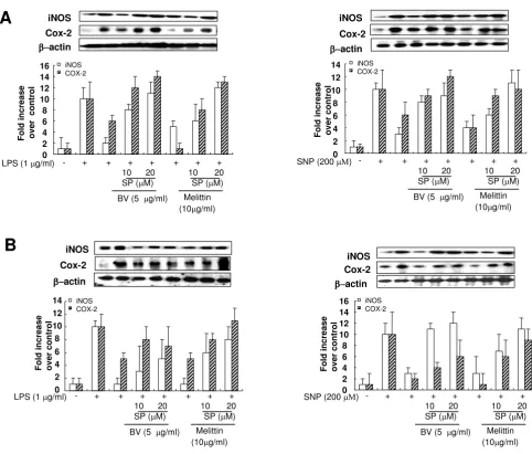
To investigate whether the suppressed effect of SP600125 on the inhibitory effect of bee venom and melittin on the inflammatory gene expression, iNOS and COX-2 expres-sion was determined. The inhibitory effect of melittin and bee venom on iNOS and COX-2 expression by LPS and SNP in Raw 264.7 cells (Fig. 5A) and in synoviocytes (Fig. 5B) were dose dependently suppressed by SP600215 (10
and 20 μM). The suppressive effect of SP600125 on the
inflammatory mediator generation was then examined. Significant concentration-dependent suppression by the pretreatment of SP600215 on the NO generation was observed in Raw 264.7 cells (Fig. 6A,C) and synoviocytes (Fig. 6B,D) treated with melittin and bee venom in
com-JNK inhibitor suppressed the inhibitory effect of melittin and bee venom on the on NF-κB-dependent luciferase induced by LPS
or SNP Figure 4
JNK inhibitor suppressed the inhibitory effect of melittin and bee venom on the on NF-κB-dependent luciferase induced by LPS or SNP. Raw 264.7 cells (A) and THP-1 cells (B) were transfected with pNF-κB-Luc plasmid (5× NF-κB), Raw 264.7 cells or THP-1 cells were pretreated with 10 and 20 μM SP600125 1 hr prior to the treatment with melittin or bee venom with or without LPS (1 μg/ml) or SNP (200 μM) at 37°C for 2 hr, and then luciferase activities were determined. All values represent means ± S.E. of three independent experiments performed in triplicate.
R e la ti v elu c if e ra s e a c ti v it y ( RL U / β -g a l ) R e la ti v elu c if e ra s e a c ti v it y ( R L U / β -g a l ) - + + + + + + + 10 20 10 20 SP (μM) SP (μM) BV (5 μg/ml) Melittin (10 μg/ml)
LPS (1 μg/ml)
- + + + + + + + 10 20 10 20 SP (μM) SP (μM) BV (5 μg/ml) Melittin (10 μg/ml)
SNP (200 μM)
R e la ti v e lu c if e ra se a c ti v it y ( R L U / β -g al ) R e la ti v e lu cif e ra s e a c ti v it y ( RL U / β -g a l ) - + + + + + + + 10 20 10 20 SP (μM) SP (μM) BV (5 μg/ml) Melittin (10 μg/ml)
LPS (1 μg/ml)
- + + + + + + + 10 20 10 20 SP (μM) SP (μM) BV (5 μg/ml) Melittin (10 μg/ml)
SNP (200 μM)
A
bination with LPS (Fig. 6A,B) and SNP (Fig. 6C,D). Signif-icant concentration-dependent suppressive effect by the
pretreatment of SP600215 (10 and 20 μM) on the PGE2
generation was also observed in Raw 264.7 cells (Fig. 7A,C) and synoviocytes (Fig. 7B,D) treated with melittin and bee venom in combination with LPS (Fig. 7A,C) and SNP (Fig. 7B,D).
Discussion
We previously found that bee venom and its major com-ponent, melittin inhibits inflammatory stimuli such as
LPS, TNF-α, and SNP-induced NF-κB activation by
pre-venting p50 translocation via an interaction between
melittin and sulfhydryl group of p50 and/or IKKα and
IKKβ, and that these inhibit inflammatory reaction in the
development of rheumatoid arthritis [4,5]. In the present study, we further found that melittin and bee venom sig-nificantly reduced inflammatory stimuli (LPS and SNP)-induced activation of JNK signal, and the JNK signal spe-cific inhibitor SP600215 suppressed the inhibitory effect
of melittin and bee venom on the NF-κB activation, and
inflammatory reaction in Raw 264.7 macrophages and synoviocytes obtained from rheumatoid arthritis patients. This data reflected that the inhibition of JNK pathway JNK inhibitor suppressed inhibitory effect of melittin and bee venom on the inflammatory gene expression induced by LPS or SNP
Figure 5
JNK inhibitor suppressed inhibitory effect of melittin and bee venom on the inflammatory gene expression induced by LPS or SNP. Raw 264.7 cells (A) or synoviocytes (B) were pretreated with 10 and 20 μM SP600125 1 hr prior to the treatment with melittin or bee venom with or without LPS (1 μg/ml) or SNP (200 μM) at 37°C for 24 hr. Equal amounts of total proteins (80 μg/lane) were subjected to 10% SDS ± PAGE, and the expression of iNOS, COX-2 and β-actin were detected by western blotting using specific antibodies. Each panel representative of three independent experiments.
Cox-2 iNOS
ββββ−−−−actin
iNOS Cox-2 ββββ−−−−actin
B
iNOSCox-2 ββββ−−−−actin
LPS (1 μg/ml) - + + + + + + +
10 20 10 20
SP (μM) SP (μM)
BV (5 μg/ml) Melittin
(10μg/ml)
A
Cox-2 iNOS
ββββ−−−−actin
F
o
ld
in
crease
o
v
er co
n
tro
l
0 2 4 6 8 10 12 14 16 iNOS
COX-2
SNP (200 μΜ) - + + + + + + +
10 20 10 20 SP (μM) SP (μM)
BV (5 μg/ml) Melittin
(10μg/ml) 0
2 4 6 8 10 12 14
F
o
ld
in
crease
o
v
er co
n
tro
l
iNOS COX-2
LPS (1 μg/ml) - + + + + + + +
10 20 10 20 SP (μM) SP (μM)
BV (5 μg/ml) Melittin
(10μg/ml)
SNP (200 μΜ) - + + + + + + +
10 20 10 20 SP (μM) SP (μM)
BV (5 μg/ml) Melittin
(10μg/ml) 0
2 4 6 8 10 12 14
F
o
ld
in
crease
ov
e
r c
ontr
o
l
iNOS COX-2
0 2 4 6 8 10 12 14 16
F
o
ld
in
crease
o
v
er co
n
tro
l
conjunction with inhibition of NF-κB pathway may also contribute to the inhibitory effect of melittin and bee venom on the inflammatory reaction of arthritis rheuma-tism.
LPS and SNP rapidly phosphorylates ERK, p38 and JNK,
which lead to NF-κB activation in macrophages [24,25].
The activation of this MAP kinase leads an increase in the production of pro-inflammatory mediators such as NO
and PGE2 [26,27]. Several studies have demonstrated the
implication of the activation of MAP kinase in LPS-induced iNOS and COX-2 expression [28-30] and the
acti-vation of NF-κB [30-33]. To demonstrate other pathway
of NF-κB inactivation by melittin and bee venom, we
investigated the relationship between NF-κB and MAP
kinase activation. Our data demonstrated that melittin and bee venom reduces LPS and SNP-induced activation of JNK signals. Even though other signals (p38 MAP kinase and ERK signal) may be also interfered by melittin and bee venom depend on the cell types and stimuli, LPS and SNP-induced JNK signal was specifically inhibited by melittin and bee venom. This finding is agreed with other data showing that JNK pathway is important signal in the
activation of NF-κB in the processes of inflammatory
reac-JNK inhibitor suppressed the inhibitory effect of melittin and bee venom on the generation of NO induced by LPS or SNP Figure 6
JNK inhibitor suppressed the inhibitory effect of melittin and bee venom on the generation of NO induced by LPS or SNP. Raw 264.7 cells (A, C) or synoviocytes (B, D) were pretreated with 10 and 20 μM SP600125 1 hr prior to the treatment with melittin or bee venom with or without LPS (1 μg/ml) or SNP (200 μM) at 37°C for 24 hr. The amounts of NO in the medium of cultured Raw264.7 cells (A, C) or synoviocytes (B, D) were determined by the methods described in the methods. Results are expressed as means ± SE of three independent experiments performed in triplicate. * indicates signifi-cantly different from the LPS or SNP treated groups (p < 0.05). # and ##, indicates significantly different from LPS or SNP + melittin or BV treated group (p < 0.05).
0 4 8 12 16 20
0 4 8 12 16 20
LPS (1 μg/ml)
10 20 10 20 BV (5 μg/ml) Melittin (10μg/ml)
SP (μM) 10 20 10 20
BV (5 μg/ml) Melittin (10μg/ml) SP (μM)
N
it
ri
te
(
μ
M
)
N
it
ri
te
(
μ
M
)
SNP (200 μM)
0 4 8 12 16 20
0 4 8 12 16 20
N
it
ri
te
(
μ
M
)
N
itri
te
(
μ
M
)
LPS (1 μg/ml)
10 20 10 20 BV (5 μg/ml) Melittin (10μg/ml)
SP (μM) 10 20 10 20
BV (5 μg/ml) Melittin (10μg/ml) SP (μM)
SNP (200 μM)
A
B
C
D
tion [29,30,34]. In more precise investigation with spe-cific JNK inhibitor SP600215, we further showed that the combination treatment of JNK inhibitor with bee venom and melittin suppressed inhibitory effects of melittin and
bee venom on the LPS and SNP-induced NO and PGE2
release with the suppressed effect on the inhibitory effect
of melittin and bee venom on the NF-κB DNA binding
and transcriptional activities. Moreover, we also showed that JNK inhibitor SP600215 abrogated the inhibitory effect of melittin and bee venom on the LPS and
SNP-induced translocation of NF-κB by western blotting as
well as translocation of p50, a subunit of NF-κB by
confo-cal microscope observation. These data show that specific inhibition of JNK pathway may be important for
inactiva-tion of NF-κB, and thus inhibitory effects of melittin and
bee venom on the LPS and SNP-induced NO and PGE2
production.
The involvement of MAPK pathways in the biological activities of melittin and bee venom has been demon-strated. Bee venom triggered the activation of p38 MAPK and JNK and increased lactate dehydrogenase (LDH) release in the bee venom-induced apoptosis of human leukemic U937 [35]. Very similar to our finding, Jang et al. showed that bee venom inhibited mRNA level of
iNOS, COX-2 and NF-κB paralleled with inhibition of
mRNA level of MAP kiase induced by LPS [36]. In addi-tion, we also found that bee venom and melittin inhibited platelet-derived growth factor BB (PDGF-BB)-induced smooth muscle cell proliferation through inactivation of
JNK inhibitor suppressed the inhibitory effect of melittin and bee venom on the generation of PGE2 induced by LPS or SNP
Figure 7
JNK inhibitor suppressed the inhibitory effect of melittin and bee venom on the generation of PGE2 induced by LPS or SNP. Raw 264.7 cells (A, C) or synoviocytes (B, D) were pretreated with 10 and 20 μM SP600125 1 hr prior to the treatment with melittin or bee venom with or without LPS (1 μg/ml) or SNP (200 μM) at 37°C for 24 hr. The amounts of PGE2 in the medium of cultured Raw264.7 cells (A, C) or synoviocytes (B, D) were determined by the methods described in the methods. Results are expressed as means ± SE of three independent experiments performed in triplicate. * indicates signifi-cantly different from the LPS or SNP treated groups (p < 0.05). # and ##, indicates significantly different from LPS or SNP + melittin or BV treated group (p < 0.05).
0 1000 2000 3000 4000 5000 6000 7000
10 20 10 20 SP(μM)
BV (5 μg/ml) Melittin (10 μg/ml)
LPS (1 μg/ml)
PGE
2
(CP
M
)
0 1000 2000 3000 4000 5000 6000 7000
10 20 10 20 SP(μM)
BV (5 μg/ml) Melittin (10 μg/ml)
SNP (200 μM)
PGE
2
(CP
M
)
10 20 10 20 SP (μM)
BV (5 μg/ml) Melittin (10 μg/ml)
LPS (1μg/ml) 0
1000 2000 3000 4000 5000 6000
PGE
2
(CP
M
)
0 1000 2000 3000 4000
PGE
2
(CP
M
)
10 20 10 20 SP(μM)
BV (5 μg/ml) Melittin (10 μg/ml)
SNP (200 μM)
A
B
C
D
NF-κB via inhibition of ERK pathway [37]. These results suggest that, the cross talking between the MAP kinase
and the NF-κB signals may be important for relaying the
biological effect of melittin and bee venom. Several stud-ies have been reported the cross talking between MAP
kinase signals and NF-κB signals. Minutoli et al.,
demon-strated the abrogation of JNK and p38 signals, but enhancement of ERK 1/2 activity by disruption of the
transcriptional factor NF-κB in the development of
testic-ular ischemia-reperfusion injury [38]. It was also found
that TNF-induced NF-κB activation was abrogated in cells
deleted of MKK4 gene which is a dual-specificity kinase that activates both JNK and p38 MAPK [39].
Differential MAPK pathways in the activation of NF-κB
can be activated depend upon cell types and stimuli as
well as biological activities. It is noteworthy that a NF-κB
inducing kinase activates NF-κB transcriptional activity
through a p38 MAPK-dependent RelA phosphorylation pathway in the induction of pro-inflammatory gene expression [40]. However, agreement with our finding, de
Haij et al., demonstrated that NF-κB mediated IL-6
pro-duction by renal epithelial cells in the tubulointerstitial inflammation, a hallmark of most renal diseases is regu-lated by JNK [41]. JNK pathway is also involved in IL-6
gene expression by enhancing NF-κB activity in human
monocytes [42], as well as induction of proinflammatory responses in macrophages by the glycosylphosphatidyli-nositols of Plasmodium falciparum [43]. Taken together, our data indicate that inhibition of JNK signal is the most involved in the inhibitory effect of melittin and bee
venom on the LPS and SNP-induced activation of NF-κB
as well as in the production of NO and PGE2. In
inflam-matory diseases, PGs and NO contribute to the patho-physiology of local and chronic inflammation [44-47]. The promoter region of the murine gene encoding iNOS
and COX-2 contains NF-κB binding sites [15,48], which
suggests that the inhibitory effect of inflammatory gene expression is related with the inhibition of the DNA
bind-ing activity of NF-κB. The therapeutic potential of the
inhibition of NF-κB activity has been recognized as an
effective anti-inflammatory treatment strategy against the progression of arthritis [49]. Therefore, the present data show that inhibition of JNK pathway may also contribute to the anti-inflammatory and anti-arthritis effects of melittin and bee venom, and cross talking between JNK
and NF-κB signals may be important anti-inflammatory
mechanism of melittin and bee venom.
Conclusion
These data show that melittin and bee venom prevent LPS
and SNP-induced NO and PGE2 production via JNK
path-way dependent inactivation of NF-κB, and suggest that
inactivation of JNK pathways may also contribute to the
anti-inflammatory and anti-arthritis effects of melittin and bee venom.
Abbreviations
COX-2: cyclooxgenase-2; cPLA2: cytosolic phospholipase
A2; DTT: dithiothreitol; ELISA: Enzyme Linked Immuno Sorbent Assay; EMSA: electrophoretic mobility shift assay; ERK: extracellular signal-regulated kinase; GSH:
glutath-ione; iNOS: inducible nitric oxide synthetase; IKK: IκB
kinase; IκB: inhibitory κB; JNK: c-Jun NH2-terminal
kinase; LPS: lipopolysaccharide; NF-κB: nuclear
factor-kappa B; NO: nitric oxide; PGE2: prostaglandin E2;
SB203580: methylsulfinylphenyl)-5-(4-pyridyl)imida-zole; SNP: sodium nitroprusside; SP600125: anthra
[1,9-cd]pyrazole-6 (2H)-one; TNF-α: Tumor necrosis factor-α;
U0126,1,4-diamino-2,3-dicyano-1,4-bis (2-aminophe-nylthio)butadiene.
Competing interests
The authors declare that they have no competing interests.
Authors' contributions
HJP carried out RAW264.7 cells culture, and participated
in the NF-κB luciferase assay, HJL carried out western blot
analysis, MSC carried out the DNA binding activity of
NF-κB, JWL carried out Synoviocyte culture, DJS participated
in Prostaglandin E2 assay, HSS KYS and LBS participated
in study design and coordination as well as editing of the manuscript, JTH participated in the design of this study and prepared the manuscript. All authors have read and approved the final manuscript.
Acknowledgements
This work was supported by the grant from Korea Research Foundation Grant funded by the Korea Government (MOCIE) (10018284200511), and by the Korea Research Foundation Grant funded by the Korean Govern-ment (MOEHRD)" (The Regional Research Universities Program/Chung-buk BIT Research-Oriented University Consortium).
References
1. Billingham ME, Morley J, Hanson JM, Shipolini RA, Vernon CA: Let-ter: An anti-inflammatory peptide from bee venom. Nature 1973, 245:163-164.
2. Kwon YB, Lee JD, Lee HJ, Han HJ, Mar WC, Kang SK, Beitz AJ, Lee JH: Bee venom injection into an acupuncture point reduces arthritis associated edema and nociceptive responses. Pain 2001, 90(3):271-280.
3. Perez-Paya E, Houghten RA, Blondelle SE: The role of amphip-athicity in the folding, self-association and biological activity of multiple subunit small proteins. J Biol Chem 1995,
270(3):1048-1056.
4. Park HJ, Lee SH, Son DJ, Oh KW, Kim KH, Song HS, Kim GJ, Oh GT, Yoon DY, Hong JT: Antiarthritic effect of bee venom: inhibi-tion of inflammainhibi-tion mediator generainhibi-tion by suppression of NF-kappaB through interaction with the p50 subunit. Arthritis Rheum 2004, 50(11):3504-3515.
5. Park HJ, Son DJ, Lee CW, Choi MS, Lee US, Song HS, Lee JM, Hong JT: Melittin inhibits inflammatory target gene expression and mediator generation via interaction with IkappaB kinase.
6. Salvemini D, Misko TP, Masferrer JL, Seibert K, Currie MG, Needle-man P: Nitric oxide activates cyclooxygenase enzymes. Proc Natl Acad Sci USA 1993, 90:7240-7244.
7. Guastadisegni C, Nicolini A, Balduzzi M, Ajmone-Cat MA, Minghetti L: Modulation of PGE(2) and TNFalpha by nitric oxide and LPS-activated RAW 264.7 cells. Cytokine 2002, 19(4):175-180. 8. Pelletier JP, Jovanovic D, Fernandes JC, Manning P, Connor JR, Currie
MG, Di Battista JA, Martel-Pelletier J: Reduced progression of experimental osteoarthritis in vivo by selective inhibition of inducible nitric oxide synthase. Arthritis Rheum 1998,
41(7):1275-1286.
9. Yang Y, Hutchinson P, Morand EF: Inhibitory effect of annexin I on synovial inflammation in rat adjuvant arthritis. Arthritis Rheum 1999, 42(7):1538-1544.
10. Longo WE, Panesar N, Mazuski J, Kaminski DL: Contribution of cyclooxygenase-1 and cyclooxygenase-2 to prostanoid for-mation by human enterocytes stimulated by calcium iono-phore and inflammatory agents. Prostaglandins Other Lipid Mediat 1994, 56(5-6):325-330.
11. Baeuerle PA: IkappaB-NF-kappaB structures: at the interface of inflammation control. Cell 1998, 95(6):729-731.
12. O'Connell MA, Bennett BL, Mercurio F, Manning AM, Mackman NJ:
Role of IKK1 and IKK2 in lipopolysaccharide signaling in human monocytic cells. J Biol Chem 1998, 273(46):30410-30214. 13. Magnani M, Crinelli R, Bianchi M, Antonelli A: The ubiquitin-dependent proteolytic system and other potential targets for the modulation of nuclear factor-κB (NF-κB). Curr Drug Targets 2000, 1:387-399.
14. Karin M, Delhase M: The I kappa B kinase (IKK) and NF-kappa B: key elements of proinflammatory signalling. Semin Immunol 2000, 12:85-98.
15. Tak PP, Gerlag DM, Aupperle KR, Geest DA, Overbeek M, Bennett BL, Boyle DL, Manning AM, Firestein GS: Inhibitor of nuclear fac-tor kappaB kinase beta is a key regulafac-tor of synovial inflam-mation. Arthritis Rheum 2001, 44(8):1897-1907.
16. Jeon KI, Byun MS, Jue DM: Gold compound auranofin inhibits IkappaB kinase (IKK) by modifying Cys-179 of IKKbeta subu-nit. Exp Mol Med 2003, 35(2):61-66.
17. Cuzzocrea S, Wayman NS, Mazzon E, Dugo L, Di Paola R, Serraino I, Britti D, Chatteriee pk, Caput AP, Thiemermann C: The cyclopen-tenone prostaglandin 15-deoxy-Delta(12,14)-prostaglandin J(2) attenuates the development of acute and chronic inflam-mation. Mol Pharmacol 2002, 61(5):997-1007.
18. Kapahi P, Takahashi T, Natoli G, Adams SR, Chen Y, Tsien RY, Karin M: Inhibition of NF-kappa B activation by arsenite through reaction with a critical cysteine in the activation loop of Ika-ppa B kinase. J Biol Chem 2002, 275:36062-36066.
19. Moon DO, Park SY, Lee KJ, Heo MS, Kim KC, Kim MO, Lee JD, Choi YH, Kim GY: Bee venom and melittin reduce proinflamma-tory mediators in lipopolysaccharide-stimulated BV2 micro-glia. Int Immunopharmacol 2007, 7(8):1092-1101.
20. Kim JH, Kim DH, Baek SH, Lee HJ, Kim MR, Kwon HJ, Lee CH:
Rengyolone inhibits inducible nitric oxide synthase expres-sion and nitric oxide production by down-regulation of NF-kappaB and p38 MAP kinase activity in LPS-stimulated RAW 264.7 cells. Biochem Pharmacol 2006, 71(8):1198-1205.
21. Kim YH, Lee SH, Lee JY, Choi SW, Park JW, Kwon TK: Triptolide inhibits murine-inducible nitric oxide synthase expression by down-regulating lipopolysaccharide-induced activity of nuclear factor-kappa B and c-Jun NH2-terminal kinase. Eur J Pharmacol 2004, 494(1):1-9.
22. Pergola C, Rossi A, Dugo P, Cuzzocrea S, Sautebin L: Inhibition of nitric oxide biosynthesis by anthocyanin fraction of black-berry extract. Nitric Oxide 2006, 15(1):30-39.
23. Arnett FC, Edworthy SM, Bloch DA, McShane DJ, Fries JF, Cooper NS, Healey LA, Kaplan SR, Lianq MH, Luthra HS: The American Rheumatism Association 1987 revised criteria for the classi-fication of rheumatoid arthritis. Arthritis Rheum 1998,
31:315-324.
24. Cario E, Rosenberg IM, Brandwein SL, Beck PL, Reinecker HC, Podol-sky DK: Lipopolysaccharide activates distinct signaling path-ways in intestinal epithelial cell lines expressing Toll-like receptors. J Immunol 2000, 164(2):966-972.
25. Zhang FX, Kirschning CJ, Mancinelli R, Xu XP, Jin Y, Faure E, Man-tovani A, Rothe M, Muzio M, Arditi M: Bacterial lipopolysaccha-ride activates nuclear factor-kappaB through interleukin-1
signaling mediators in cultured human dermal endothelial cells and mononuclear phagocytes. J Biol Chem 1999,
274(12):7611-7614.
26. Kuprash DV, Udalova IA, Turetskaya RL, Kwiatkowski D, Rice NR, Nedospasov SA: Similarities and differences between human and murine TNF promoters in their response to lipopolysac-charide. J Immunol 1999, 162(7):4045-4052.
27. Rudders S, Gaspar J, Madore R, Voland C, Grall F, Patel A, Pellacani A, Perrella MA, Libermann TA, Oettgen P: ESE-1 is a novel tran-scriptional mediator of inflammation that interacts with
NF-κB to regulate the inducible nitric-oxide synthase gene. J Biol Chem 2001, 276(5):3302-3309.
28. Ban HS, Suzuki K, Lim SS, Jung SH, Lee SH, Ji J, Lee HS, Lee YS, Lee YS, Shin KH, Ohuchi K: Inhibition of lipopolysaccharide-induced expression of inducible nitric oxide synthase and tumor necrosis factor-αby 2'-hydroxychalcone derivatives in RAW 264.7 cells. Biochem Pharmacol 2004, 67(8):1549-1557.
29. Chen BC, Chen YH, Lin WW: Involvement of p38 mitogen-acti-vated protein kinase in lipopolysaccharide-induced NOS 2 and COX-2 expression in J774 macrophages. Immunology 1999,
97(1):124-129.
30. Lee SY, Son DJ, Lee YK, Lee JW, Lee HJ, Yun YW, Ha TY, Hong JT:
Inhibitory effect of sesaminol glucosides on lipopolysaccha-ride-induced NF-kappaB activation and target gene expres-sion in cultured rat astrocytes. Neurosci Res 2006,
56(2):204-212.
31. Tsao LT, Tsai PS, Lin RH, Huang LJ, Kuo SC, Wang JP: Inhibition of lipopolysaccharide-induced expression of inducible nitric oxide synthase by phenolic (3E)-4-(2-hydroxyphenyl)but-3-en-2-one in RAW 264.7 macrophages. Biochem Pharmacol 2005,
70(4):618-626.
32. Zhao Q, Lee FS: Mitogen-activated protein kinase/ERK kinase kinases 2 and 3 activate nuclear factor-κB through IκB kinase-α and IκB kinase-β. J Biol Chem 1999, 274(13):8355-8358. 33. Jeon YJ, Kim YK, Lee M, Park SMS, Han B, Kim HM: Radicicol sup-presses expression of inducible nitric-oxide synthase by blocking p38 kinase and nuclear factor-kappaB/Rel in lipopol-ysaccharide-stimulated macrophages. J Pharmacol Exp Ther 2000, 294(2):548-554.
34. Lee JS, Jung ES, Park JH, Jung KS, Lee SY, Hong ST, Park JN, Park EY, Kim JE, Park SH, Park DH: Anti-inflammatory effects of Mag-nolol and Honokiol are mediated through inhibition of the downstream pathway of MEKK-1 in NF-B activation signal-ing. Planta Med 2005, 71(4):338-343.
35. Moon DO, Park SY, Heo MS, Kim KC, Park C, Ko WS, Choi YH, Kim GY: Key regulators in bee venom-induced apoptosis are Bcl-2 and caspase-3 in human leukemic U937 cells through down regulation of ERK and Akt. Int Immunopharmacol 2006,
6(12):1796-1807.
36. Jang SI, Kim HJ, Kim YJ, Jeong SI, You YO: Tanshinone IIA inhibits LPS-induced NF-kappaB activation in RAW 264.7 cells:pos-sible involvement of the NIK-IKK, ERK1/2, p38 and JNK path-ways. Eur J Pharmacol 2006, 542(1-3):1-7.
37. Son DJ, Ha SJ, Song HS, Lim Y, Yun YP, Lee JW, Moon DC, Park YH, Park BS, Song MJ, Hong JT: Melittin inhibits vascular smooth muscle cell proliferation through induction of apoptosis via suppression of nuclear factor-kappaB and Akt activation and enhancement of apoptotic protein expression. J Pharmacol Exp Ther 2006, 317(2):627-634.
38. Minutoli L, Antonuccio P, Polito F, Bitto A, Fiumara T, Squadrito F, Nicotina PA, Arena S, Marini H, Romeo C, Altavilla D: Involvement of mitogen-activated protein kinases (MAPKs) during testic-ular ischemia-reperfusion injury in nuclear factor-kappaB knock-out mice. Life Sci 2007, 81(5):413-422.
39. Sethi G, Ahn KS, Xia D, Kurie JM, Aggarwal BB: Targeted deletion of MKK4 gene potentiates TNF-induced apoptosis through the down-regulation of NF-kappa B activation and NF-kappa B-regulated antiapoptotic gene products. J Immunol 2007,
179(3):1926-1933.
40. Jijon H, Allard B, Jobin C: NF-kappaB inducing kinase activates NF-kappaB transcriptional activity independently of IkappaB kinase gamma through a p38 MAPK-dependent RelA phos-phorylation pathway. Cell Signal 2004, 16(9):1023-1032. 41. de Haij S, Bakker AC, Geest RN van der, Haegeman G, Vanden
Publish with BioMed Central and every scientist can read your work free of charge "BioMed Central will be the most significant development for disseminating the results of biomedical researc h in our lifetime."
Sir Paul Nurse, Cancer Research UK
Your research papers will be:
available free of charge to the entire biomedical community peer reviewed and published immediately upon acceptance cited in PubMed and archived on PubMed Central yours — you keep the copyright
Submit your manuscript here:
http://www.biomedcentral.com/info/publishing_adv.asp
BioMedcentral
c-jun NH2-terminal kinase. J Am Soc Nephrol 2005,
16(6):1603-1611.
42. Tuyt LM, Dokter WH, Birkenkamp K, Koopmans SB, Lummen C, Kruijer W, Vellenga E: Extracellular-regulated kinase 1/2, Jun N-terminal kinase, and c-Jun are involved in NF-kappa B-dependent IL-6 expression in human monocytes. J Immunol 1999, 162(8):4893-4902.
43. Zhu J, Krishnegowda G, Gowda DC: Induction of proinflamma-tory responses in macrophages by the glycosylphosphatidyli-nositols of Plasmodium falciparum: the requirement of extracellular signal-regulated kinase, p38, c-Jun N-terminal kinase and NF-kappaB pathways for the expression of proin-flammatory cytokines and nitric oxide. J Biol Chem 2005,
4(9):8617-8627.
44. Warner TD, Mitchell JA: Cyclooxygenases: new forms, new inhibitors, and lessons from the clinic. FASEB J 2004,
18(7):790-804.
45. Bertolini A, Ottani A, Sandrini M: Selective COX-2 inhibitors and dual acting anti-inflammatory drugs: critical remarks. Curr Med Chem 2002, 9(10):1033-1043.
46. Nguyen HX, Tidball JG: Expression of a muscle-specific, nitric oxide synthase transgene prevents muscle membrane injury and reduces muscle inflammation during modified muscle use in mice. J Physiol 2003, 550(Pt 2):347-356.
47. Abramson SB, Attur M, Amin AR, Clancy R: Nitric oxide and inflammatory mediators in the perpetuation of osteoarthri-tis. Curr Rheumatol Rep 2001, 3(6):535-541.
48. Xie QW, Kashiwabara Y, Nathan C: Role of transcription factor NF-kappa B/Rel in induction of nitric oxide synthase. J Biol Chem 1994, 269(7):4705-4708.
49. Baeuerle PA, Baltimore D: NF-kappaB: ten years after. Cell 1996,



![Absence of Genetic Differences among G10P[11] Rotaviruses Associated with Asymptomatic and Symptomatic Neonatal Infections in Vellore, India](data:image/gif;base64,R0lGODlhAQABAIAAAP///wAAACH5BAEAAAAALAAAAAABAAEAAAICRAEAOw==)