R E S E A R C H
Open Access
Clinical aspects of
TP53
gene inactivation in
diffuse large B-cell lymphoma
Elena N. Voropaeva
1*, Tatyana I. Pospelova
2, Mikhail I. Voevoda
1,2, Vladimir N. Maksimov
1,2, Yuriy L. Orlov
1,3and Olga B. Seregina
2From11th International Multiconference“Bioinformatics of Genome Regulation and Structure\Systems Biology”- BGRS\SB-2018
Novosibirsk, Russia. 20-25 August 2018
Abstract
Background:The knowledge about specific mechanisms generatingTP53dysfunction in diffuse large B-cell lymphoma is limited. The aim of the current study was to comprehensively exploreTP53gene variability resulting from somatic mutations, promoter methylation, and allelic imbalance in tumorous tissue of diffuse large B-cell lymphoma (DLBCL).
Methods:DNA samples from 74 patients with DLBCL were used. Genomic DNA was isolated from paraffin blocks of lymph nodes or from extranodal biopsies of tumors by the phenol–chloroform extraction method with guanidine. Analysis of coding sequences of the TP53 gene was based on Sanger’s direct sequencing method. The methylation status of the TP53 promoter was analyzed using by methylation-specific PCR on bisulfite-converted DNA. Assessment of the detected mutations was carried out in the IARCTP53Database and theTP53UMD mutation database of human cancer.
Results:The mutations in regions coding for the DNA-binding domain were prevalent (95%). In the analyzed sample of patients, codons 275, 155, 272, and 212 were hotspots of mutations in theTP53gene. In addition, functionally significant intron mutations (IVS6-36G > C and IVS5 + 43G > T) were detected. Instances ofTP53 promoter methylation were observed only in a few samples of diffuse large B-cell lymphoma tissue. Furthermore, loss of heterozygosity was revealed only in the subgroup of patients with altered status of the gene (mutations were detected in five patients and promoter methylation in one case).
Conclusions:Thus, the results suggest that there are two sequential events in the formation of diffuse large B-cell lymphoma in at least some cases. The first event is mutation or methylation of theTP53promoter, leading to appearance of a cell with increased risk of malignant transformation. The second event is the loss of an intact allele of the gene; this change is necessary for tumorigenesis. We identifiedTP53mutation patterns in a Russian cohort of patients with de novo DLBCL who were treated with R-CHOP and R-CHOP-like regimens and confirmed thatTP53 mutation status is a valuable prognostic biomarker.
Keywords:TP53gene, Diffuse large B-cell lymphoma, Methylation, Allelic imbalance, Intron mutations, Sequencing
* Correspondence:vena.81@mail.ru
1Institute of Internal and Preventive Medicine, Branch of Institute of Cytology
and Genetics, Siberian Branch of Russian Academy of Sciences, Novosibirsk, Russia
Full list of author information is available at the end of the article
Introduction
Diffuse large B-cell lymphoma (DLBCL) is characterized by diffuse proliferation of atypical large lymphocytes containing a vesicular nucleus, prominent nucleoli, and basophilic cytoplasm. DLBCL occurs in one third of cases of non-Hodgkin’s lymphoma among adults: up to 25–30% in developed countries and 30–40% in develop-ing countries; these statistics make it one of the most frequent types of lymphoma in the world [1,2].
An important mechanism underlying the development of DLBCL is the genetic instability of lymphoid cells as part of normal maturation of B cells; this instability can lead to precancerous genetic lesions. As a result, a disturb-ance of B-cell homeostasis with unregulated proliferation, differentiation blockage, and B-cell immortalization oc-curs at one of the stages of lymphoid-cell maturation [3].
Genetic factors that disrupt DNA repair or apoptosis may increase the risk of precancerous events. The le-sions in the B-lymphocyte genome that had not been repaired or had not been eliminated by apoptosis may be modulated in the future by environmental influences, epigenetic factors (hypo- or hypermethylation), con-comitant (autoimmune) diseases, and/or genetic poly-morphism and may promote further tumorigenesis [4].
The protein p53 is a nuclear phosphoprotein playing a crucial role in rapid elimination of damaged and potentially dangerous cells [5]. Its tumor-suppressing function results from participation in such processes as cell cycle control, DNA repair, apoptosis, aging, and autophagy through transcription-dependent and -independent mechanisms [6]. Lymphocytes under stress tend to go through p53-dependent apoptosis, in contrast to other cell types, which undergo cell cycle arrest as well as p53-independent apoptosis or necrosis under stressful conditions [7]. For this reason, dysfunction of theTP53gene is a basis for initiation and progression of lymphoproliferative disorders [7,8].
An increase in genetic instability that promotes further tumor progression and allows malignant cells to escape immunosurveillance and therapeutic interventions has been observed in B lymphocytes with an inactivated
TP53gene. Acceleration of the pace of polyclonal evolu-tion of B cells—with various genetic abnormalities, such as changes in chromosome numbers, chromosomal rear-rangements, gene mutations, and amplification of some regions of the genome—takes place under conditions of p53 dysfunction [9].
Dysfunction of the p53 protein may be due to distur-bances in the structure of the gene, changes in the tran-scription process and stability of mRNA or malfunction of post-translational modifications or of interactions of the p53 protein. Probably, molecular mechanisms in-volving DNA and leading to dysfunction of p53 include gene mutations, promoter methylation, allelic imbalance, and genetic polymorphism [4].
Materials and methods
The aim of this study was to comprehensively describe the frequency of promoter methylation and that of loss of heterozygosity as well as the frequency, diversity, and functional significance of mutations in coding and intron regions of theTP53gene among patients with DLBCL in Novosibirsk, Russia.
Study population
The study population included 74 patients with DLBCL (35 men and 39 women), aged 21–78 years (52.8 ± 14.3, mean ± SD), who were admitted to Novosibirsk Hematological Center during 2012–2015. As many as 91% of these patients had advanced (III–IV) stages of the disease and two-thirds of them had a poor prognosis according to the International Prognostic Index (IPI). All patients underwent 6–8 cycles of R-CHOP-21 and R-CHOP–like regimens. The Table 1. presents summar-izing patients characteristics.
Genomic DNA isolation
Genomic DNA was isolated from paraffin blocks of lymph nodes or from extranodal biopsies of tumors by the phenol–chloroform extraction method with guan-idine. The tissue sections containing at least 70–80% of tumor cells were chosen for analysis.
TP53gene sequencing and mutation analysis
A prescreening of mutations was not performed. Ana-lysis of coding sequences of theTP53gene (from exon 3 to exon 10) and of adjacent intron regions was carried out by Sanger’s direct sequencing method, according to the IARC protocol (2010 update) [10]. At the first stage, single fragments of DNA were produced by PCR, with the genomic DNA as a template. The obtained ampli-cons were desalted and cleaned up from unincorporated primers and deoxynucleotide triphosphates on microcol-umns with SephadexТМ G-50 resin (GE Healthcare Bio-Sciences AB).
The sequencing of samples was carried out by the method of capillary electrophoresis on a Hitachi 3500 Genetic Analyzer (Applied Biosystems) with the BigDye® Terminator v.3.1 Kit (Applied Biosystems). Analysis of the sequencing results and alignment and comparison of the obtained data with a reference sequence were con-ducted in software packages Chromas, SeqScape v.2.7, and Sequence Scanner.
Assessment of biological significance of the detected mutations was carried out in the IARC TP53 Database and theTP53UMD mutation database of human cancer [11,12].
the biological significance of substitutions in introns, the NetGene2 software was employed [14].
Methylation-specific PCR
The bisulfite conversion of DNA samples was performed by means of the EZ DNA Methylation Kit (Zymo Re-search, USA). Three hundred to 500 ng of DNA was used per reaction. Analysis of the methylation status of the TP53 promoter was carried out by methylation-specific PCR on bisulfite-converted DNA in
two microtubes with primers specific to methylated and unmethylated alleles, in accordance with the method de-scribed above [15]. For methodological reasons, detec-tion of the methyladetec-tion status of theTP53 promoter was performed on tumor samples from 69 patients with DLBCL by the methylation-specific PCR method (Fig.1).
Microsatellite analysis
Assessment of the loss of heterozygosity of TP53 was carried out at microsatellite locus D17S796 by a PCR method [16]. In this analysis, 24 pairs of samples of nor-mal and tumorous tissue from patients with DLBCL were used (Fig.2).
Statistical analysis
A comparison of type frequencies of nucleotide substitu-tions in TP53 in DLBCL between the examined sample of patients and data in the IARC TP53 mutation data-base was performed by statistical methods: Pearson’sχ2 test and Fisher exact test. Clinical and laboratory fea-tures were compared using the Fisher exact test. Differ-ences were considered statistically significant at p < 0.05.
Results
Mutations in coding and intron sequences of theTP53 gene
Overall, 33 mutations were revealed: 21 in coding and 12 in intron sequences of TP53 (Fig. 3). The following distribution of mutations was observed (Table2): 1 (3%) mutation causing a defect of RNA splicing, 11 (33%) in-tron mutations with an unknown effect, 12 (37%) mis-sense mutations, 6 (18%) mis-sense mutations, 2 (6%) nonsense mutations, and 1 (3%) frameshift mutation in the TP53 gene. Except for A189Pfs, all these mutations (96.9%) were single-nucleotide substitutions, 5 (15.6%) of which were mutations of type GC > AT in CpG islands. Substitutions GC > AT constituted 34.4%, GC > CG 3.1%, GC > TA 9.4%, AT>GC 12.5%, AT>CG 12.5%, and AT>TA substitutions represented 12.5%; these
Table 1Clinical features of DLBCL patients at the time of diagnosis (n= 74)
All group (n= 74)
TP53mut (n= 12)
TP53wt (n= 62)
P-value (TP53mut vs.TP53wt)
Mean age (yrs) 52.8 ± 14.3 50.3 ± 10.6 58.6 ± 18.5 0.347
Sex 0.403
M 35 7 28
F 39 5 34
B-symptoms 0.016
No 36 2 34
Yes 38 10 28
Performance score 0.352
0 + 1 57 8 49
2–3 17 4 13
Stage 0.264
I-II 7 0 7
III-IV 67 12 55
Extranodal foci 0.074
No 42 4 38
Yes 32 8 24
Splenomegaly 0.044
No 59 7 52
Yes 15 5 10
Bone marrow involvement
0.028
No 51 4 47
Yes 23 8 15
S-LDH 0.296
Normal 41 5 36
Elevated 33 7 26
IPI score 0.018
0–2 21 0 21
3–5 53 12 41
Therapy response
CR 58 7 51 0.066
Abbreviations:Mmale,Ffemale,S-LDHserum lactate dehydrogenase,IPI International Prognostic Index,CRcomplete remission
results did not significantly differ from the data in the IARCTP53mutation database (Fig.2).
All mutations in the coding sequences of TP53 that were identified in our sample of patients with DLBCL had been described earlier in the IARCTP53 mutations database [11] in other oncological diseases, and these mutations (with the exception of р.A307A) are lo-cated in exons 5–8 coding for the DNA-binding do-main p53. In the examined sample, 4 (6.8%) patients had multiple mutations, and some findings were
revealed repeatedly (each in two cases) (in coding se-quences: p.W146R, p.T155I, p.V272E, and p.R213Х; in intron regions: IVS7 + 31G >С, IVS9 + 12Т>С, and IVS8 + 10С>А; see Table 1).
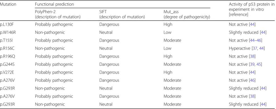
Evaluation of biological significance of all the revealed missense mutations ofTP53in our patients with DLBCL yielded the following results (Table 3): All three prog-nostic software tools (PolyРhen-2, SIFT and Mut_ass) classified mutations p.L130F, p.T155I, p.R196Q, p.G244S, p.V272E, and p.A276V (which lead to appear-ance of a functionally inactive protein) as dangerous, probably pathogenic substitutions, or substitutions with a high/moderate degree of danger. In contrast, mutations р.W146R and p.G293R (slightly decreasing the activity of p53) and mutation p.R156C (hyper-active p53) were regarded as non-pathogenic, neutral substitutions or substitutions with a low/moderate de-gree of danger.
Undoubtedly, mutations p.R213Х and p.A189Pfs have biological significance because each causes emergence of a nonfunctional truncated protein. It is more compli-cated to evaluate the effect of sense mutations detected in our sample of patients because such mutations are considered synonymous substitutions, i.e., keeping the sense of a codon. According to the prognosis of TP53 Mutant Assessor, among the sense mutations, substitu-tion р.A307A is noteworthy because codon 307 is near the end of an exon and potentially may be located in a splicing site of an RNA molecule [17].
Fig. 2The analysis of microsatellite instability in locus D17S796: the results of PCR with flanking primers, electrophoresis in an 8% polyacrylamide gel (length of the product: 144–174 bp); B: DNA from blood, T: DNA of a tumor tissue, 1–3: the ID numbers of cases; 1, 3: loss of heterozygosity; 2: normal
The functional effects of the majority of intronic mu-tations revealed in our group of patients with DLBCL are unknown. One of the biologically significant muta-tions of theTP53 gene (IVS6-36G > C) is located in in-tron 6 of the gene. According to the TP53 UMD mutation database, in human cancer, this mutation means changes that influence splicing [11]. An in vitro experiment indicates that this substitution in the ab-sence of a change in gene coding sequence leads to the survival of cells after chemotherapy and inhibits apop-tosis for a long period [18]. According to NetGene2 prognosis, in the group of patients with DLBCL in our study, among the substitutions detected within introns, mutation IVS5 + 43G >Т caused formation of an add-itional acceptor site of splicing, which is absent under normal conditions. It may lead to inclusion of a part of intron 5 in the sequence of mRNA, premature formation of a stop codon at position 189, and the synthesis of a truncated p53 protein lacking functional activity. Be-sides, IVS4-30Т>С is outstanding among intron
mutations because the alternative gene promoter is placed in intron 4 of TP53. This promoter takes part in the synthesis of the delta133 isoform, which is expressed in lymphoid tissue under normal conditions [19].
Analysis of allelic imbalance andTP53methylation status
The frequency of TP53 promoter methylation in the sample of 69 patients with DLBCL was 4 (5.8%). Pro-moter methylation frequency did not significantly differ in the subgroups with the mutant and wild-type gene se-quence [1 (4.2%) out of 24 vs 3 (6.7%) out of 45, р= 0.5663).
Detection of the loss of heterozygosity of TP53 using microsatellite marker D17S796 was performed on tumor samples from 24 patients with DLBCL, among which 13 patients had mutations, and 11 patients did not have changes inTP53sequence. Six (25%) cases of loss of het-erozygosity were revealed, among which 5 (83.3%) cases of loss of heterozygosity were detected by the sequen-cing method in DLBCL tissue samples with mutations in
Table 2General characteristics of sequencing results
Intron mutations In coding sequence ofТР53gene
With unknown effect Influence on splicing Nonsense Frame-shift mutations Missense Samesense
IVS4-30Т>С p.L130F
IVS5 + 43G > T p.W146Ra
IVS5-17Т>С p.T155Ia р.V157 V
IVS7 + 31G >Сa p.R156C р.H179H
IVS8 + 10С>Аa IVS6-36G > C p.R213Хa p.A189Pfs p.R196Q p.L252 L
IVS8 + 20A>G p.G244S р.V272 V
IVS8 + 37A > G p.V272Ea p.G302G
IVS9 + 12Т>Сa p.A276V р.A307A
(rs1800899) p.G293R
Note.a
Mutations occurring twice in the study population
Table 3The results of functional analysis of missense mutations ofТР53
Mutation Functional prediction Activity of p53 protein in
experiment in vitro [reference] PolyРhen-2
(description of mutation)
SIFT
(description of mutation)
Mut_ass
(degree of pathogenicity)
p.L130F Probably pathogenic Dangerous High Not active [44]
p.W146R Non-pathogenic Neutral Low Slightly reduced [44]
p.T155I Probably pathogenic Dangerous Moderate Not active [44–46]
p.R156C Non-pathogenic Neutral Low Hyperactive [37,44]
p.R196Q Probably pathogenic Dangerous High Not active [38]
p.G244S Probably pathogenic Dangerous Moderate Not active [39,45]
p.V272E Probably pathogenic Dangerous High Not active [44]
p.A276V Probably pathogenic Dangerous Moderate Not active [46]
p.G293R Non-pathogenic Neutral Moderate Slightly reduced [44]
p.A276V Probably pathogenic Dangerous Moderate Not active [38]
exons 5–8 and adjacent intron regions of theTP53gene. Gene promoter methylation was uncovered in one case of the loss of heterozygosity in a DLBCL tissue sample.
TP53mutation status and clinical features of DLBCL
The study cohort was divided into two subgroups: with and without functional TP53 gene mutations (TP53mut andTP53wt, accordingly). TheTP53mut subgroup com-prised 12 patients: patients with p.L130F, p.T155I,
р.A307A, p.R196Q, p.G244S, IVS6-36G > C and p.A276V mutations, two patients with p.R213Х muta-tion, two patients with p.V272E mutation and one pa-tient with multiple mutations (p.T155, p.A189Pfs and IVS5 + 43G > T). The clinical features of patient’s sub-groups are compared and summarized in Table 1. The analysis demonstrate that TP53mut correlated with B-symptoms (P= 0.016), splenomegaly (P= 0.044) and bone marrow involvement (P= 0.028), as well as IPI score of > 2 (P= 0.018).
DLBCL patients withTP53wt tended to had complete remission more often (P= 0.066, Table 1) and had better overall survival (OS) (P= 0.026, Fig. 4) compared with DLBCL patients withTP53mut. The 5-year OS was 69.4% for patients with TP53wt versus 41.7% for those with
TP53mut DLBCL. The median OS of DLBCL patients withTP53mut was 20 months. In contrast, median OS of DLBCL patients withTP53wt was not achieved. Univari-ate analysis showed, that extranodal foci, IPI score of > 2 andTP53mutations predicted decrease OS of DLBCL pa-tients. Multivariate analysis showed that IPI score were the only prognostic factors that independently predicted worse OS of DLBCL patients treated with R-CHOP and
R-CHOP-like regiments. Patients with IPI score of > 2 had a three times hazard for OS (P= 0.005) compared with pa-tients with IPI score of≤2 (Table4).
Because of the small cohort of DLBCL patients with other TP53 aberrations in our study, we do not present the analysis of prognostic and predictive impact of loss of heterozygosity and methylation.
Discussion
Analysis of literature data has shown that deletion of 17p13.1 leading to the loss of heterozygosity ofTP53has been registered at different frequencies in various stud-ies: from 30.4 to 42% according to Chinese authors [20– 22], from 40 to 50.4% in the Czech population [23, 24], 30.4% in the Arab population [25], and 22.5% in the Austrian population [26]. The lowest frequency of 17p13.1 deletion (22.2%) is reported in the International DLBCL Rituximab-CHOP Consortium Program Study [27], combining the samples of patients from 16 hematological centers in the USA, Switzerland, Holland, Germany, Italy, and Spain.
The most actively studied topic onTP53gene variabil-ity in DLBCL is the analysis of its coding sequences re-vealing the presence of mutations. It has been shown that mutation frequency in the TP53 gene in DLBCL is 20% or higher [28].
In 1990, the IARC TP53 mutation database was cre-ated for documenting the mutations in this gene and contains information on more than 30,000 somatic and 700 germ-line mutations at present [24].Research in this database has revealed that in DLBCL, more than 120 mutations of TP53 have been described; 95% of them are single-nucleotide substitutions: 88% are missense
Fig. 4Overall Survival of DLBCL patients withTP53mut and
TP53wt status
Table 4Univariate and multivariate analysis of OS predictors of patients with DLBCL
HR 95% CI P-value
Univariate analysis
B-symptoms 2.401 0.530–0.876 0.256
Performance score 2–3 1.938 0.905–4.149 0.088
Stage III-IV 2.025 0.576–7.116 0.271
Extranodal foci 3.233 1.140–9.951 0.029
Bone marrow involvement 1.539 0.501–4.722 0.451
Elevated S-LDH 1.073 0.900–9.438 0.068
IPI score > 2 2.844 1.384–5.842 0.004
TP53mut 2.707 1.077–6.800 0.034
Multivariate analysis
IPI score > 2 2.994 1.405–6.381 0.005
TP53mut 2.128 0.627–7.217 0.226
Extranodal foci 2.405 0.787–7.369 0.125
mutations, 7% are nonsense mutations, and 5% are frame-shift mutations. More than 95% of mutations have been detected in exons 5–8, but mutations in exons 9– 11 have not been described.
The following codons are hotspots of mutations in
TP53 in relation to DLBCL (in the order of decreasing frequency): 248, 273, 175, 245, 281, 244, 305, 249, and 297 (http://p53.iarc.fr/DownloadDataset.aspx). Nonethe-less, the prevalence of so-called hotspot mutations may change depending on the type of cancer and the ethnic origin of patients [29]. Comparative analysis of TP53 mutations indicates that their diversity and frequencies may significantly vary too, depending on the population under study [30]. There is no information about a Rus-sian population in the current version of the IARCTP53 mutation database.
The vast majority of studies on the role of changes in the nucleotide sequence ofTP53 have focused on exons 5–8. The intron regions have hardly been researched. Nevertheless, these sequences may potentially influence not only splicing of RNA but also gene expression by disturbing the processing autoregulation, normal post-transcriptional mRNA modifications, and post-translational protein modifications [18,31,32].
The relation between hypermethylation of the TP53 promoter and downregulation of gene transcription has been revealed in some tumors [4,33, 34]. Despite thor-ough exploration of this gene’s methylation in cancer, this topic has been insufficiently studied in hematological cancers and hardly addressed in lympho-proliferative diseases [4]. For example, methylation of the TP53 promoter is observed in one-third of patients with acute lymphoblastic leukemia [35] and in one-fifth of patients with chronic lymphocytic leukemia [4]. There is only anecdotal evidence on the frequency of methyla-tion of theTP53promoter in DLBCL [36]. Comprehen-sive characterization of TP53 gene variability in DLBCL has not been carried out yet.
The aim of this study was to comprehensively describe the frequency of promoter methylation and that of loss of heterozygosity as well as the frequency, diversity, and functional significance of mutations in coding and intron regions of theTP53gene among patients with DLBCL in Novosibirsk, Russia.
The diversity of single-nucleotide substitutions de-tected in tumor samples from our group of patients with DLBCL did not significantly differ from that in the IARC
TP53 mutation database. Mutations in regions encoding the DNA-binding site were predominant (95%). Muta-tion p.G293R is the only revealed missense substituMuta-tion that does not affect the functionally significant DNA-binding domain of p53.
Searches in the IARCTP53mutation database showed that all the functionally significant mutations detected in
our study population had been described previously in a wide range of human cancers. Codons 196 and 213 in various cancers and codon 244 in hematological cancers in general and in DLBCL in particular are hotspots of mu-tations inTP53(http://p53.iarc.fr/DownloadDataset.aspx). Moreover, Li-Fraumeni syndrome (http://p53.iarc.fr/ DownloadDataset.aspx) has been described, which is characterized by the development of cancer in different parts of the body because of a germline mutation, p.R213Х, р.G244S, p.L130F, or p.T155, similar to the mutations described in our study.
In this study, mutations were not detected in the ma-jority of codons (248, 273, 175, 245, 281, 305, 249, and 297, except codon 244) for which most of the mutations in TP53 have been described in DLBCL in the IARC
TP53mutation database. Codons 275, 155, 272, and 212 were found to be hotspots of mutations in the analyzed sample of patients.
Review of the published literature [37–40] about the consequences of mutations in the TP53 gene showed that each of these mutations may have multidirectional effects on different functions of p53. These effects may be tentatively subdivided into the consequences for pro-tein structure, its biochemical properties, and biological activity and, in some cases, lead to the emergence of new functions of p53, absent in the wild-type protein.
Thus, each of the mutant forms of the protein is a unique product of a mutation and may combine an in-crease and/or dein-crease in a particular type of p53 activ-ity and may alter its structure or generate new properties.
The emergence of a functionally inactive protein p53 in the analyzed group of patients with DLBCL was caused by one of missense mutations—p.L130F, p.T155I, p.R196Q, p.G244S, p.V272E, or p.A276V—together with mutation p.A189Pfs, leading to frameshift mutations, nonsense substitution p.R213Х, or splicing mutation IVS6-36G > C.
All the above events in the coding part of the gene— that affect the sequence carrying information about highly conserved sites of the DNA-binding domain of p53—were fixed during evolution (in phylogeny) and occur in most isoforms of p53 and in the structure of homologous proteins p63 and p73.
Mutations p.R213Х and р.G244S as well as p.V272E have already been described in DLBCL [41, 42], and
р.T155I (detected in our study) has been previously de-tected in tumor samples from some patients with chronic lymphocytic leukemia and is known to be associated with poor prognosis and a weak response to treatment [43].
All these findings are suggestive of selection of p.L130F, p.T155I, p.R196Q, p.G244S, p.V272E, p.A276V, p.R213Х, and p.A189Pfs at various stages of cancer pro-gression [37–39, 44–46]; therefore, their detection in tumor samples from patients with DLBCL is not coincidental.
Our findings indicate that only two missense substitu-tions (p.W146R and p.G293R)—among all the revealed cases in the analyzed group of patients with DLBCL— did not significantly influence the function of p53. In contrast, p.R156C led to the appearance of a hyperactive mutant protein.
Out of all the mutations detected in our study, only two may influence splicing of RNA. These include same-sense substitutions р.A307A and IVS6-36G > C. The functional significance of IVS6-36G > C has been proved in an in vitro experiment [18]. According to the predic-tion of TP53 Mutant assessor (release 1.00, 2012),
р.A307A is also located at a splicing site of RNA [17]. Even though in sense mutations, the new codon con-tinues encoding the same amino acid, it is believed that this type of mutations may change splicing, transcrip-tion, and/or stability of RNA [11].
The functional effects of most intron and sense muta-tions detected in our group of patients with DLBCL (but not discussed) remain unknown. It is likely that intron mutations may influence not only RNA splicing but also gene expression control, by causing gain- or loss-of-function regulatory elements in TP53 region, thereby creating or disrupting binding sites for certain DNA-sequence-specific transcription factors that inter-fere with normal activation or autoregulation of TP53, and possibly transcriptional regulation of other potential downstream genes [31, 47]. Furthermore, intron muta-tion IVS4-30Т>С is noteworthy because an alternative gene promoter is placed in intron 4 of TP53 and takes part in the synthesis of delta133 and delta160 isoforms of p53 [19]. Thus, whether these newly identified intron mutations of TP53 are functional warrants further investigation.
Some published reports about low frequencies ofTP53 promoter methylation in DLBCL were verified in our group of patients. The frequency of TP53 promoter methylation in the analyzed group of patients with DLBCL was 5.8% and did not differ significantly from subgroups with mutant (4.2%) and wild-type (6.7%) gene structure or from the findings of K. Amara and coau-thors (3.7%) [36].
Analysis of the loss of heterozygosity ofTP53was per-formed on 24 tumor samples of DLBCL in our study population, and 13 of them were found to have muta-tions, whereas 11 did not have changes in the sequences of TP53. Our investigation of microsatellite marker D17S796, which is located near TP53, detected 6 (25%)
cases of loss of heterozygosity, in agreement with the lit-erature data [27].
Furthermore, loss of heterozygosity was observed only in the subgroup of patients with a alteration of theTP53 gene (mutations were revealed in 5 tumor samples, and promoter methylation in one tumor), which accounted for 6 (42.9%) out of 14 versus 0% in the group of 10 pa-tients with the intact gene (р= 0.0223).
Our comprehensive assessment of gene variability in-dicates that the dysfunction of p53 in DLBCL may emerge via a two-hit mechanism. According to this model, two sequential events are necessary for trans-formation of a normal B cell into a cancerous cell during carcinogenesis of at least some cases of DLBCL. The first event is a mutation or methylation in theTP53 pro-moter, giving rise to a cell with increased risk of malig-nant transformation. The second event is the loss of an intact allele of TP53in the cell; this change is necessary for tumorigenesis.
Thus, in DLBCL, our results proved the selection of functionally significant mutations in the gene regions en-coding the DNA-binding domain of p53.
In the analyzed sample of patients with DLBCL, the location of hotspots of mutations (in contrast to the set of single-nucleotide substitutions) is different from the data listed in the IARCTP53Mutation database.
The presence of important intronic and samesense substitutions was demonstrated and confirmed the im-portance of studying the noncoding gene regions adja-cent to exons and of bioinformatic analysis of the uncovered synonymous substitutions. In total, our re-sults show thatTP53mutation status is a prognostic fac-tor that stratifies DLBCL patients treated with R-CHOP and R-CHOP-like regimens. This observation is a further supports the crucial role of p53 in death of tumor cells and tumor suppression. In Russian cohort of de novo DLBCL patients, we show that TP53mut are correlated with B-symptoms, splenomegaly and bone marrow in-volvement, as well as adverse IPI prognostic groups. Nonetheless, DLBCL patients with TP53wt tended to had complete remission more often (P= 0.066).
It is known, that despite the addition of rituximab to therapy, TP53 mutation is an independent prognostic factor that predicts poor survival in patients with DLBCL [28]. To eliminate the possible impact of
TP53mut on OS, we also performed survival analysis for patients study cohort and found that patients with
TP53wt had significantly better OS (P= 0.026). These data are in agreement with a recent study [27].
show that the impact on survival DLBCL patients of IPI score of > 2 is more pronounced than the impact of
TP53mut. This might be because of the small cohort of DLBCL patients withTP53mut in our study.
Conclusions
In conclusion, in the present study, we identified TP53 mutation patterns in a Russian cohort of patients with de novo DLBCL who were treated with R-CHOP and R-CHOP-like regimens and confirmed that TP53 muta-tion status is a valuable prognostic biomarker. Therefore, therapeutic approaches targeting the inactivated TP53 pathway may further improve clinical outcomes of pa-tients with DLBCL.
Because of the small cohort of DLBCL patients with other TP53 aberrations in our study we do not present the analysis of prognostic and predictive impact of LOH and methylation. However, the comprehensive analysis of TP53 status gives us better insights into the possible mechanisms behind participation of this gene’s variabil-ity in the pathogenesis of DLBCL. It was shown here that dysfunction of p53 in DLBCL may emerge accord-ing to the two-hit principle.
Abbreviations
CI:Confidence Interval; CR: Complete Remission; DLBCL: Diffuse Large B-cell Lymphoma; HR: Hazard Ratio; IARC: International Agency for Research on Cancer; IPI: International Prognostic Index; OS: Overall Survival; R-CHOP: Rituximab, Cyclophosphamide, Hydroxydaunorubicin, Oncovin, Prednisone; SIFT: Sorting Intolerant From Tolerant; S-LDH: Serum Lactate Dehydrogenase
Acknowledgements
We ate grateful to BGRS\SB Organizing Committee and to the reviewers for valuable comments.
Funding
The publication cost was covered by Russian Ministry of Education and Science Project No.28.12487.2018/12.1. The study was supported by Budgetary Project No.0541–2014-0002. YLO was supported by ICG SB RAS budget project 0324–2019-0040.
Availability of data and materials
All data generated or analysed during this study are included in this published article [and its supplementary information files].
About this supplement
This article has been published as part ofBMC Medical Genomics Volume 12 Supplement 2, 2019: Selected articles from BGRS\SB-2018: medical genomics.
The full contents of the supplement are available online athttps:// bmcmedgenomics.biomedcentral.com/articles/supplements/volume-12-supplement-2.
Authors’contributions
ENV - performed analysis and data interpretation and was a major contributor in writing the manuscript. TIP supervised research. MIV -supervised research. VNM - performed genetic testing. YLO - performed statistical analysis. OBS - collected the patients data. All the authors read and approved the final manuscript.
Ethics approval and consent to participate
The study was approved by the Local Ethics Committee of Novosibirsk State Medical University. All participating patients signed an informed consent form before the study started.
Consent for publication
Not applicable.
Competing interests
The authors declare that they have no competing interests.
Publisher’s Note
Springer Nature remains neutral with regard to jurisdictional claims in published maps and institutional affiliations.
Author details
1Institute of Internal and Preventive Medicine, Branch of Institute of Cytology
and Genetics, Siberian Branch of Russian Academy of Sciences, Novosibirsk, Russia.2Novosibirsk State Medical University, Novosibirsk, Russia.3Novosibirsk State University, Novosibirsk, Russia.
Published: 13 March 2019
References
1. Swerdlow S.H., Campo E., Harris N.L., Jaffe E.S., Pileri S.A., Stain H., et al. WHO classification of tumors of Haematopoetic and lymphoid tissues. Lyon; 2008. 2. Niroula R, Butera J. Genetics and diffuse large B-cell lymphoma. R I Med J.
2015;98(11):23–6.
3. Skibola CF, Curry JD, Nieters A. Genetic susceptibility to lymphoma. Haematologica. 2007;92(7):960–9.
4. Xu-Monette ZY, Medeiros LJ, Li Y, Orlowski RZ, Andreeff M, Bueso-Ramos CE, et al. Dysfunction of the TP53 tumor suppressor gene in lymphoid malignancies. Blood. 2012;119(16):3668–83. https://doi.org/10.1182/blood-2011-11-366062.
5. Hollstein M, Hainaut P. Massively regulated genes: the example of TP53. J Pathol. 2010;220(2):164–17.https://doi.org/10.1002/path.2637.
6. Cheung KJ, Horsman DE, Gascoyne RD. The significance of TP53 in lymphoid malignancies: mutation prevalence, regulation, prognostic impact and potential as a therapeutic target. Br J Haematol. 2009;146(3):257–69.
https://doi.org/10.1111/j.1365-2141.2009.07739.x.
7. Gudkov AV, Komarova EA. The role of p53 in determining sensitivity to radiotherapy. Nat Rev Cancer. 2003;3(2):117–29.
8. Peller S, Rotter V. TP53 in hematological cancer: low incidence of mutations with significant clinical relevance. Hum Mutat. 2003;21(3):277–84. 9. Kopnin B.P., Kopnin P.B., Khromova N.V., Agapova L.S. Multifaced p53: variety
of forms, functions, tumor-supressive and oncogenic activities. Clinical Oncohematology. Basic Research and Clinical Practice. Russian Journal (Klinicheskaya onkogematologiya). 2008; 1(1): 2--9. (in Engl.)
10. http://p53.iarc.fr/download/tp53_directsequencing_iarc.pdf(access date 31/ 01/2019).
11. Edlund K, Larsson O, Ameur A, Bunikis I, Gyllensten U, Leroy B, et al. Data-driven unbiased curation of the TP53 tumor suppressor gene mutation database and validation by ultradeep sequencing of human tumors. Proc Natl Acad Sci U S A. 2012;109(24):9551–6.
12. Petitjean A, Mathe E, Kato S, Ishioka C, Tavtigian SV, Hainaut P, et al. Impact of mutant p53 functional properties on TP53 mutation patterns and tumor phenotype: lessons from recent developments in the IARC TP53 database. Hum Mutat. 2007;28(6):622–9.
13. Adzhubei I., Jordan D.M., Sunyaev S.R. Predicting functional effect of human missense mutations using PolyPhen-2. Curr Protoc Hum Genet. 2013; Ch.7: Unit 7.20. doi:https://doi.org/10.1002/0471142905.hg0720s76.
14. Brunak S, Engelbrecht J, Knudsen S. Prediction of human mRNA donor and acceptor sites from the DNA sequence. J Mol Biol. 1991;220(1):49–65. 15. Almeida LO, Custódio AC, Pinto GR, Santos MJ, Almeida JR, Clara CA, et al.
Polymorphisms and DNA methylation of gene TP53 associated with extra-axial brain tumors. Genet Mol Res. 2009;8(1):8–18.
16. Grebe SK, McIver B, Hay ID, Wu PS, Maciel LM, Drabkin HA, et al. Frequent loss of heterozygosity on chromosomes 3p and 17p without VHL or p53 mutations suggests involvement of unidentified tumor suppressor genes in follicular thyroid carcinoma. J Clin Endocrinol Metab. 1997;82(11):3684–91. 17. Leroy B, Fournier JL, Ishioka C, Monti P, Inga A, Fronza G, Soussi T. The TP53
website: an integrative resource Centre for the TP53 mutation database and TP53 mutant analysis. Nucleic Acids Res. 2013;41(Database issue):D962–9.
18. Lehman TA, Haffty BG, Carbone CJ, Bishop LR, Gumbs AA, Krishnan S, et al. Elevated frequency and functional activity of a specific germ-line p53 intron mutation in familial breast cancer. Cancer Res. 2000;60(4):1062–9. 19. Mondal AM, Horikawa I, Pine SR, Fujita K, Morgan KM, Vera E, Mazur SJ,
Appella E, Vojtesek B, Blasco MA, Lane DP, Harris CC. p53 isoforms regulate aging- and tumor-associated replicative senescence in T lymphocytes. J Clin Invest. 2013;123(12):5247–57.
20. Lu JT, Cen L, Zhou M. Prognostic value of P53 aberrations in diffuse large B-cell lymphoma. Zhongguo Shi Yan Xue Ye Xue Za Zhi. 2012;20(1):100–2. 21. Sun GX, Cao XS, Li Q, Wang ZL. Correlation of BCL-6, MYC and p53 gene abnormalities with immunological subtypes andprognosis of diffuse large B-cell lymphoma. Zhonghua Yi Xue Yi Chuan Xue Za Zhi. 2012;29(5):576–81.
22. Gao P, Li Q, Wang Z, Yan F, Lu C, Cao X. Significance of BCL-6, MYC, P53 genes abnormalities for the prognosis of diffuse large B-cell lymphoma. Zhonghua Yi Xue Yi Chuan Xue Za Zhi. 2014;31(5):628–31.https://doi.org/ 10.3760/cma.j.issn.1003-9406.2014.01.020.
23. Stefancikova L, Moulis M, Fabian P, Vasova I, Zedek F, Ravcukova B, et al. Prognostic impact of p53 aberrations for R-CHOP-treated patients with diffuse large B-cell lymphoma. Int J Oncol. 2011;39(6):1413–20. 24. Stocklein H, Smardova J, Macak J, Katzenberger T, Holler S, Wessendorf S,
et al. Detailed mapping of chromosome 17p deletions reveals HIC1 as a novel tumor suppressor gene candidate telomeric to TP53 in difuse large B-cell lymphoma. Oncogene. 2008;27(18):2613–25.
25. Tamimi Y, Al-Harthy S, Al-Haddabi I, Al-Kindi M, Babiker H, Al-Moundhri M, Burney I. The p53 mutation/deletion profle in a small cohort of the Omani population with diffuse large B-cell lymphoma. Sultan Qaboos Univ Med J. 2014;14(1):e50–8.
26. Simonitsch-Klupp I, Hauser I, Ott G, Drach J, Ackermann J, Kaufmann J, et al. Diffuse large B-cell lymphomas with plasmablastic/plasmacytoid features are associated with TP53 deletions and poor clinical outcome. Leukemia. 2004; 18(1):146–55.
27. Xu-Monette ZY, Wu L, Visco C, Tai YC, Tzankov A, Liu WM, et al. Mutational profile and prognostic significance of TP53 in diffuse large B-cell lymphoma patients treated with R-CHOP: report from an international DLBCL rituximab-CHOP consortium program study. Blood. 2012;120(19):3986–96. 28. Young KH, Leroy K, Møller MB, Colleoni GW, Sánchez-Beato M, Kerbauy FR,
Haioun C, et al. Structural profiles of TP53 gene mutations predict clinical outcome in diffuse large B-cell lymphoma: an international collaborative study. Blood. 2008;112(8):3088–98.
29. Frebourg T, Barbier N, Kassel J, Ng YS, Romero P, Friend SH. A functional screen for germ line p53 mutations based on transcriptional activation.
Сancer Res. 1992;52(24):6976–8.
30. Tennis M, Krishnan S, Bonner M, Ambrosone CB, Vena JE, Moysich K, et al. p53 mutation analysis in breast tumors by a DNA microarray method. Cancer Epidemiol Biomark Prev. 2006;15(1):80–5.
31. Glick BR, Pasternak JJ. Eds. Moleculer biotechnology. Principles and applications of recombinant DNA. 2nd ed. Washington: ASM Press; 1994. 32. Agirre X, Novo FJ, Calasanz MJ, Larrayoz MJ, Lahortiga I, Valganon M, et al.
TP53 is frequently altered by methylation, mutation, and/or deletion in acute lymphoblastic leukaemia. Mol Carcinog. 2003;38(4):201–8.
33. Pogribny IP, James SJ. Reduction of p53 gene expression in human primary hepatocellular carcinoma is associated with promoter region methylation without coding region mutation. Cancer Lett. 2002;176(2):169–74. 34. Kang JH, Kim SJ, Noh DY, Park IA, Choe KJ, Yoo OJ, Kang HS. Methylation in
the p53 promoter is a supplementary route to breast carcinogenesis: correlation between CpG methylation in the p53 promoter and the mutation of the p53 gene in the progression from ductal carcinoma in situ to invasive ductal carcinoma. Lab Investig. 2001;81(4):573–9.
35. Garcia-Delgado M, Larrayoz MJ, Novo FJ. Methylation of CpG dinucleotides and/or CCWGG motifs at the promoter of TP53 correlates with decreased gene expression in a subset of acute lymphoblastic leukemia patients. Oncogene. 2003;22(7):1070–2.
36. Amara K, Trimeche M, Ziadi S, Laatiri A, Hachana M, Sriha B, et al. Presence of simian virus 40 DNA sequences in diffuse large B-cell lymphomas in Tunisia correlates with aberrant promoter hypermethylation of multiple tumor suppressor genes. Int J Cancer. 2007;121(12):2693–702. 37. Kakudo Y, Shibata H, Otsuka K, Kato S, Ishioka C. Lack of correlation
between p53-dependent transcriptional activity and the ability to induce apoptosis among 179 mutant p53s. Cancer Res. 2005;65(6):2108–14.
38. Monti P, Campomenosi P, Ciribilli Y, Iannone R, Aprile A, Inga A, et al. Characterization of the p53 mutants ability to inhibit p73 beta transactivation using a yeast-based functional assay. Oncogene. 2003;22(34):5252–60. 39. Dearth LR, Qian H, Wang T, Baroni TE, Zeng J, Chen SW, et al. Inactive
full-leng thеp53 mutants lacking dominant wild-type p53 inhibition highlight loss of heterozygosity as an important aspect of p53 status in human cancers. Carcinogenesis. 2007;28(2):289–98.
40. Dekairelle AF, Tombal B, Cosyns JP, Gala JL.Аssessment of the transcriptional activity of p53 improves the prediction of recurrence in superficial transitional cell carcinoma of the bladder. Clin Cancer Res. 2005;11(13):4724–32. 41. Young KH, Weisenburger DD, Dave BJ, Smith L, Sanger W, Iqbal J, et al.
Mutations in the DNA-binding codons of TP53, which are associated with decreased expression of TRAIL receptor-2, predict for poor survival in diffuse large B-cell lymphoma. Blood. 2007;110(13):4396–405.
42. Stefancikova L, Moulis M, Fabian P, Ravcukova B, Vasova I, Muzik J, Malcikova J, Falkova I, Slovackova J, Smardova J. Loss of the p53 tumor suppressor activity is associated with negative prognosis of mantle cell lymphoma. Int J Oncol. 2010;36(3):699–706.
43. Zenz T, Eichhorst B, Busch R, Denzel T, Habe S, Winkler D, et al. TP53 mutation and survival in chronic lymphocytic leukemia. J Clin Oncol. 2010; 28(29):4473–9.
44. Shiraishi K, Kato S, Han SY, Liu W, Otsuka K, Sakayori M, et al. Isolation of temperature-sensitive p53 mutations from a comprehensive missense mutation library. J Biol Chem. 2004;279(1):348–55.
45. Monti P, Campomenosi P, Ciribilli Y, Iannone R, Inga A, Abbondandolo A, et al. Tumour p53 mutations exhibit promoter selective dominance over wild type p53. Oncogene. 2002;21(11):1641–8.
46. Campomenosi P, Monti P, Aprile A, Abbondandolo A, Frebourg T, Gold B, et al.Р53 mutants can often transactivate promoters containin gap21 but not Bax or PIG3 responsive elements. Oncogene. 2001;20(27):3573–9. 47. Gao P, Xia JH, Sipeky C, Dong XM, Zhang Q, Yang Y, et al. Biology and