R E S E A R C H A R T I C L E
Open Access
Interplay between estrogen receptor and AKT in
Estradiol-induced alternative splicing
Poornima Bhat-Nakshatri
1, Eun-Kyung Song
1, Nikail R Collins
1, Vladimir N Uversky
3,7, A Keith Dunker
3,4,
Bert W O
’
Malley
5, Tim R Geistlinger
6,8, Jason S Carroll
6,9, Myles Brown
6and Harikrishna Nakshatri
1,2*Abstract
Background:Alternative splicing is critical for generating complex proteomes in response to extracellular signals. Nuclear receptors including estrogen receptor alpha (ERα) and their ligands promote alternative splicing. The endogenous targets of ERα:estradiol (E2)-mediated alternative splicing and the influence of extracellular kinases that phosphorylate ERαon E2-induced splicing are unknown.
Methods:MCF-7 and its anti-estrogen derivatives were used for the majority of the assays. CD44 mini gene was used to measure the effect of E2 and AKT on alternative splicing. ExonHit array analysis was performed to identify E2 and AKT-regulated endogenous alternatively spliced apoptosis-related genes. Quantitative reverse transcription polymerase chain reaction was performed to verify alternative splicing. ERαbinding to alternatively spliced genes was verified by chromatin immunoprecipitation assay. Bromodeoxyuridine incorporation-ELISA and Annexin V labeling assays were done to measure cell proliferation and apoptosis, respectively.
Results:We identified the targets of E2-induced alternative splicing and deconstructed some of the mechanisms surrounding E2-induced splicing by combining splice array with ERαcistrome and gene expression array. E2-induced alternatively spliced genes fall into at least two subgroups: coupled to E2-regulated transcription and ERα binding to the gene without an effect on rate of transcription. Further, AKT, which phosphorylates both ERαand splicing factors, influenced ERα:E2 dependent splicing in a gene-specific manner. Genes that are alternatively spliced include FAS/CD95, FGFR2, and AXIN-1. E2 increased the expression of FGFR2 C1 isoform but reduced C3 isoform at mRNA level. E2-induced alternative splicing of FAS and FGFR2 in MCF-7 cells correlated with resistance to FAS activation-induced apoptosis and response to keratinocyte growth factor (KGF), respectively. Resistance of MCF-7 breast cancer cells to the anti-estrogen tamoxifen was associated with ERα-dependent overexpression of FGFR2, whereas resistance to fulvestrant was associated with ERα-dependent isoform switching, which correlated with altered response to KGF.
Conclusion:E2 may partly alter cellular proteome through alternative splicing uncoupled to its effects on transcription initiation and aberration in E2-induced alternative splicing events may influence response to anti-estrogens.
Background
Estradiol (E2) signaling primarily involves activation of nuclear receptors, estrogen receptors alpha (ERα) and beta (ERβ), which function as transcription factors that regulate gene expression through either DNA binding or through protein-protein interaction [1,2]. E2 signaling in
cells is further controlled by several post-transcriptional modifications of ERα and ERβ including phosphoryl-ation, acetylphosphoryl-ation, and ubiquitination. These post-transcriptional events influence the ability of ERα to interact with co-regulator molecules, its stability, and localization. Kinases known to phosphorylate ERα in-clude MAPK, IKKα, RSK, AKT/PKB, p38 kinase, PKA, Src, cyclin A/cdk2, and cdk7 [1,3-7]. It is suggested that changes in the phosphorylation status of the receptor contribute to ERα dysfunction in various pathological conditions including breast cancer.
* Correspondence:[email protected]
1
Departments of Surgery, Indiana University School of Medicine, 980 West Walnut Street, Indianapolis, IN 46202, USA
2
Biochemistry and Molecular Biology, Indiana University School of Medicine, Indianapolis, IN 46202, USA
Full list of author information is available at the end of the article
Alternative splicing is an important post-transcriptional mechanism that permits the generation of multiple pro-tein products from a single gene. 92-94% of human genes undergo alternative splicing and 70-90% of these spliced RNAs are translated into proteins [8,9]. A recent genome-wide sequencing indicated 22,000 tissue-specific alterna-tive splicing events [8]. Two families of splicing factors have been identified: heterogeneous nuclear ribonucleo-protein (hnRNP) related ribonucleo-proteins and the serine-arginine rich (SR) proteins [9,10]. Phosphorylation controls activity of these proteins [9,10]. For example, RS domains of SR proteins contain multiple copies of consensus AKT phos-phorylation site RXRXXS and AKT controls their activity in the nucleus and cytoplasm [11].
Defects in alternative splicing are linked to various dis-eases including spinal muscular atrophy, neurofibroma-tosis type 1, cystic fibrosis, breast cancer, and ovarian cancer [12,13]. For example, alternative splicing can change CC3, which codes for a protein with anti-metastatic and pro-apoptotic properties, to TC3, which codes for a protein with pro-metastatic and anti-apoptotic properties [14]. Alternative splicing of the transcription factor FOXP1 influences pluripotency and differentiation of embryonic stem cells [15]. At least 15% of human genetic diseases arise from mutations either in consensus splice sites or in splicing silencer or enhancer elements [12]. Cancer-associated alternative splicing, which is regulated by FOX1, FOX2, and Nova proteins, has been reported [16,17]. These alternative splicing events empower cancer cells to express developmentally regulated proteins [13]. Breast cancer subtypes show dis-tinct splicing pattern, which may partly be related to FOX1/FOX2 expression [16,18]. Genetic alterations in splicing machinery is linked to myelodysplasia [19]. Re-cent studies using bioinformatics tools including intrin-sic disorder predictions have shown that alternative splicing is a non-random event and often involves the region of the protein engaged in protein-protein inter-action [20]. Thus, loss or gain of exons through alterna-tive splicing has major functional implications.
A clear link between steroid regulated transcription and alternative splicing has been established [21]. At least 25 proteins have been described to have both tran-scription coregulator and splicing activity [22]. Proteins involved in steroid-regulated alternative splicing events include U2AF65-related proteins CAPERαand CAPERβ, ASC-1, ASC-2, and CoAA [23]. Several of these mole-cules interact directly with ERα. ERα:E2 controls transcription-coupled alternative splicing of the first in-tron in a gene specific manner, which impacts the rate of co-transcriptional RNA processing [24]. However, whether these ERα:splicing factor interactions contribute to breast cancer subtype-enriched alternative splicing or whether ERα:E2 induces alternative splicing uncoupled
to transcription initiation activity, is yet to be deter-mined. Additionally, there are limited number of reports that have examined the effect of ERα phosphorylation on alternative splicing [25]. ERαphosphorylated at S118 by MAPKs interacts with the splicing factor SF3a, a component of U2 snRNP complex, and promotes exon skipping [25].
We had previously shown the effects of AKT on ERα: E2 mediated transcription, which correlated with the phosphorylation of S167 residue of ERα [6]. Crosstalk between ERαand AKT is likely to play a significant role in ERα-positive breast cancer because recent genome analyses have observed frequent activating PI3K muta-tion in ERα-positive breast cancer, which often leads to increased AKT activity [26,27]. Using chromatin immu-noprecipitation coupled microarray (ChIP-on-chip) and gene expression analysis, we demonstrated specific effects of AKT on ERα binding to the genome and regulated transcription [28]. The effect of AKT on E2-induced alternative splicing is unknown. To test the effect of AKT on E2-induced alternative splicing, we first performed CD44 minigene splicing assay and observed a dominant effect of AKT on E2-induced alternative spli-cing in two cell types examined. To identify endogenous targets of E2-regulated alternative splicing and the influ-ence of AKT on this process, we performed ExonHit Apoptosis Splice Array analysis of ERα -positive/E2-dependent MCF-7 breast cancer cells (MCF-7p) and the same cell line overexpressing constitutively active AKT (MCF-7AKT) with or without E2 treatment. Combined analysis of splice array and other published cancer-specific splicing data with E2-induced ERαcistrome and gene expression arrays revealed two distinct categories of E2-regulated alternatively spliced genes [17,28-30]. E2 induced alternative splicing of FAS/CD95 in MCF-7 cells, which correlated with resistance to FAS-activation induced apoptosis. In addition, E2 increased the expres-sion of fibroblast growth factor receptor 2 (FGFR2) C1 isoform without significantly altering C2 isoform levels in MCF-7p cells. Consistent with the effect of E2 on FGFR2 splicing, MCF-7 cells showed differential re-sponse to keratinocyte growth factor (KGF), a ligand for FGFR2 C1 and C2 isoforms, in the presence of E2 and tamoxifen, an anti-estrogen. Furthermore, acquired re-sistance to anti-estrogens tamoxifen and fulvestrant was associated with distinct expression pattern of FGFR2 isoforms and response to KGF. Collectively, these results suggest a role for E2 induced alternative splicing in anti-estrogen response of breast cancer cells.
Results
AKT influences E2 induced alternative splicing
CAPERβ contain several putative AKT phosphorylation sites [23]. Both CAPERα and CAPERβ associate with ERαand modulate E2-dependent transcription as well as alternative splicing [23]. Thus, AKT can potentially influ-ence E2-induced alternative splicing by phosphorylating multiple components of the splicing machinery. We used the CD44 minigene splicing system containing vari-able exons 4 and 5 (v4,5) of CD44 gene under the con-trol of estrogen response element driven promoter (ERE-CD44) and measured the exon inclusion/skipping to determine the effect of AKT on E2-induced alterna-tive splicing [21]. Two cell systems were used: 1) 293 cells requiring transfection of both the CD44 minigene and ERα or its mutants; 2) MCF-7p and MCF-7AKT cells transfected with the CD44 minigene. Studies in 293 cells allowed us to study phosphorylation defective mu-tants of ERαon splicing as these cells lack ERα. Studies in MCF-7 cells allowed us to study E2-induced splicing in the context of E2 responsive breast cancer cells, en-dogenous ERα and to distinguish ERE-dependent and ERE-independent effects of E2 on alternative splicing. We designed unique primers spanning exon-exon junc-tions to measure exon-included and exon-skipped prod-ucts by quantitative reverse transcription polymerase chain reaction (qRT-PCR).
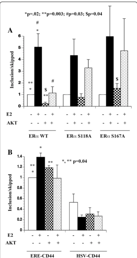
In 293 cells, only exon skipped transcripts from ERE-CD44 minigene could be reliably measured in the ab-sence and preab-sence of E2 by qRT-PCR (exon-included CT >30, exon-skipped ~25). In contrast, robust ERE-CD44 gene activity with expression of both exon-included (~27 CT) and exon-skipped products (~24 CT) was observed when co-transfected with ERα(Figure 1A). The exon-inclusion/exon-skipped ratio changed dramat-ically in cells treated with E2 for 24 hours (range 1.7 to 6 fold in favor of included product, Figure 1A). We next compared E2-induced alternative splicing in the pres-ence of MAPK-phosphorylation defective (S118A) and AKT phosphorylation defective (S167A) mutants of ERα. ERαmutants were effective as wild type ERαin modulat-ing alternative splicmodulat-ing in the presence of E2 (Figure 1A).
We then compared ERα:E2-induced alternative spli-cing in the presence of constitutively active AKT. AKT had a dominant effect on alternative splicing of ERE-CD44 minigene-derived transcripts. Compared to con-trol vector-transfected cells, exon-included/exon-skipped transcript ratio was dramatically lower indicating the ability of AKT to promote exon skipping and E2 failed to reverse this effect of AKT (Figure 1A, p=0.003). Please note that AKT did not influence transcription of ERE-CD44 minigene in the absence of ERα, as only exon-skipped transcripts under this transfection condi-tion could be reliably measured (data not shown). Inter-estingly, AKT failed to influence alternative splicing events orchestrated by ERα phosphorylation defective
mutants. In particular, the ability of AKT to promote exon skipping was completely lost in cells transfected with S167 mutant, which is defective in AKT-mediated phosphorylation (p=0.04; exon inc/skip ratio in wild type vs S167A mutant ERα transfected cells with AKT). These results indicate that AKT alters ERα:E2 induced alternative splicing in ERα S167 phosphorylation-dependent manner to a larger extent and S118 to a minor extent.
We performed similar assay in parental MCF-7 cells (MCF-7p) and cells overexpressing AKT (MCF-7AKT) with ERE-CD44 minigene. Additionally, we included CD44 minigene driven by herpes simplex virus enhancer-promoter (HSV-CD44) to account for second-ary effects of ERα:E2 on alternative splicing. As with 293 cells, E2 increased exon-inclusion/exon-skipped ratio in cells transfected with ERE-CD44 minigene (Figure 1B). Although alternative splicing of HSV-CD44 minigene in E2 treated cells was different from untreated cells, none of the differences reached statistical significance (Figure 1B). Similarly, AKT had minimum effect on alter-native splicing of transcripts from HSV-CD44 minigene (Figure 1B). Taken together, these results suggest that E2 has distinct effects on alternative splicing of genes with ERE compared to genes without ERE. In addition, AKT has dominant effect on E2-induced alternative splicing or confers ligand-independent splicing activity to ERα, de-pending on the cell type.
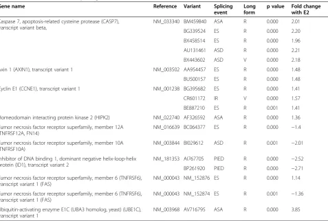
E2 induced alternative splicing of apoptosis-related genes To identify endogenous targets of E2-induced alternative splicing and the potential influence of AKT in this process, we performed microarray analysis that detects alternative splicing of 893 genes related to apoptosis. Array analysis was performed in quadruplicate using RNA from MCF-7p and MCF-7AKT cells with or with-out E2 treatment for three hours. Three hours time point was selected to enrich for genes that are primary targets of E2-regulated transcription and alternative spli-cing. Data from both cell types together with or without E2 treatment were analyzed using a more comprehensive ANOVA model after dye bias and cell type effects were removed. Four hundred sixty three splicing events out of possible 10659 splice events (p<0.001) (~4%) targeting 154 genes (17% of genes) were altered in E2 treated cells compared to untreated cells. These events were common to both cell types. Individual cell types also showed E2-dependent changes in splicing patterns (527 in MCF-7p and 451 in MCF-7AKT) although statistical analysis is not as robust as combined analysis because comparison was only between two cell types (with and without E2 treatment). A gene list is provided as Additional file 1, Additional file 2, Additional file 3 available online and a partial list of the genes, few of which were verified
further by qRT-PCR assays, is provided in Table 1 along with type of splicing events and fold change in response to E2 treatment.
Genes that undergo E2-induced alternative splicing are enriched for ERαbinding sites
E2 induced alternative splicing could involve E2-regulated transcription-coupled splicing, ERα binding to the gene that permits co-recruitment of splicing factors without an effect on the rate of transcription initiation, or a secondary event due to E2-mediated induction/re-pression of a splicing factor(s). To test these possibilities, we examined alternatively spliced genes for ERαbinding sites from our previous ChIP-on-chip study datasets of MCF-7p and MCF-7AKT cells as well as a published study involving ChIP-seq of untreated, E2-treated or tamoxifen treated MCF-7 cells [28,29]. Among 154 genes that under-went alternative splicing in E2 treated cells, 89 genes contained ERαbinding sites in regions within or ~20-kb 5’ or 3’ of the coding unit (Additional file 1). A 20-kb window has previously been shown to be optimal for ERα enhancer-promoter interactions [31]. However, we cannot rule out the possibility of ERα binding sites located at 50–100 KB distance from 5’and 3’of the coding units and ERαbinding to these sites influencing gene expres-sion or alternative splicing through looping mechanism. We had previously shown that 299 of 837 E2-regulated genes in MCF-7p cells and 363 of 1063 E2-regulated genes in MCF-7AKT cells (~35%) contain ERα binding sites [28]. In comparison, it appears that ERα binding sites are enriched on genes that underwent E2-induced alternative splicing (89 out of 154, ~60%, p=0.0001, Fisher’s exact test).
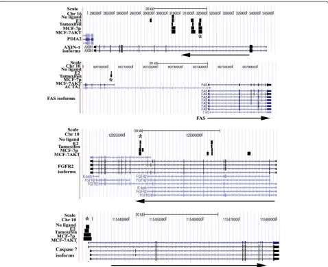
binding to specific regions of the above genes upon E2 treatment was observed in MCF-7p, MCF-7AKT, or both cell types. E2 had distinct effect on ERαbinding to FAS gene in MCF-7p and MCF-7AKT cells. Unliganded ERα bound to previously identified ChIP-seq region in FAS gene in MCF-7p cells, which was reduced upon E2 treatment. However, in MCF-7AKT cells, E2 increased ERα binding, which correlated with repression of E2-regulated expression of total FAS transcripts in these cells (see below). ChIP with control IgG did not display any E2-dependent increase in amplification of ERα bind-ing regions (data not shown). Taken together, the results presented above demonstrate a correlation between ERα binding to genes whose transcripts undergo alternative splicing in response to E2 treatment.
We used two resources to determine whether E2-induced alternative splicing is coupled to ERαbinding as well as changes in overall rate of transcription. First, we determined the effect of E2 on overall mRNA levels of alternatively spliced genes with ERα binding sites in MCF-7p or MCF-7AKT cells using gene expression array data from same cells treated with E2 for four hours [28].
Among 89 alternatively spliced genes with ERα binding sites, E2 induced the expression of eight genes but re-pressed the expression of two genes in MCF-7p cells. In MCF-7AKT cells, E2 increased the expression of 12 genes but repressed the expression of four genes (Additional file 1). Therefore, ~80% of genes with ERα binding sites undergo alternative splicing upon E2 treat-ment without changing overall levels of transcription. The second set of data derived from the publicly available data-base (www.nursa.org/gems/) was used to independently verify this possibility. This database contains gene expres-sion meta-signature of MCF-7 cells treated with E2 for three hours and 24 hours, which permitted us to meas-ure early and late effects of E2 on overall transcription of alternatively spliced genes [30]. Among 89 genes with ERα binding sites, 18 genes were early-response inducible genes (3–4 hours) whereas 15 genes were late-response inducible genes (24 hours) with q-value of <0.05 (Additional file 1, Additional file 4). 16 and 22 genes were repressed at early and late time points, re-spectively. Remaining genes with ERα binding sites (60%) underwent alternative splicing without an effect Table 1 Selected E2-induced splicing events
Gene name Reference Variant Splicing
event
Long form
p value Fold change with E2
Caspase 7, apoptosis-related cysteine protease (CASP7), transcript variant beta,
NM_033340 BM459840 ASA R 0.000 2.01
BG339524 ES R 0.000 2.20
BX458514 ES R 0.000 1.96
AU131461 ASD R 0.000 2.21
BX443602 ASD V 0.000 2.18
Axin 1 (AXIN1), transcript variant 1 NM_003502 AA954457 ES R 0.000 1.48
BU500157 ES R 0.000 1.48
Cyclin E1 (CCNE1), transcript variant 1 NM_001238 BG395682 ES R 0.000 1.41
CR601172 IR V 0.000 1.57
BE887210 ES R 0.001 1.41
Homeodomain interacting protein kinase 2 (HIPK2) NM_022740 AF326592 ASA R 0.000 1.36
Tumor necrosis factor receptor superfamily, member 12A (TNFRSF12A, FN14)
NM_016639 BC064377 ES R 0.000 −1.4
Tumor necrosis factor receptor superfamily, member 10A (TNFRSF10A)
NM_003844 BI029612 ASD R 0.001 −2.01
Inhibitor of DNA binding 1, dominant negative helix-loop-helix protein (ID1), transcript variant 2
NM_181353 AI767705 PIED R 0.000 −2.52
BP261920 PIED R 0.000 −2.71
Tumor necrosis factor receptor superfamily, member 6 (TNFRSF6), transcript variant 1 (FAS)
NM_000043 NM_152876 ES R 0.000 1.14
Tumor necrosis factor receptor superfamily, member 6 (TNFRSF6), transcript variant 1 (FAS)
NM_000043 NM_152874 ES R 0.001 −1.36
Ubiquitin-activating enzyme E1C (UBA3 homolog, yeast) (UBE1C), transcript variant 1
NM_003968 AV716795 ASA R 0.000 3.85
on overall transcription after E2 treatment. Among 65 alternatively spliced genes without ERα binding sites, 13 and 19 genes were early and late E2 induced genes, respectively, whereas four and 10 were early and late repressed genes, respectively. Based on these two inde-pendent analyses, we conclude that E2 induced alterna-tive splicing need not have to be coupled to the effects of E2 on the rate of transcription initiation. In fact, E2 had no effect on transcription of the majority of genes (85 out of 154 were unaffected at the level of tran-scripts) that underwent E2-regulated alternative spli-cing with or without ERαbinding sites.
E2 regulated expression of splicing factors
Since ~40% of genes lacking ERα binding sites within 20 kb of 5’and 3’ends of the coding unit underwent E2
dependent alternative splicing, we examined the effect of E2 on the expression of genes of the alternative splicing machinery including those linked to cancer. First, we an-alyzed ChIP-on-chip for ERα binding and gene expres-sion array data of E2 treated MCF-7p and MCF-7AKT cells for E2-regulated expression of splicing factors [28] and observed E2-dependent increase in the SRSF7 (also called 9G8) by more than 2-fold. Indeed, E2 increased the expression of SRSF7 in both 7p and MCF-7AKT cells by more than 2-fold, despite lacking ERα binding sites based on ChIP-on-Chip and ChIP-seq data (Figure 3B). Therefore, SRSF7 may contribute to E2-regulated alternative splicing of genes without ERα bind-ing sites. FOX2 (also called RBM9) downregulation is responsible for cancer-associated alternative splicing [16]. However, FOX2 gene lacks ERα binding sites and
E2 had no effect on its expression in 7p and MCF-7AKT cells [28]. Genome-wide RNAi screen for genes involved in alternative splicing of apoptosis-related genes revealed ASF/SF2/SRSF1 as the main splicing factor that regulates apoptosis [35]. The regulatory regions of SF2
contain an ERα binding site; however, E2 did not alter the expression of this gene in both 7p and MCF-7AKT cells. The regulatory regions of Polypyrimidine Tract Binding protein 1 (PTB), another cancer-associated splicing factor [13], bind to ERα in tamoxifen but not E2
treated cells and E2 induces its expression only after 24 hours. Thus, PTB is less likely to be a major player in E2-induced alternative splicing. The regulatory regions of SRp20, another splicing factor overexpressed in cancer [36], contain ERα binding sites and SRp20 is induced by E2 at transcript level (1.36-fold, q value <0.01). However, we did not observe elevated SRp20 protein levels in cells treated with E2 compared to untreated cells possibly due to higher abundance and stability of the protein (data not shown). Furthermore, analysis of more than 20 known splicing factors did not reveal significant effects of E2 on their expression [37,38]. Thus, SRSF7 is the only documented E2-inducible splicing factor, which may cause alternative splicing as a secondary event in E2 treated cells.
Characterization of E2-induced alternative splicing at mRNA level
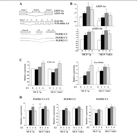
A schematic view of some of the genes that underwent alternative splicing in response to E2 treatment is given in Figure 4A. Alternative splicing pattern of the FGFR2 C-terminus is also shown. FGFR2 was included in this analysis because it is one among 19 genes that undergo breast cancer-specific alternative splicing, which results in proteins with distinct functional domains [17]. To visualize and measure splice variants, we employed qRT-PCR using primer sets that measure wild type or isoform mRNA levels. We selected AXIN-1, FAS, and FGFR2 for further analysis because of their known role in breast cancer and apoptosis.
AXIN-1a and AXIN-1b showed distinct expression pattern in MCF-7p and MCF-7AKT cells. In MCF-7p cells, E2 increased the expression of both AXIN-1a and AXIN-1b although there was a difference in the expres-sion levels of these isoforms at given time after E2 treat-ment (Figure 4B). Basal AXIN-1a but not AXIN-1b levels were lower in MCF-7AKT cells compared to MCF-7p cells (p=0.006) suggesting the ability of AKT to alter AXIN-1 splicing independent of ERαand E2. Con-sequently, overall levels of AXIN-1a but not AXIN-1b were lower in E2 treated MCF-7AKT cells compared to MCF-7p cells. AXIN-1b lacks exon-8, which codes for a 30 amino acid region containing a putative casein kinase I phosphorylation site, oligomerization site, and two po-tential nuclear export signals [39].
Several isoforms of FAS have been characterized [40]. FAS lacking exon 6, which is a more frequent event in cancer, yields a soluble FAS protein with anti-apoptotic activity [13,40]. ExonHit array experiments identified E2-inducible expression of an isoform lacking exons 3, 4, and 6 (NM_152876) but repression of another isoform lacking both exons 4 and 7 (NM_152874) in MCF-7p cells (Table 1 and Additional file 2). We designed mul-tiple qRT-PCR primers to measure the levels of total
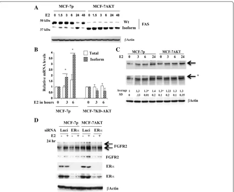
FAS transcripts (primers spanning exon 9) and splice variants alone. Only primers spanning total FAS and the splice variant lacking exons 3 and 4 (forward primer designed to exon 2 and 5 junction and reverse primer spanning exon 6) gave reproducible results. E2 did not change the levels of total FAS transcripts but signifi-cantly increased the levels of the splice variant lacking exons 3 and 4 in MCF-7p cells (Figure 4C) (please see Figure 5 below for even higher induction of the splice variant). E2 decreased the levels of full length but not the splice variant in MCF-7AKT cells after one-hour treatment suggesting a dominant effect of AKT in redirecting ERαto suppress total FAS.
The effect of E2 on FAS and FGFR2 proteins
To further confirm E2-induced alternative splicing of above genes leads to measurable expression of distinct protein species, we performed western blotting. These studies were not intended to show a direct correlation between alternatively spliced mRNA and translation of the specific spliced mRNA into a protein. Instead, the
purpose was to show that the alternative splicing creates translatable mRNAs. Indeed, E2 treated cells showed a faster migrating FAS protein of ~38 kDa in MCF-7p cells and the basal level of this faster migrating protein was constantly higher in MCF-7AKT cells (Figure 5A). We do note that there were experimental variations in the number of faster migrating bands of FAS in E2
treated cells as well as detection by Western blot ana-lysis possibly reflecting experimental and/or cell ana-lysis conditions altering the stability of the isoforms (data not shown). In addition, since FAS undergoes multiple alter-native splicing [40], it is impossible to assign a splice variant transcript to a protein species.
We further characterized the role of AKT in E2-induced alternative splicing of FAS by generating MCF-7 cells overexpressing dominant negative AKT (KD-AKT) [6]. While E2 increased alternatively spliced FAS in par-ental cells, this effect of E2 was completely abolished in cells overexpressing KD-AKT (Figure 5B).
We observed an effect of E2 on two FGFR2 protein species in MCF-7p cells (Figure 5C). One is an abun-dantly expressed faster migrating protein, which was in-duced by E2 (indicated by an arrow). The basal level of this FGFR2 protein was significantly higher in MCF-7AKT cells compared to MCF-7p cells. Another minor slower migrating FGFR2 protein was detected in both cells and levels of this protein declined initially with E2 treatment but restored/increased after 24 hours (indi-cated by an arrow with star). To determine the require-ment of ERα for the expression of these isoforms of FGFR2, we knocked down ERαusing siRNA. ERαsiRNA
Figure 5E2-induced changes in FAS and FGFR2 proteins. A) MCF-7p and MCF-7AKT cells express FAS proteins of different mobility with or without E2 treatment. Cells were treated with E2 for indicated time (in hours) and cell lysates were subjected to western blotting.B) Dominant-negative AKT (KD-AKT) inhibits E2-induced alternative splicing of FAS as evident from qRT-PCR assays. *p<0.03, total vs isoform at 3 and 6 hour E2 treated condition in control cells.C) The effect of E2 on FGFR2 protein in MCF-7p and MCF-7AKT cells. *p=0.0004 untreated vs six hour E2 treated MCF-7p cells (for faster migrating FGFR2). *p=0.03 untreated MCF-7p versus untreated MCF-7AKT cells. Two exposures of FGFR2 blot are shown.
significantly reduced basal and E2-inducible expression of both FGFR2 protein species in MCF-7AKT cells (Figure 5D). We are unsure why ERαsiRNA had lower effect on FGFR2 expression in MCF-7p cells. Residual ERαin siRNA treated cells may be sufficient for FGFR2 expression in these cells.
Anti-estrogen resistance is associated with differential FGFR2 C isoform expression
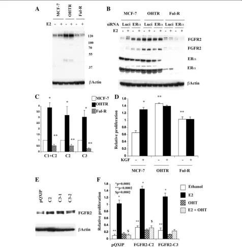
We next investigated whether resistance to anti-estrogens is associated with altered expression of FGFR2 isoforms. For this purpose, we used a single cell-derived clone of MCF-7 cell line that has acquired resistance to tamoxifen (OHTR) or fulvestrant (Ful-R). MCF-7 and OHTR cells express similar levels of ERα, whereas Ful-R cells express very little ERα protein compared to other cell types [44]. Western blot analysis was performed to determine whether FGFR2 isoforms are differentially expressed in these cell lines. Similar to results presented in Figure 5, two FGFR2 proteins, one abundant faster migrating species and one minor slower migrating spe-cies were detected in these cell lines and E2-dependency varied between cell lines (Figure 6A). The basal expres-sion of the faster migrating band was significantly higher in OHTR cells compared to parental cells and was not influenced by E2 in this cell line. Note that OHTR cells do not express slower migrating FGFR2 indicating iso-form switching in resistant cells. In contrast, the minor slower migrating FGFR2 was E2-inducible in the paren-tal cell line but constitutively elevated in Ful-R cells. Therefore, anti-estrogen resistance is associated with alternative splicing of FGFR2 and corresponding change in protein levels. We used siRNA against ERαto deter-mine which among the FGFR2 isoform expression requires ERα. ERα siRNA completely abolished the E2-inducible expression of slower migrating minor FGFR2 in parental cells and even in Ful-R cells, which has lower levels of ERα compared to other cell lines (Figure 6B). Therefore, elevated basal expression of slower migrating FGFR2 in Ful-R cells is dependent on residual ERα levels. Basal expression of faster migrating FGFR2 in OHTR cells was also ERα dependent. For unknown rea-son, we were only able to knockdown <50% of ERα in this single cell-derived MCF-7 clone and OHTR cells. Despite discrepancy with respect to ERαdependency of faster migrating FGFR2 in parental MCF-7 cells and clonal MCF-7 (Figure 5 and Figure 6), results support the conclusion that slower migrating FGFR2 is E2 and/ or ERαdependent across cell lines.
To confirm that overexpression of FGFR2 protein in OHTR cells and lower expression in Ful-R cells correlate with mRNA changes, we performed qRT-PCR analysis (Figure 6C). OHTR cells expressed higher levels of all three FGFR2 isoforms tested, whereas Ful-R cells expressed
lower levels of all three isoforms tested compared to parental cells. Therefore, FGFR2 protein changes in anti-estrogen resistant cells correlate changes in mRNAs but it is difficult to assign these changes to specific isoforms.
KGF serves as a ligand to FGFR2 C1 and C2 isoforms, whereas C3 is active independent of ligand [41,42]. Since OHTR cells expressed higher levels of FGFR2 protein compared to parental cells and Flu-R cells expressed dif-ferent levels of FGFR2 isoforms compared to parental or OHTR cells, it is possible that anti-estrogen resistant cells respond differently to KGF compared to parental cells. While KGF stimulated the growth of parental cells, OHTR cells displayed elevated basal proliferation, which was unaffected upon KGF stimulation. Ful-R cells also displayed elevated basal proliferation and were unre-sponsive to KGF stimulation (Figure 6D). These results indicate that resistance to anti-estrogens is associated with differential FGFR2 isoform expression and response to KGF.
FGFR2 C2 and FGFR2 C3 alter basal proliferation rate and consequently response to E2
Since FGFR2 C2 isoform expression was not E2 respon-sive, whereas C3 expression was repressed by E2, we examined how constitutive overexpression of these isoforms alters cellular response to E2 and tamoxifen. Expression levels of overexpressed FGFR2 isoforms are
shown in Figure 6E. Both FGFR2 C2 and C3
overexpressing cells showed elevated basal proliferation compared to parental cells with retrovirus vector alone and E2 further stimulated proliferation (Figure 6F). Simi-lar to parental cells, tamoxifen reduced E2-stimulated proliferation of FGFR2 C2 and C3 overexpressing cells.
These results suggest that FGFR2 C2 and C3
overexpression provides proliferative advantage mimick-ing increased proliferation observed with anti-estrogen resistant cells compared to parental cells. Overall, prolif-eration rate of C2 or C3 overexpressing cells was much higher than parental cells (p<0.0002). Proliferation rate of FGFR2 C2 overexpressing cells remained higher than parental cells when cells were treated with E2 plus tam-oxifen. Thus, individual isoforms of FGFR2 can influence basal proliferation and consequently response to E2 and anti-estrogens. Since it is difficult to knockdown individ-ual isoforms of FGFR2 due to limited sequence diver-gence between isoforms and to develop isoform specific inhibitors, our observation linking specific isoforms of FGFR2 to anti-estrogen response remains correlative.
Estrogen-induced alternative splicing of FAS and FGFR2 correlates with resistance to FAS activation-induced cell death and KGF-induced proliferation, respectively
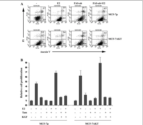
induced cell death, we treated MCF-7p and MCF-7AKT cells with an antibody that triggers FAS-dependent apoptosis. MCF-7p and MCF-7AKT cells maintained in charcoal-stripped serum containing media displayed basal apoptosis, which was reduced upon E2-treatment (Figure 7A). FAS antibody treatment increased apoptosis in both cell types, which was more pronounced in MCF-7p compared to MCF-7AKT cells. Lower sensitivity of MCF-7AKT cells compared to MCF-7p cells is consist-ent with diminished basal full-length FAS protein levels in these cells. This observation is also consistent with
the known anti-apoptotic function of AKT [45]. E2 sub-stantially reduced the FAS-antibody triggered apoptosis in both cell types. Thus, E2-induced alternative splicing of FAS may have an impact on FAS-activation mediated cell death.
We next examined the effect of KGF on growth of MCF-7p and MCF-7AKT cells in the presence of E2, low dose 4-hydroxy-tamoxifen, or both. (Figure 7B). At this dose, tamoxifen reduced only E2-induced growth of both cell types. KGF increased the proliferation of both cell types (untreated versus KGF treated p=0.006 for
MCF-7p and 0.0127 for MCF-7AKT). KGF plus E2 treated MCF-7p cells but not MCF-7AKT cells showed significant increase in proliferation (MCF-7p cells E2 versus E2 plus KGF, p=0.015). The proliferation rate of MCF-7p cells treated with a combination of KGF, E2, and tamoxifen were higher than untreated, tamoxifen-treated, and tamoxifen plus E2 treated cells (p<0.0002), indicating the ability of activated FGFR2 to overcome the effects of tamoxifen on E2-induced proliferation. Consistent with our previous results [6], MCF-7AKT cells treated with E2 plus tamoxifen were more prolifera-tive than MCF-7p cells treated with E2 plus tamoxifen (p=0.021) and KGF did not additionally increase the pro-liferation of E2 plus tamoxifen treated MCF-7AKT cells. These results indicate E2-mediated increase in FGFR2 C1 isoform provides additional receptors for KGF allowing higher proliferation rate as well as diminished response to tamoxifen.
Discussion
Role and regulation of ERαin E2-regulated alternative splicing
Extracellular signal-dependent alternative splicing is a major mechanism responsible for diversification of mo-lecular repertoire by affecting several cellular functions including apoptosis, stem cell maintenance and differen-tiation, cell migration, and invasion [9,46,47]. Apart from growth factor-induced signaling pathways, which can modulate alternative splicing through phosphorylation of splicing factors, steroid hormones couple transcription to splicing by recruiting splicing factors to the transcrip-tion machinery through nuclear receptors [21]. It is not known whether steroid hormones increase splicing effi-ciency without altering the rate of transcription. Genome wide analysis of ERα binding patterns has revealed that the largest fraction (38%) of ERαbinding sites is mapped to intragenic regions including introns and several of these interactions have previously been described as un-productive [48-50]. It is possible that the primary func-tion of ERα bound to intragenic regions is to recruit splicing factors to modulate alternative splicing without changing overall transcription rates. ERα and associated co-factors recruited to these sites may induce specific histone modifications that favor exon skipping or exon inclusion [51].
It is interesting that several of the genes that under-went E2-regulated alternative splicing have intragenic ERα binding sites (Figure 2). In fact, ~60% of the genes that bound to ERα and underwent alternative splicing without change in the rate of transcription (Additional file 1 and Additional file 4) may utilize ERα for local recruitment of histone modifying enzymes that favor al-ternative splicing events. We also observed both E2-induced and repressed genes undergoing alternative
splicing. Thus, E2-induced alternative splicing is unre-lated to rate of transcription but could be reunre-lated to the ability of E2:ERα to recruit histone-modifying enzymes along the path of transcript elongation. In this context, rate of transcription elongation has greater influence on alternative splicing [51]. Alternatively, E2 inducible spli-cing factors such as SRSF7 is responsible for E2-induced alternative splicing without an effect of E2 on rate of transcription. SRSF7 has previously been shown to in-crease alternative splicing of exon 6 of BRCA1 variant with exon skipping mutation c.231G>T [52]. Therefore, genetic variation, in addition to SRSF7 induction, may contribute to diversity in E2-regulated alternative spli-cing events.
A large number of splicing factors associate with ERα including splicing factors of the hnRNPA1 complex (CoAA for example) and SR proteins (CAPER and SF2) [22,23,53]. Select RNA-recognition motifs (RRMs) present in these splicing factors are involved in inter-action with ERα, transactivation, and splicing [53,54]. RS domains, which are present in SR proteins including SRSF7, are heavily phosphorylated by AKT [11]. Our re-sults suggest that AKT has dominant but gene-specific effect on E2-induced alternative splicing. Activating mu-tation of AKT is mostly observed in ERα-positive breast cancer [26]. Therefore, it is possible that the dominant gene-specific effect of AKT on ERα:E2 induced alterna-tive splicing is one of many functions of AKT that con-tribute to tumorigenesis. However, mechanistic aspect of ERα:E2 induced alternative splicing, which is additionally controlled by AKT, remain to be investigated.
E2-regulated alternative splicing and cell signaling To date, the specific contribution of ERα:E2induced, -repressed, and -alternatively spliced genes on breast tumorigenesis has not been established. However, our analyses show that ERα:E2 contributes to breast cancer-enriched alternative splicing. Cancer cells often use al-ternative splicing as a mechanism to produce proteins that promote growth and survival [13]. Genes that we have characterized in this study for E2-regulated alterna-tive splicing provide glimpse of how these alternaalterna-tive- alternative-spliced products may alter signal transduction as well as response to therapy (Figures 6 and 7).
the transforming ability, C3 isoform is the most potent and imparts invasive capabilities [41,55]. Our results showed that E2 increases the expression of C1 isoform but reduces C3 expression in parental cells. AKT in-creased C1 in ERα-dependent but E2-independent man-ner as siRNA against ERαreduced FGFR2 levels at both basal and E2 treated condition (Figure 5). FGFR2 overexpression and isoform switching are involved in anti-estrogen resistance as FGFR2 protein species in tamoxifen and fulvestrant resistant cells are different from each other as well as from parental cells. At present, there are no assays or reagents to verify whether FGFR2 proteins differentially expressed in anti-estrogen resistant cells correspond to C1, C2, or C3 isoforms characterized in this study. Presence of at least 11 differ-ent FGFR2 isoforms, most affecting open reading frames, complicates interpretation. Nonetheless, data presented in this study suggest FGFR2 is a target for overcoming anti-estrogen resistance in breast cancer. FGFRs (both 1 and 2) are upregulated in breast cancer and cooperate with Wnt signaling to enhance mammary tumorigenesis [56]. Whether E2-induced splicing of FGFR2 and AXIN-1 (a negative regulator of Wnt) con-tributes to this cooperative tumorigenesis and how aber-rant splicing events contribute to response to therapy remains to be investigated.
Alternative splicing of FAS is linked to apoptosis re-sistance in cancer [40]. FAS itself regulates alternative splicing through dephosphorylation of SR proteins [57]. FAS-activated signaling is essential for bone protective function of E2 as it is responsible for E2-induced apop-tosis of osteoclasts [58,59]. Paradoxically, FAS is essen-tial in tumor growth, although whether this function of FAS is dependent on a particular isoform is unknown [60]. The effect of E2 in preventing FAS-activation dependent apoptosis in breast cancer cells will have an impact on therapeutic response as active Fas/FasL can amplify the pro-apoptotic effects of chemotherapeutic drugs.
Conclusions
This study identified E2 regulated alternative splicing of genes involved in apoptosis and breast epithelial cell transformation. E2-regulated splicing may be coupled to ERα:E2 regulated transcription or independent of tran-scription but involves ERα:E2 mediated recruitment of splicing factors/histone-modifying enzymes and/or E2 regulated expression of splicing factors such as SRSF7. Extracellular signal activated kinases such as AKT, which are often activated/overexpressed in therapy resistant breast cancers, may additionally alter these functions of E2 to modify the course of the disease. Furthermore, FGFR2 overexpression and/or alternative splicing may be associated with anti-estrogen resistance.
Methods
Plasmid constructs, reagents, and antibodies
CD44 minigene constructs have been described previ-ously [21]. The constitutively active AKT (CA-AKT) and the dominant negative AKT (KD-AKT) have been de-scribed previously [6] and retrovirus constructs with CA-AKT and KD-AKT sequences were generated using the bicistronicpQCXIP vector (Clontech) [28]. FGFR2 C2 and C3 cDNAs in pBabe retrovirus vector were a generous gift from Dr. Channing Der and have been de-scribed previously [41]. ER ab10 (Neomarker, Fremont, CA) and ERα sc-543 (Santa Cruz Biotechnology, Santa Cruz, CA) were used in combination for ChIP assay. The antibody against FGFR2 was from Santa Cruz Biotechnology. Antibody against FAS (clone CH11) was from Millipore (Temecula, CA). Keratinocyte Growth Factor (KGF) was purchased from R&D Systems (Minneapolis, MN).
Cell culture, retrovirus generation, and transfection MCF-7 cells were grown in MEM with 10% fetal calf serum (FCS), whereas Amphophenix cells were maintained in DMEM plus 10% FCS. Retrovirus packaging and gener-ation of MCF-7 cells overexpressing CA-AKT, KD-AKT, FGFR2 C2, and FGFR2 C3 have been described previously [28]. In all studies involving E2 treatment, cells were maintained in phenol red-free MEM plus 5% dextran char-coal treated serum (CCS) for at least four days prior to ex-periments and all assays were done in this media. Single cell-derived MCF-7 clone and its tamoxifen and fulvestrant resistant variants of MCF-7 cells have been described pre-viously and were gift from Dr. Ken Nephew [44].
ExonHit splice array
A microarray analysis of alternative splicing events cov-ering a possible 10659 splice events of 893 apoptosis re-lated genes was done using the array services of ExonHit Therapeutics, Inc. (Gaithersburg, MD). The splicing events analyzed include alternative splice acceptor (ASA), alternative splice donor (ASD), exon skipped (ES), exons skipped (EsS), intron retained (IR), novel exon (NE), novel exons (NEs), and partial internal exon deletion (PIED). RNA from untreated or E2 treated (10-10M for 3 hours) MCF-7p and MCF-7AKT cells were assayed in quadruplicate and those probes which gave difference between untreated and E2-treated cells with a p value from the ANOVA analysis of less than or equal to 0.001 were considered for fur-ther analysis.
RNA isolation and quantitative reverse transcription polymerase chain reaction (qRT-PCR)
performed to measure the expression levels of wild type and splice variants. All qRT-PCR reactions were done using 2–5 independent RNA preparations. Mean and standard error of the mean (SE) are presented in the figures. Statistical evaluations were done using unpaired t test (Graphpad.com). In some cases, results from experiments that showed extreme variability were ex-cluded. Sequences of primers used for qRT-PCR are listed in Additional file 5.
Chromatin immunoprecipitation assay (ChIP)
ChIP-on-chip assay of MCF-7p and MCF-7AKT cells for ERα binding site has been described previously [28]. ERα binding to select genes that were alternatively spliced in response to E2 was verified by ChIP followed by real time PCR (ChIP-qPCR) as described previously [48]. The results presented are from two or more experi-ments. Since ChIP-qPCR is a very sensitive assay with wide experimental variability with respect to levels of E2-induced ERα binding, results of experiments that showed lower variability were selected for presentation.
SiRNA transfection and western blotting
siRNA against ERα and the control luciferase were pur-chased from Dharmacon (Lafayette, CO) and transfected into cells using lipofectamine reagent. Protein extracts from transfected cells were prepared four days after transfection. Whole cell lysates from cells with or with-out E2 treatment for 24 hours were prepared in RIPA buffer and western blotting was preformed as described previously [61].
Cell proliferation and cell death assays
For cell proliferation, 2000 cells maintained in phenol red-free plus 5% CCS containing media for four days were plated in the same media in each well of a 96-well plate overnight and then treated with indicated reagents for 2– 6 days depending on the experiment. Bromodeoxyuridine-ELISA (EMD Chemicals, Gibbstown, NJ) was used to measure cell proliferation. For cell death assay, cells were plated in a 60 mm plate and treated with indicated re-agents for 48 hours. Cell death assay was performed using Annexin V labeling followed by flow cytometry as sug-gested by the manufacturer (Invitrogen).
Additional files
Additional file 1:E2-induced splice events common to MCF-7p and MCF-7AKT cells.This file describes E2-regulated splicing events common to MCF-7p and MCF-7AKT cells. In addition, this file also indicates ERα binding sites in the genes that undergo E2-regulated alternative splicing as well as the effect of E2 on expression of these genes in MCF-7p and MCF-7AKT cells. Furthermore, the early (three hours) and the late effects (24 hours) of E2 treatment on overall transcription of genes (extracted from NURSA website) are also shown. Longer version (LF) of the
transcript in the array [reference (R) or the variant (V)] is also indicated. Positive number in fold change column indicates E2-induced splicing generating higher levels of the variant whereas negative number indicates repression of splicing event leading to generation of the variant. ASA, alternative splice acceptor; ASD; Alternative splice donor; ES, exon skipping; IR, intron retention; PEID, partial internal exon deletion.
Additional file 2:E2-induced splicing events observed in MCF-7p cells.This file is similar to Additional file 1 except that splicing events unique to MCF-7p cells are indicated.
Additional file 3:E2-induced splicing events observed in MCF-7AKT cells.This file is similar to Additional file 1 except that splicing events unique to MCF-7AKT cells are shown.
Additional file 4:Summary of ERαbinding and E2-regulated expression of genes described in Additional file 1.
Additional file 5:Primer sequences used for qRT-PCR analyses. Abbreviations
AXIN-1:Axis inhibition protein 1; CCS: Charcoal stripped serum;
ChIP: Chromatin immunoprecipitation; ER: Estrogen receptor; ERE: Estrogen response element; FGFR2: Fibroblast growth factor receptor 2;
hnRNP: Heterogeneous nuclear ribonuclear protein; KGF: Keratinocyte growth factor; PTB: Polypyrimidine tract binding protein; RRM: RNA-recognition motif; RT-PCR: Reverse transcription polymerase chain reaction; SMRT: Silencing mediator of retinoic acid receptor and thyroid hormone receptor; SF2: Splicing factor 2; SR proteins: Serine/arginine-rich proteins; SRSF7: Serine/arginine-rich splicing factor 7.
Competing interests
Authors declare that they have no competing interests.
Authors’contribution
PN performed several of the experiments described in Figures 1, 3, 4, 5, 6 and 7. E-K.S. performed experiments described in Figure 5 and RT-PCR analysis of CD44 minigene assay. NRC characterized AXIN-1 isoforms. VNU and AKD conducted intrinsic disorder prediction of splice isoforms. BWO provided reagents used in Figure 1 and provided intellectual input for these experiments. TRG, JSC, and MB were involved in ChIP-on-chip assay of MCF-7p and MCF-7AKT cells and the analysis of data derived from this assay. HN designed most of the experiments presented in this manuscript and wrote the manuscript. All authors read and approved the final manuscript.
Acknowledgements
We thank Drs. Frank Costantini for AXIN-1a and AXIN-1b expression vectors, Channing Der for FGFR2 cDNAs, and Ken Nephew for OHTR and Ful-R cells. HN is the Marian J. Morrison Professor of Breast Cancer Research.
Funding
This work is supported by a grant from the National Institutes of Health (CA89153) and Susan G. Komen for the Cure grant SAC110025. E-K. Song was supported by a post-doctoral fellowship from the Korea Science &
Engineering Foundation.
Author details 1
Departments of Surgery, Indiana University School of Medicine, 980 West Walnut Street, Indianapolis, IN 46202, USA.2Biochemistry and Molecular Biology, Indiana University School of Medicine, Indianapolis, IN 46202, USA. 3Center for Computational Biology and Bioinformatics, Indiana University
Received: 29 January 2013 Accepted: 3 June 2013 Published: 11 June 2013
References
1. Ali S, Coombes RC:Endocrine-responsive breast cancer and strategies for combating resistance.Nat Rev Cancer2002,2:101–112.
2. Musgrove EA, Sutherland RL:Biological determinants of endocrine resistance in breast cancer.Nat Rev Cancer2009,9:631–643.
3. Rayala SK, Talukder AH, Balasenthil S, Tharakan R, Barnes CJ, Wang RA, Aldaz M, Khan S, Kumar R:P21-activated kinase 1 regulation of estrogen receptor-alpha activation involves serine 305 activation linked with serine 118 phosphorylation.Cancer Res2006,66:1694–1701.
4. Lee H, Bai W:Regulation of estrogen receptor nuclear export by ligand-induced and p38-mediated receptor phosphorylation.Mol Cell Biol2002, 22:5835–5845.
5. Park KJ, Krishnan V, O'Malley BW, Yamamoto Y, Gaynor RB:Formation of an IKKalpha-dependent transcription complex is required for estrogen receptor-mediated gene activation.Mol Cell2005,18:71–82.
6. Campbell RA, Bhat-Nakshatri P, Patel NM, Constantinidou D, Ali S, Nakshatri H:Phosphatidylinositol 3-kinase/AKT-mediated activation of estrogen receptor alpha: a new model for anti-estrogen resistance.J Biol Chem
2001,276:9817–9824.
7. Kato S, Endoh H, Masuhiro Y, Kitamoto T, Uchiyama S, Sasaki H, Masushige S, Gotoh Y, Nishida E, Kawashima H,et al:Activation of the estrogen receptor through phosphorylation by mitogen- activated protein kinase.
Science1995,270:1491–1494.
8. Wang ET, Sandberg R, Luo S, Khrebtukova I, Zhang L, Mayr C, Kingsmore SF, Schroth GP, Burge CB:Alternative isoform regulation in human tissue transcriptomes.Nature2008,456:470–476.
9. Shin C, Manley JL:Cell signalling and the control of pre-mRNA splicing.
Nat Rev Mol Cell Biol2004,5:727–738.
10. Matlin AJ, Clark F, Smith CW:Understanding alternative splicing: towards a cellular code.Nat Rev Mol Cell Biol2005,6:386–398.
11. Blaustein M, Pelisch F, Tanos T, Munoz MJ, Wengier D, Quadrana L, Sanford JR, Muschietti JP, Kornblihtt AR, Caceres JF,et al:Concerted regulation of nuclear and cytoplasmic activities of SR proteins by AKT.Nat Struct Mol Biol2005,12:1037–1044.
12. Caceres JF, Kornblihtt AR:Alternative splicing: multiple control mechanisms and involvement in human disease.Trends Genet2002, 18:186–193.
13. David CJ, Manley JL:Alternative pre-mRNA splicing regulation in cancer: pathways and programs unhinged.Genes Dev2010,24:2343–2364. 14. Whitman S, Wang X, Shalaby R, Shtivelman E:Alternatively spliced
products CC3 and TC3 have opposing effects on apoptosis.Mol Cell Biol
2000,20:583–593.
15. Gabut M, Samavarchi-Tehrani P, Wang X, Slobodeniuc V, O'Hanlon D, Sung HK, Alvarez M, Talukder S, Pan Q, Mazzoni EO,et al:An alternative splicing switch regulates embryonic stem cell pluripotency and reprogramming.
Cell2011,147:132–146.
16. Venables JP, Klinck R, Koh C, Gervais-Bird J, Bramard A, Inkel L, Durand M, Couture S, Froehlich U, Lapointe E,et al:Cancer-associated regulation of alternative splicing.Nat Struct Mol Biol2009,16:670–676.
17. Venables JP, Klinck R, Bramard A, Inkel L, Dufresne-Martin G, Koh C, Gervais-Bird J, Lapointe E, Froehlich U, Durand M,et al:Identification of alternative splicing markers for breast cancer.Cancer Res2008,68:9525–9531. 18. Lapuk A, Marr H, Jakkula L, Pedro H, Bhattacharya S, Purdom E, Hu Z,
Simpson K, Pachter L, Durinck S,et al:Exon-level microarray analyses identify alternative splicing programs in breast cancer.Mol Cancer Res
2010,8:961–974.
19. Yoshida K, Sanada M, Shiraishi Y, Nowak D, Nagata Y, Yamamoto R, Sato Y, Sato-Otsubo A, Kon A, Nagasaki M,et al:Frequent pathway mutations of splicing machinery in myelodysplasia.Nature2011,478:64–69. 20. Romero PR, Zaidi S, Fang YY, Uversky VN, Radivojac P, Oldfield CJ, Cortese
MS, Sickmeier M, LeGall T, Obradovic Z, Dunker AK:Alternative splicing in concert with protein intrinsic disorder enables increased functional diversity in multicellular organisms.Proc Natl Acad Sci USA2006, 103:8390–8395.
21. Auboeuf D, Honig A, Berget SM, O'Malley BW:Coordinate regulation of transcription and splicing by steroid receptor coregulators.Science2002, 298:416–419.
22. Auboeuf D, Dowhan DH, Dutertre M, Martin N, Berget SM, O'Malley BW:A subset of nuclear receptor coregulators Act as coupling proteins during synthesis and maturation of RNA transcripts.Mol Cell Biol2005, 25:5307–5316.
23. Dowhan DH, Hong EP, Auboeuf D, Dennis AP, Wilson MM, Berget SM, O'Malley BW:Steroid hormone receptor coactivation and alternative RNA splicing by U2AF65-related proteins CAPERalpha and CAPERbeta.Mol Cell2005,17:429–439.
24. Bittencourt D, Dutertre M, Sanchez G, Barbier J, Gratadou L, Auboeuf D: Cotranscriptional splicing potentiates the mRNA production from a subset of estradiol-stimulated genes.Mol Cell Biol2008,28:5811–5824. 25. Masuhiro Y, Mezaki Y, Sakari M, Takeyama K, Yoshida T, Inoue K, Yanagisawa
J, Hanazawa S, O'Malley BW, Kato S:Splicing potentiation by growth factor signals via estrogen receptor phosphorylation.Proc Natl Acad Sci USA
2005,102:8126–8131.
26. Koboldt DC:Comprehesive molecular portraits of humna breast cancers.
Nature2012,490:61–70.
27. Ellis MJ, Ding L, Shen D, Luo J, Suman VJ, Wallis JW, Van Tine BA, Hoog J, Goiffon RJ, Goldstein TC,et al:Whole-genome analysis informs breast cancer response to aromatase inhibition.Nature2012,486:353–360. 28. Bhat-Nakshatri P, Wang G, Appaiah H, Luktuke N, Carroll JS, Geistlinger TR,
Brown M, Badve S, Liu Y, Nakshatri H:AKT Alters genome-wide estrogen receptor alpha binding and impacts estrogen signaling in breast cancer.
Mol Cell Biol2008,28:7487–7503.
29. Welboren WJ, van Driel MA, Janssen-Megens EM, van Heeringen SJ, Sweep FC, Span PN, Stunnenberg HG:ChIP-Seq of ERalpha and RNA polymerase II defines genes differentially responding to ligands.EMBO J2009, 28:1418–1428.
30. Ochsner SA, Steffen DL, Hilsenbeck SG, Chen ES, Watkins C, McKenna NJ: GEMS (Gene Expression MetaSignatures), a Web resource for querying meta-analysis of expression microarray datasets: 17beta-estradiol in MCF-7 cells.Cancer Res2009,69:23–26.
31. Fullwood MJ, Liu MH, Pan YF, Liu J, Xu H, Mohamed YB, Orlov YL, Velkov S, Ho A, Mei PH,et al:An oestrogen-receptor-alpha-bound human chromatin interactome.Nature2009,462:58–64.
32. Haugsten EM, Wiedlocha A, Olsnes S, Wesche J:Roles of fibroblast growth factor receptors in carcinogenesis.Mol Cancer Res2010,8:1439–1452. 33. Hunter DJ, Kraft P, Jacobs KB, Cox DG, Yeager M, Hankinson SE, Wacholder
S, Wang Z, Welch R, Hutchinson A,et al:A genome-wide association study identifies alleles in FGFR2 associated with risk of sporadic
postmenopausal breast cancer.Nat Genet2007,39:870–874.
34. Easton DF, Pooley KA, Dunning AM, Pharoah PD, Thompson D, Ballinger DG, Struewing JP, Morrison J, Field H, Luben R,et al:Genome-wide association study identifies novel breast cancer susceptibility loci.Nature2007, 447:1087–1093.
35. Moore MJ, Wang Q, Kennedy CJ, Silver PA:An alternative splicing network links cell-cycle control to apoptosis.Cell2010,142:625–636.
36. He X, Ee PL, Coon JS, Beck WT:Alternative splicing of the multidrug resistance protein 1/ATP binding cassette transporter subfamily gene in ovarian cancer creates functional splice variants and is associated with increased expression of the splicing factors PTB and SRp20.Clin Cancer Res2004,10:4652–4660.
37. Chen M, Manley JL:Mechanisms of alternative splicing regulation: insights from molecular and genomics approaches.Nat Rev Mol Cell Biol
2009,10:741–754.
38. Busch A, Hertel KJ:Evolution of SR protein and hnRNP splicing regulatory factors.Wiley Interdiscip Rev RNA2012,3:1–12.
39. Salahshor S, Woodgett JR:The links between axin and carcinogenesis.
J Clin Pathol2005,58:225–236.
40. Tawara M, Maeda T, Yamada Y, Harasawa H, Tsuruda K, Sugahara K, Moriuchi R, Tomonaga M, Kamihira S:Aberrant processing of Fas transcripts in adult T-cell leukemia: a possible role in tumor cell survival.
Cancer Lett2003,193:235–242.
41. Cha JY, Lambert QT, Reuther GW, Der CJ:Involvement of fibroblast growth factor receptor 2 isoform switching in mammary oncogenesis.
Mol Cancer Res2008,6:435–445.
42. Moffa AB, Ethier SP:Differential signal transduction of alternatively spliced FGFR2 variants expressed in human mammary epithelial cells.
J Cell Physiol2007,210:720–731.
generates a K-sam messenger RNA with higher transforming activity.
Cancer Res1994,54:3237–3241.
44. Fan M, Yan PS, Hartman-Frey C, Chen L, Paik H, Oyer SL, Salisbury JD, Cheng AS, Li L, Abbosh PH,et al:Diverse gene expression and DNA methylation profiles correlate with differential adaptation of breast cancer cells to the antiestrogens tamoxifen and fulvestrant.Cancer Res2006, 66:11954–11966.
45. Datta SR, Brunet A, Greenberg ME:Cellular survival: a play in three Akts.
Genes Dev1999,13:2905–2927.
46. Schwerk C, Schulze-Osthoff K:Regulation of apoptosis by alternative pre-mRNA splicing.Mol Cell2005,19:1–13.
47. Pritsker M, Doniger TT, Kramer LC, Westcot SE, Lemischka IR:Diversification of stem cell molecular repertoire by alternative splicing.Proc Natl Acad Sci USA2005,102:14290–14295.
48. Carroll JS, Meyer CA, Song J, Li W, Geistlinger TR, Eeckhoute J, Brodsky AS, Keeton EK, Fertuck KC, Hall GF,et al:Genome-wide analysis of estrogen receptor binding sites.Nat Genet2006,38:1289–1297.
49. Lin CY, Vega VB, Thomsen JS, Zhang T, Kong SL, Xie M, Chiu KP, Lipovich L, Barnett DH, Stossi F,et al:Whole-genome cartography of estrogen receptor alpha binding sites.PLoS Genet2007,3:e87.
50. Lupien MMC, Bailey ST, Eeckhoute J, Cook J, Westerling T, Zhang X, Carroll JS, Rhodes DR, Liu XS, Brown M:Growth factor stimulation induces a distinct ERalpha cistrome underlying breast cancer endocrine resistance.
Genes Dev2010,24. in press.
51. Luco RF, Allo M, Schor IE, Kornblihtt AR, Misteli T:Epigenetics in alternative pre-mRNA splicing.Cell2011,144:16–26.
52. Raponi M, Kralovicova J, Copson E, Divina P, Eccles D, Johnson P, Baralle D, Vorechovsky I:Prediction of single-nucleotide substitutions that result in exon skipping: identification of a splicing silencer in BRCA1 exon 6.
Hum Mutat2011,32:436–444.
53. Auboeuf D, Dowhan DH, Kang YK, Larkin K, Lee JW, Berget SM, O'Malley BW:Differential recruitment of nuclear receptor coactivators may determine alternative RNA splice site choice in target genes.Proc Natl Acad Sci USA2004,101:2270–2274.
54. Jung DJ, Na SY, Na DS, Lee JW:Molecular cloning and characterization of CAPER, a novel coactivator of activating protein-1 and estrogen receptors.J Biol Chem2002,277:1229–1234.
55. Moffa AB, Tannheimer SL, Ethier SP:Transforming potential of alternatively spliced variants of fibroblast growth factor receptor 2 in human mammary epithelial cells.Mol Cancer Res2004,2:643–652.
56. Pond AC, Herschkowitz JI, Schwertfeger KL, Welm B, Zhang Y, York B, Cardiff RD, Hilsenbeck S, Perou CM, Creighton CJ,et al:Fibroblast growth factor receptor signaling dramatically accelerates tumorigenesis and enhances oncoprotein translation in the mouse mammary tumor virus-Wnt-1 mouse model of breast cancer.Cancer Res2010,70:4868–4879. 57. Chalfant CE, Ogretmen B, Galadari S, Kroesen BJ, Pettus BJ, Hannun YA:FAS
activation induces dephosphorylation of SR proteins; dependence on the de novo generation of ceramide and activation of protein phosphatase 1.J Biol Chem2001,276:44848–44855.
58. Nakamura T, Imai Y, Matsumoto T, Sato S, Takeuchi K, Igarashi K, Harada Y, Azuma Y, Krust A, Yamamoto Y,et al:Estrogen prevents bone loss via estrogen receptor alpha and induction of Fas ligand in osteoclasts.
Cell2007,130:811–823.
59. Krum SA, Miranda-Carboni GA, Hauschka PV, Carroll JS, Lane TF, Freedman LP, Brown M:Estrogen protects bone by inducing Fas ligand in osteoblasts to regulate osteoclast survival.EMBO J2008,27:535–545. 60. Chen L, Park SM, Tumanov AV, Hau A, Sawada K, Feig C, Turner JR, Fu YX,
Romero IL, Lengyel E, Peter ME:CD95 promotes tumour growth.Nature
2010,465:492–49.
61. Bhat-Nakshatri P, Campbell RA, Patel NM, Newton TR, King AJ, Marshall MS, Ali S, Nakshatri H:Tumour necrosis factor and PI3-kinase control oestrogen receptor alpha protein level and its transrepression function.
Br J Cancer2004,90:853–859.
doi:10.1186/1755-8794-6-21
Cite this article as:Bhat-Nakshatriet al.:Interplay between estrogen receptor and AKT in Estradiol-induced alternative splicing.BMC Medical Genomics20136:21.
Submit your next manuscript to BioMed Central and take full advantage of:
• Convenient online submission
• Thorough peer review
• No space constraints or color figure charges
• Immediate publication on acceptance
• Inclusion in PubMed, CAS, Scopus and Google Scholar
• Research which is freely available for redistribution