Effect of athletic fatigue damage and the
associated bone targeted remodeling
in the rat ulna
Li Hao
1, Li Rui‑Xin
1, Han Biao
1, Zhao Bin
2, Hao Bao‑Hui
2, Liu Ying‑Jie
1and Zhang Xi‑Zheng
1*Background
Bones experience various mechanical environments in daily human activities, with exceptional mechano-sensitivity resulting from bone remodeling by an activity “balance” between osteoblasts (OBs) and osteoclasts (OCs) [1–3]. A large proportion (approxi-mately 70%) of bone remodeling is stochastic, participating in biochemical regulation and balancing calcium salt, and the other 30% is targeted remodeling, which is spe-cialized to repair damage or microcracks in bones according to their load situation
and demands [4–6]. There are common or universal mechanisms of bone damage and
Abstract
Background: Fatigue damage of the long bones is prevalent in running athletes and
military recruits due to vigorous mid‑ and long‑term physical activity. The current study attempted to know the features of bony athletic fatigue damage and to explore the mechanism of fatigue damage repair through bone targeted remodeling process.
Methods: Right ulnae of the Wistar rats were fatigue loaded on an INSTRON 5865 to
construct the athletic fatigue damage model, and several time points (i.e. experimental days: 0, 7, 13 and 19) were selected to simulate physiological status, preliminary, mid‑ term and perennial stage during continuous high‑intensive training, respectively. The multi‑level responses of rat ulnae under the athletic fatigue loading, including cellular protein expression, micro damage or micro‑crack and macro mechanical properties, were tested and statistically analyzed.
Results: Wistar rats, subjected to the athletic fatigue loading protocol, experienced a decrease of ulna fatigue mechanical properties and an active bone resorption of the loaded ulnae in the early stage, whereafter, a hyperactive bone formation and signifi‑ cant improvements of ulnae fatigue mechanical properties were detected. However, a deterioration of quasi‑static mechanical properties in the subsequent period implied limitations of bone remodeling to maintain the bearing capacity of bone during long‑ term strenuous exercise.
Conclusions: In summary, after athletic fatigue loading, bone targeted remodeling is
activated and proceeds to repair fatigue damage, but only to a certain extent.
Keywords: Athletic injuries, Fatigue, Bone remodeling, Microcracks, Mechanical
property
Open Access
© The Author(s) 2017. This article is distributed under the terms of the Creative Commons Attribution 4.0 International License (http://creativecommons.org/licenses/by/4.0/), which permits unrestricted use, distribution, and reproduction in any medium, provided you give appropriate credit to the original author(s) and the source, provide a link to the Creative Commons license, and indicate if changes were made. The Creative Commons Public Domain Dedication waiver (http://creativecommons.org/publicdo‑ main/zero/1.0/) applies to the data made available in this article, unless otherwise stated.
RESEARCH
*Correspondence: [email protected]
1 Institute of Medical
Equipment, Academy of Military Medical Sciences, Tianjin, China
targeted remodeling processes among species that are based on the local strain or stress distribution [1, 7].
Long bone fatigue damage is prevalent in running athletes and military recruits due to vigorous mid- and long-term physical activity [8, 9]. It is characterized as generation, accumulation and coalescence of microcracks; the deterioration of mechanical proper-ties; or even stress fractures [10, 11]. Loading intensity, frequency (f), and number of cycles (N) are critical factors that influence bone fatigue damage [12, 13], and there also seems to be a threshold of local peak strain that leads to destructive bone damage and bearing invalidation [14–16].
In the present study, we hypothesized that 8000 cycles of dynamic loading producing a 3000 με peak strain on the rat ulna could imitate athletic fatigue damage and activate the bone targeted remodeling process, and based on our earlier experiments, we con-structed an athletic fatigue damage model of the rat ulna, and attempted to explore the features of bony athletic fatigue damage and the mechanism of fatigue damage repair through bone targeted remodeling process.
Methods
Animals and materials
Female Wistar rats (3 months old) were obtained from the laboratory animal center of the Academy of Military Medical Sciences in Beijing, China. The experiments per-formed were within the animal welfare regulations and guidelines for the Academy of Military Medical Sciences. ELISA Kits for rat estradiol (E2), bone gamma-carboxyglu-tamic-acid-containing protein (BGP) and tartrate-resistant acid phosphatase 5b (TRAP-5b) were purchased from Cloud-Clone Corporation in Wuhan, China.
Experimental design
A total of 33 female Wistar rats were randomly divided into four groups by athletic fatigue loading duration, including a physiological intensity group (i.e. Group Day 0 or the Control group, 9 rats) and three high-intensity groups (Group Day 7, Day 13 and Day 19; 8 rats/group). Rats in the control group were fed normally with no fatigue loading, while the right ulnae of the rats in the high-intensity groups were fatigue-loaded on an INSTRON 5865 with general anesthesia every other day for 8000 cycles at a frequency of 1.5 Hz and a constant strain amplitude of 3000 με over a period of 20 days (a total of nine loading days starting from the second day), and data of σ–ε were recorded every 500 loading cycles (Fig. 1). Samples of rat ulnae and serum were obtained from the rats sacrificed on day 7 (Group Day 7), day 13 (Group Day 13) and day 19 (Group Day 19). Histological and morphometric analyses were performed using hematoxylin–eosin (HE) staining on non-decalcified ulna slices. Concentrations of serum proteins (E2, BGP and TRAP-5b) and mechanical properties were also analyzed.
Development of the athletic fatigue damage model in the rat ulna
ulna under a specific loading intensity (εmax = 3000 με) was simulated using 3D recon-struction and finite element analysis (FEA) [17]. The percent decrease in the secant modulus (Es) and the associated cyclic energy dissipation (Hc) were common indices of material fatigue behavior [12, 18, 19]. In the present study, the number of fatigue loading cycles was determined by an in vitro fatigue test of rat ulnae, and we considered a 25%
a
0.1 0.2 0.3 0.4 0.5 0.6
0 1000 2000 3000 4000
Compressive
stress
/ MP
a
Compressive strain / με
1000 cycels 1500 cycles 2000 cycles
b
Fig. 1 The athletic fatigue loading of rat ulna (a) and compressive σ–ε curves recording (b) on an INSTRON
5865
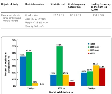
Table 1 Determination of the athletic fatigue loading frequency based on Chinese sports statistics
Objects of study Basic information Stride (A, cm) Stride frequency
(f, steps/min) Loading frequency on the right leg (fL, Hz)
Chinese middle‑dis‑ tance athletes and military recruits
Gender: Male 150.2 ± 3.3 179.7 ± 3.9 1.50 ± 0.03 Age: 18.7 ± 1.4 years
Height: 173.8 ± 5.1 cm Velocity: 16.2 km/h
44.7%
17.0% 20.1%
51.2%
60.1%
22.0%
4.1%
17.1%
31.1%
0.0%
5.9%
26.7%
0% 10% 20% 30% 40% 50% 60% 70%
1500 με 3000 με 5000 με
la
col
fo
tn
ecr
eP
st
ra
in
distribuon
/
%
Global axial strain / με
<1000 1000-3000 3000-5000 >5000
decrease in the Es [20–22] with significant Hc changes as validation of the athletic fatigue damage model, which could activate bone targeted remodeling.
Histology and morphometric analysis
Hematoxylin–eosin staining on non-decalcified slices of the middle sections of the ulnae were performed by XueBang Pathology Corporation in Beijing, China. A qualitative observation of ulnar micromorphology was performed on an optical microscope (Olym-pus, Japan), and the percentage of empty osteocyte lacunae was quantitatively analyzed and compared between different groups.
Tests of serum protein expression and ulnar mechanical behavior
ELISA kits for TRAP-5b and BGP were used to detect bone resorption and formation, respectively. The influence of estrogen on bone remodeling [17, 23, 24] was also deter-mined by assaying serum estradiol with an E2 ELISA Kit. The quasi-static mechanical properties of the ulnae were tested on an INSTRON 5865 (Instron Corporation, Eng-land) with the following parameters: (1) 0.5 N preload, 5 uniaxial loading cycles of dis-placement with a loading rate of 1%/min to a maximum of 0.4%; (2) a loading rate of 5%/ min to fracture. Compressive modulus (E), strength (σb) and fracture energy (Hf) were determined. Fatigue mechanical properties, including cyclic Es, Hc and cycles to fatigue (Nf), were evaluated based on the stress–strain (σ–ε) data recorded during fatigue load-ing. Es was the slope of the line through the start and end points in the loading stage of the σ–ε loop. Hc was the area of the σ–ε loop, determined by integration, and Nf was the number of fatigue loading cycles at 25% Es decrease, determined by interpolation.
Statistical analysis
Data are presented as the mean ± SD and were from at least three independent
experi-ments. The research was designed and simplified from a two-independent-variable experiment based on the theory of “mechanostat” [1]. Significant differences were evalu-ated with a one-way analysis of variance (ANOVA) followed by a least significant differ-ence (LSD) t test. Significance was defined as p < 0.05.
Results
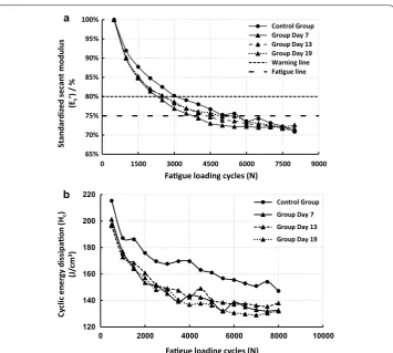
Values of Es were calculated and standardized (Es’) based on the 500th cycle,and curves of Es’–N and Hc–N are shown in Fig. 3. The ulnae in all groups experienced fatigue suc-cessively, with Es’ falling to the fatigue line (25% decrease) (Fig. 3a), and there were obvi-ous decreases in Hc with increasing fatigue loading cycles (Fig. 3b). Therefore, the model of athletic fatigue damage was validated.
Qualitative observation of the HE-stained, non-decalcified ulnae slices from different groups showed deterioration of the bone microstructure under athletic fatigue load-ing (Fig. 4). Microcrack generation (Fig. 4b), growth (Fig. 4c, d) and even coalescence (Fig. 4e) in the interstitial bone reflected an aggravation of bone fatigue damage. Signifi-cant increases in the percent of empty osteocyte lacunae were also detected (Fig. 4f) in the high intensity groups, suggesting apoptosis of osteocytes.
the possible influence of estrogen on bone targeted remodeling could be disregarded in our study. A significant increase in serum TRAP-5b in Day 7 suggested fatigue dam-age and the activation of bone targeted remodeling, and reduced serum TRAP-5b and increased serum BGP from Day 7 to Day 19 demonstrated a process of continuous bone targeted remodeling.
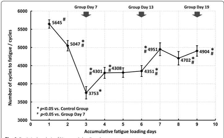
Fatigue mechanical behavior of the ulnae (Es and Hc) were sensitive to fatigue load-ing, and significant changes were detected from Day 7, while the quasi-static mechani-cal properties (E, σb and Hf) seemed to be hysteretic and did not experience significant changes until Day 13 or even Day 19 (Fig. 5). Nf was also statistically analyzed and the results are shown in Fig. 6. Decreases in Nf were detected in all fatigue loading groups, with the valley value on Day 7, which implied a rapid deterioration of the fatigue mechanical properties after fatigue loading and effective improvements by subsequent bone targeted remodeling.
Finally, we summarized the relative changes of several key indices of bone targeted remodeling at different levels (Fig. 7) and achieved a systematic understanding of the bone targeted remodeling process under athletic fatigue loading. This understand-ing could likely offer effective insights regardunderstand-ing to the trainunderstand-ing of athletes and military recruits.
120 140 160 180 200 220
0 2000 4000 6000 8000 10000
Cy
clic
e
nergy dissipaon
(Hc
)
(J
/c
m
3)
Fague loading cycles (N)
Control Group Group Day 7 Group Day 13 Group Day 19 65%
70% 75% 80% 85% 90% 95% 100%
0 1500 3000 4500 6000 7500 9000
sul
ud
o
mt
na
ce
s
dez
idr
ad
nat
S
(Es
’) / %
Fague loading cycles (N)
Control Group Group Day 7 Group Day 13 Group Day 19 Warning line Fague line
a
b
Fig. 3 Changes of standardized secant modulus (Es’) (a) and cyclic energy dissipation (Hc) (b) in different
Discussion
The basic principles of bone remodeling are universal regardless of bone dimensions [1, 7], but there are huge differences in the metabolism rates of rats and humans [23]. When
50 µm a
50 µm b
50 µm c
50 µm d
50 µm e
0 10 20 30 40 50 60
Control Group Group Day 7 Group Day 13 Group Day 19
et
yc
oet
so
yt
p
me
fo
tn
ecr
eP
lacunae
/
%
*
*
*
* p<0.05 vs. Control Group f
Fig. 4 Non‑decalcified slices of rat ulnae and empty osteocyte lacunae statistics. Few empty osteocyte lacu‑
nae and almost no obvious microcracks were detected in the Control Group (a), while generation, growth and coalescence of microcracks (black arrows) were observed sequentially in Group Day 7 (b), Group Day 13 (c) and Group Day 19 (d, e). Significant increases of empty osteocyte lacunae, especially around microcracks, were also detected (white arrows) and statistically verified (f)
Table 2 ELISA assay of serum E2, BGP and TRAP-5b
* p < 0.05 vs. Control group
#p < 0.05 vs. Group Day 7
Groups Concentration of serum proteins (pg/ml)
E2 BGP TRAP-5b
Day 7 448.58 ± 94.03 2504.26 ± 120.73* 1452.48 ± 35.38* Day 13 512.07 ± 65.97 3669.96 ± 206.59*/# 1215.62 ± 52.94*/#
Day 19 478.53 ± 62.68 4321.60 ± 102.73*/# 1177.62 ± 20.71*/#
0.5 0.6 0.7 0.8 0.9 1.0 1.1 1.2 Elasc Modulus
(E) strength (σb)Compressive Fracture energy(Hf) Es(3000th cycle)Secant Modulus Energy dissipaonHc(3000th cycle)
Chan
ge
s of ulnar
m echanical propere s (Standardi ze d
to the Control
G
roup
)
Control Group Group Day 7 Group Day 13 Group Day 19 * P<0.05 vs. Control Group
*
*
*
*
*
* *
*
*
*
*
Fig. 5 Histogram of ulnar mechanical properties in different fatigue loading groups
5645
5047
3753
4301 4308 4351
4951 4702 4904 3000 3500 4000 4500 5000 5500 6000
0 1 2 3 4 5 6 7 8 9 10
sel cy c / eu git af ot sel cy cf or eb mu N
Accumulave fague loading days
* p<0.05 vs. Control Group
#p<0.05 vs. Group Day 7
# #
*
*
#*
#*
#*
#*
#*
#*
Group Day 7 Group Day 13 Group Day 19
Fig. 6 Statistical analysis of Nf on each loading day
Cellular Response Mechanical Properes
Serum Proteins Expression Stac Tests Fague Tests Hormone
(E2) BGP Trap-5b Modulus(E)Elasc Compression Strength(σb)
Fracture Energy(Hf)
Cyclic Secant Modulus(Es)
Cyclic Energy Dissipaon(Hc)
Cycles to Fague(Nf)
Increase
Decrease
Fague Loading Unchanged
Group Day 7 (3rd load)
Group Day 13 (6th load)
Group Day 19 (9th load)
>40% 20%-40% <20%
we determined the athletic fatigue loading parameters (loading duration, amplitude and sampling intervals), its effect on the bone targeted remodeling process had been fully considered.
We originally designed the experiment with two independent variables (periods and exercise), and simplified it (during sampling, assay and analysis) as a single variable (ath-letic fatigue loading duration) experiment based on Frost’s theory of the “mechanostat” [1]. Despite of the weight increase, daily activities of the 3-month-old rats in the con-trol group could produce a physiological deformation of ulnae (with a 200–1000 με axial peak strain [25]), which would not disturb the metabolism balance between bone resorption (osteoclasts activities) and bone formation (osteoblasts activities), or lead to a detectable change of bone mechanical properties within 1 month.
Dynamic compressive loading on the long bones is common during daily activities [14, 19], and the decrease in the secant modulus is mostly a result of bone damage or microc-racks [22]. The athletic fatigue loading protocol used in the present study resulted in sig-nificant decreases in bone fatigue mechanical properties (Figs. 3, 5 and 6); the generation of microcracks in the interstitial bone (Fig. 4b); and an imbalance between bone resorp-tion and bone formaresorp-tion (Table 2) in the early stage (Group Day 7). Our hypothesis on bone fatigue damage and the associated targeted remodeling was therefore confirmed.
An osteon in the cortical bone acts not only as a stress concentrator that accounts for microcrack generation [25–27] but also as a guard against microcrack growth to main-tain the load-bearing capacity of the bone during daily activity [28, 29]. During continu-ous vigorcontinu-ous activity, however, microcracks will grow and even develop to the macro scale, leading to a stress fracture [9, 21, 30]. Our study supported these results. From Day 7 to Day 13, small microcracks were observed (Fig. 4b, c), and fatigue mechani-cal properties also improved (Figs. 5, 6). At Day 19, larger microcracks grew and coa-lesced (Fig. 4d, e), and significant decreases in quasi-static mechanical properties were detected (Fig. 5), which affected the load-bearing capacity and increased the risk of stress fractures.
The integrity of osteocytes is vital for the structural and functional stability of bone [31]. There seems to be a threshold of microdamage or local strain [16] over which fatigue loading or the necessity of bone targeted remodeling could lead to the apopto-sis of osteocytes [23, 24, 32]. In the current study, we confirmed the stable apoptosis of osteocytes in the high-intensity groups during fatigue loading through the signifi-cant increase in the percentage of empty osteocyte lacunae (Fig. 4f) along the microc-racks near osteons (Fig. 4b–d) when compared with the Control group. However, there might be different mechanisms of osteocyte apoptosis in different groups. The apoptosis observed at Day 7 and Day 13 might result from the necessity of bone targeted remod-eling at the early stage because small microcracks and active bone resorption were detected. By contrast, at Day 19, larger linear microcracks were observed despite hyper-active bone formation; therefore, it was likely the severe fatigue damage in the interstitial bone that sequentially prompted osteocyte apoptosis.
would cause rapid microdamage, decreases in fatigue mechanical properties and obvi-ous bone resorption in the long bones. With increasing time, the risks of stress frac-ture would increase with the deterioration of the quasi-static mechanical properties of the bone despite a continuous bone targeted remodeling process, suggesting a limita-tion or a maximum ability to repair excessively damaged bone. For this reason, excessive early-stage training or long-term intensive training without appropriate rests should be avoided to prevent the risk of accumulative fatigue damage or even stress fractures.
Conclusion
An athletic fatigue damage model of rat ulna was successfully established. Fatigue dam-age was aggravated in the loading process, with osteocyte apoptosis, microcrack accu-mulation, and a decrease in mechanical properties. Bone targeted remodeling was activated after athletic fatigue loading and progressively leaned towards bone formation and away from bone resorption to repair fatigue damage, though to some extent.
Abbreviations
OBs: osteoblasts; OCs: osteoclasts; E2: estradiol; BGP: bone gamma‑carboxyglutamic‑acid‑containing protein; TRAP‑5b: tartrate‑resistant acid phosphatase 5b; FEA: finite element analysis.
Authors’ contributions
LH and LRX made substantial contributions to conception and design, analysis and interpretation of data, and manuscript drafting; HB carried out the ulnar fatigue loading and mechanical property tests; ZB and HBH conducted assays of the selected biochemistry index; LYJ was responsible for manuscript revising and polishing; ZXZ, who was the corresponding author, gave financial support and final approval of the version to be submitted. All authors read and approved the final manuscript.
Author details
1 Institute of Medical Equipment, Academy of Military Medical Sciences, Tianjin, China. 2 Department of Orthopaedics
Trauma, First Hospital of Jilin University, Changchun, China.
Acknowledgements
This research is supported by the Major Program of the National Natural Science Foundation of China (No. 11432016) and the General Programs of the National Natural Science Foundation of China (No. 31370942 and No. 31470935).
Competing interests
The authors declare that they have no competing interests.
Data availability statements
The datasets generated during and/or analyzed during the current study are available from the corresponding author on reasonable request.
Statement on ethics approval
The experiments on the Wistar rats performed in the current study were within the animal welfare regulations and guidelines for the Academy of Military Medical Sciences, China.
Publisher’s Note
Springer Nature remains neutral with regard to jurisdictional claims in published maps and institutional affiliations.
Received: 16 December 2016 Accepted: 21 July 2017
References
1. Frost HM. Bone ‘‘mass’’ and the ‘‘mechanostat’’: a proposal. Anat Rec. 1987;219(1):1–9.
2. Burger EH, Klein NJ, Smit TH. Strain‑derived canalicular fluid flow regulates osteoclast activity in a remodeling osteons—a proposal. J Biomech. 2003;36(10):1453–9.
3. Noble BS, Peet N, Stevens HY, Brabbs A, Mosley JR, Reilly GC, Reeve J, Skerry TM, Lanyon LE. Mechanical loading: biphasic osteocytes survival and targeting of osteoclasts for bone destruction in rat cortical bone. Am J Physiol‑Cell Physiol. 2003;284(4):C934–43.
• We accept pre-submission inquiries
• Our selector tool helps you to find the most relevant journal • We provide round the clock customer support
• Convenient online submission • Thorough peer review
• Inclusion in PubMed and all major indexing services • Maximum visibility for your research
Submit your manuscript at www.biomedcentral.com/submit
Submit your next manuscript to BioMed Central
and we will help you at every step:
6. Burr DB. Targeted and nontargeted remodeling. Bone. 2002;30(1):2–4.
7. Frost HM. The mechanostat: a proposed pathogenetic mechanism of osteoporoses and the bone mass effects of mechanical and nonmechanical agents. Bone Miner. 1987;2(2):73–85.
8. Chapurlat RD, Delmas PD. Bone microdamage: a clinical perspective. Osteoporos Int. 2009;20(8):1299–308. 9. Green JO, Wang J, Diab T, Vidakovic B, Guldberg RE. Age‑related differences in the morphology of micro‑damage
propagation in trabecular bone. J Biomech. 2011;44(15):2659–66.
10. Danova NA, Colopy SA, Radtke CL, Kalscheur VL, Markel MD Jr, Vanderby R, McCabe RP, Escarcega AJ, Muir P. Degra‑ dation of bone structural properties by accumulation and coalescence of microcracks. Bone. 2003;33(2):197–205. 11. Chapurlat RD. Bone microdamage. Osteoporos Int. 2009;20(6):1033–5.
12. Yamamoto E, Paul CR, Chan DD, Keaveny TM. Development of residual strains in human vertebral trabecular bone after prolonged static and cyclic loading at low load levels. J Biomech. 2006;39(10):1812–8.
13. Pasqualini M, Lavet C, Elbadaoui M, Vanden BA, Laroche N, Gnyubkin V, Vico L. Skeletal site‑specific effects of whole body vibration in mature rats: from deleterious to beneficial frequency‑dependent effects. Bone. 2013;55(1):69–77. 14. Kotha SP, Hsieh YF, Strigel RM, Müller R, Silva MJ. Experimental and finite element analysis of the rat ulnar loading
model‑correlations between strain and bone formation following fatigue loading. J Biomech. 2004;37(4):541–8. 15. Nagaraja S, Couse TL, Guldberg RE. Trabecular bone microdamage and microstructural stresses under uniaxial
compression. J Biomech. 2005;38(4):707–16.
16. McNamara LM, Prendergast PJ. Bone remodeling algorithms incorporating both strain and microdamage stimuli. J Biomech. 2007;40(6):1381–91.
17. Li H, Li RX, Wan ZM, Xu C, Li JY, Hao QX, Guo Y, Liu L, Zhang XZ. Counter‑effect of constrained dynamic loading on osteoporosis in ovariectomized mice. J Biomech. 2013;46(7):1242–7.
18. Haddock SM, Yeh OC, Mummaneni PV, Rosenberg WS, Keaveny TM. Similarity in the fatigue behavior of trabecular bone across site and species. J Biomech. 2004;37(2):181–7.
19. Sobelman OS, Gibeling JC, Stover SM, Hazelwood SJ, Yeh OC, Shelton DR, Martin RB. Do microcracks decrease or increase fatigue resistance in cortical bone? J Biomech. 2004;37(9):1295–303.
20. Burr DB, Turner CH, Naick P, Forwood MR, Ambrosius W, Hasan MS, Pidaparti R. Does microdamage accumulation affect the mechanical properties of bone? J Biomech. 1998;31(4):337–45.
21. Diab T, Vashishth D. Effects of damage morphology on cortical bone fragility. Bone. 2005;37(1):96–102.
22. Muir P, Sample SJ, Barrett JG, McCarthy J Jr, Vanderby R, Markel MD, Prokuski LJ, Kalscheur VL. Effect of fatigue load‑ ing and associated matrix microdamage on bone blood flow and interstitial fluid flow. Bone. 2007;40(4):948–56. 23. Tomkinson A, Gevers EF, Wit JM, Reeve J, Noble BS. The role of estrogen in the control of rat osteocyte apoptosis. J
Bone Miner Res. 1998;13(8):1243–50.
24. Verborgt O, Gibson GJ, Schaffler MB. Loss of osteocyte integrity in association with microdamage and bone remod‑ eling after fatigue in vivo. J Bone Miner Res. 2000;15(1):60–7.
25. Burr DB, Milgrom C, Fyhrie D, Forwood M, Nyska M, Finestone A, Hoshaw S, Saiag E, Simkin A. In vivo measurement of human tibial strains during vigorous activity. Bone. 1996;18(5):405–10.
26. Akkus O, Rimnac CM. Cortical bone tissue resists fatigue fracture by deceleration and arrest of microcrack growth. J Biomech. 2001;34(6):757–64.
27. Park HC, Lakes RS. Cosserat micromechanics of human bone: strain redistribution by a hydration sensitive constitu‑ ent. J Biomech. 1986;19(5):385–97.
28. Mosekilde L. Consequences of the remodelling process for vertebral trabecular bone structure: a scanning electron microscopy study (uncoupling of unloaded structures). Bone Miner. 1990;10(1):13–35.
29. Vashishth D, Behiri JC, Bonfield W. Crack growth resistance in cortical bone: concept of microcrack toughening. J Biomech. 1997;30(8):763–9.
30. Turnbull TL, Baumann AP, Roeder RK. Fatigue microcracks that initiate fracture are located near elevated intracortical porosity but not elevated mineralization. J Biomech. 2014;47(12):3135–42.
31. Herman BC, Cardoso L, Majeska RJ, Jepsen KJ, Schaffler MB. Activation of bone remodeling after fatigue: differential response to linear microcracks and diffuse damage. Bone. 2010;47(4):766–72.