R E S E A R C H
Open Access
Anti-CRISPR-based biosensors in the yeast
S. cerevisiae
Jing Li
†, Zengliang Xu
†, Aleksandr Chupalov
†and Mario Andrea Marchisio
*Abstract
Background: Anti-CRISPR proteins are expressed by phages as a reaction to the bacterial CRISPR–Cas defense system. Recently, the structures of anti-CRISPR proteins have been determined, and their diverse functions have been clarified. Anti-CRISPR proteins such as LmAcrIIA2 and LmAcrIIA4 interact with the SpCas9:gRNA system and occlude the protospacer adjacent motif (PAM) recognition site, thereby preventing SpCas9:gRNA from binding to the DNA. Hence, anti-CRISPR proteins represent a powerful means to control and modulate the activity of SpCas9 and its nuclease-deficient version dSpCas9. LmAcrIIA2 and LmAcrIIA4 have been shown to be efficient inhibitors of SpCas9 in
Escherichia coli,Saccharomyces cerevisiae, and mammalian cells. To date, there have been no reports of anti-CRISPR-based synthetic gene circuits engineered into yeast cells.
Results: We constructed in the yeastS. cerevisiaesynthetic biosensors based on the anti-CRISPR–dSpCas9:gRNA interaction. Upon induction with galactose orβ-estradiol, anti-CRISPR proteins (LmAcrIIA4, LmAcrIIA2, and StAcrIIA5) produced an enhancement in fluorescence expression by preventing the dSpCas9–Mxi1:gRNA complex from binding to the DNA. We found that LmAcrIIA2 performed as well as LmAcrIIA4 inS. cerevisiae, whereas StAcrIIA5, which had previously been tested in bacteria only, had non-negligible negative effects on yeast cell growth. The efficiency of anti-CRISPR-based biosensors was strongly dependent on the means by which the guide RNAs were produced. The best performance, as measured by the increase in fluorescence, was achieved using a “ribozyme–gRNA–ribozyme” expression cassette under the control of the yeast constitutiveADH1promoter.
Conclusions: This work demonstrates that anti-CRISPR proteins are effective dSpCas9 suppressors in yeast cells. In particular, LmAcrIIA2 and LmAcrIIA4 could be employed as new components of yeast synthetic gene circuits.
Keywords: Synthetic biology, Biosensors, Guide RNA, dSpCas9, Anti-CRISPR
Background
Anti-CRISPR proteins are used by phages to neutralize the CRISPR–Cas system, a component of the bacterial (and archeal) immune system [25]. Anti-CRISPR proteins bind to the Cas:CRISPR RNA (crRNA) complex and prevent it from binding to and/or cleaving the targeted sequence on the DNA [3,18]. Among the various anti-CRISPR proteins studied so far, the AcrIIA family has attracted particu-lar interest, as some of its members have been proven to inhibit the type II-A CRISPR–Cas9 system, which is widely used in biotechnology and synthetic biology appli-cations [22].
*Correspondence:marchisio@hit.edu.cn;mamarchisio@yahoo.com †Jing Li, Zengliang Xu and Aleksandr Chupalov contributed equally to this
work.
School of Life Science and Technology, 2 Yikuang Street, Nan Gang District, 150080 Harbin, People’s Republic of China
Rauch et al. [30] identified four anti-CRISPR pro-teins in the bacteriumListeria monocytogenes, referred to as LmAcrIIA1–LmAcrIIA4. LmAcrIIA2 and LmAcrIIA4 were shown to block the activity ofStreptococcus pyogenes
Cas9 (SpCas9) in human cells and its nuclease-deficient version (dSpCas9) in Escherichia coli cells. LmAcrIIA4 exhibited better performance than LmAcrIIA2 in both organisms. InE. coli, Rauch and co-authors put the syn-thesis of a red fluorescent protein (RFP) under the neg-ative control of dSpCas9. LmAcrIIA4 was able to almost completely re-establish full RFP expression, whereas with LmAcrIIA2 the red fluorescence level reached only about 25% of that in the absence of a guide RNA (gRNA) targeting RFP. In re-engineered human cells, a green fluorescent protein gene was cleaved by SpCas9. Here, LmAcrIIA4 only slightly outperformed LmAcrIIA2 in inhibiting SpCas9.
Subsequent works [8, 33, 36] reported that both LmAcrIIA2 and LmAcrIIA4 could achieve repression of SpCas9:crRNA activity by mimicking the proto-spacer adjacent motif (PAM; NGG) recognized by the SpCas9:crRNA complex. These anti-CRISPR proteins occupy the SpCas9 PAM binding site and prevent recogni-tion of the DNA. Interestingly, this takes place only when SpCas9 is associated with crRNA.
More recently, during the peer-review process for this work, Basgall et al. [1] successfully used both LmAcrIIA2 and LmAcrIIA4 as inhibitors of SpCas9
in a gene drive system for the yeast Saccharomyces
cerevisiae.
Another AcrAII protein was identified inStreptococcus thermophilus and termed StAcrIIA5. This anti-CRISPR protein could inhibit SpCas9 inLactococcus lactisbut not inE. coli[19]. It was not tested in eukaryotic cells or, in general, on dSpCas9.
We evaluated the functions of these three anti-CRISPR proteins in the yeastS. cerevisiae. Each of them acted on a yeast codon-optimized version of dSpCas9 (fused to a nuclear localization sequence–NLS) [10] targeting a tran-scription unit that encoded the yeast enhanced green fluo-rescent protein (yEGFP [32]). Specifically, we constructed galactose- andβ-estradiol-biosensing devices (buffer, or YES, gates in logic terms) based on dSpCas9:gRNA–anti-CRIPSR interactions. YES gates are essential components in the design of complex gene digital circuits. They trans-duce input signals (chemicals) into proteins or small RNAs. In this way, the inputs are transmitted through the internal gates to the output gates that return fluores-cence or activate/repress a cellular pathway [24]. Digital circuits have several applications in synthetic biology, from bio-computing [2] to pollutant detection and disease recognition [23]. We chose galactose andβ-estradiol for a proof-of-concept experiment, since the former activates
GAL1promoter directly, while the latter exerts its action on the anti-CRISPR protein upon its fusion to a proper hormone binding domain (see below). Hence, these two chemicals did not require our circuits to include more than four genes, as there are genetically modified aux-otrophic markers in the CEN.PK2-1C yeast strain used in this work (see Methods).
In order to construct the buffer gates, we first evalu-ated the efficiency of transcriptional repression by means of dSpCas9:gRNA. In particular, we used different modes of gRNA expression, namely via the RNA polymerase III-dependent promotersSNR52[7] andRPR1[10] (both on integrative and multicopy plasmids), and using the RGR cassette HH ribozyme–gRNA–HDV ribozyme (HH, ham-merhead; HDV, hepatitis delta virus) under the RNA poly-merase II-dependentADH1promoter [12]. Moreover, we tested both the bare dSpCas9 and dSpCas9 fused to the repression domain Mxi1 [11,15].
After identifying the best solution for repressing the production of yEGFP from the synthetic promoter Tsynth8_pCYC1noTATA [35] employed in our circuits, we constructed several small networks (four genes overall, as mentioned above), each containing one of the anti-CRISPR proteins LmAcrIIA2, LmAcrIIA4, and StAcrIIA5. They were expressed upon induction with galactose. Both LmAcrIIA2 and LmAcrIIA4 worked well inS. cerevisiae, resulting in a significant increase (more than three-fold) in cell fluorescence via inhibiting the action of dSp-Cas9:gRNA on Tsynth8_pCYC1noTATA. Finally, we con-structed two hormone biosensors by fusing LmAcrIIA2 to the hormone binding domain of the human estro-gen receptor, HBD(ER) [20]. In the presence of 1μM β-estradiol, both circuits were able to prompt an approxi-mately two-fold enhancement in fluorescence.
Results and discussion
The output of our biosensing circuits, which were based on dSpCas9:gRNA–AcrAII (anti-CRISPR) interactions, was yEGFP. This reporter protein is expressed by the synthetic promoter Tsynth8_pCYC1noTATA, produced by fusing a synthetic terminator (Tsynth8 [5]) to the
minimal CYC1 promoter stripped of its TATA boxes
(pCYC1noTATA [35]). Thus, the efficiency element of Tsynth8 became the TATA box of the synthetic promoter. Tsynth8_pCYC1noTATA is down-regulated by the com-plex dSpCas9:gRNA. The gRNA contained a 20-nt spacer ATAAACTCATTTACTTATGT that overlapped the pro-moter TATA box. We verified with CRISPRdirect [27] that this 20-mer is absent from the yeastS. cerevisiaegenome; this was as expected, since it is a portion of a synthetic ter-minator. Moreover, the spacer seed region (i.e., the last 12 nucleotides [7]) had no perfect match in the budding yeast genome either.
Anti-CRISPR proteins, which interact with and inhibit the dSpCas9:gRNA complex, are produced upon induc-tion with galactose or translocate into the nucleus upon induction with β-estradiol. We considered a working biosensor to be any circuit where the fluorescence level expressed in the presence of the input signal was significantly different, in statistical terms, from that measured without the input (two-sided Welch’s t-test,
p-value<0.05).
dSpCas9:gRNA-based transcriptional control inS. cerevisiae
The first step in the construction of our biosens-ing devices was to test the efficiency of the dSp-Cas9:gRNA system in repressing transcription from the Tsynth8_pCYC1noTATA synthetic promoter.
terminator, SUP4t [7]); and (2) flanking the gRNA with two ribozymes [11,12] (RGR cassette). In the latter case, an RNA polymerase II-type promoter leads to the produc-tion of long mRNA chains. Upon mRNA formaproduc-tion, the two ribozymes autocleave and release a gRNA molecule.
dSpCas9 was used either in its natural conformation (the sequence was usually codon optimized for expression inS. cerevisiae) or fused to the repression domain Mxi1. The presence of Mxi1 has been reported to enhance the performance of dSpCas9 in transcription regulation [15].
Under the assumption that RNA polymerase III-dependent promoters are weak [11], transcription units containing RNA polymerase III elements for gRNA syn-thesis were generally placed into multicopy (or cen-tromeric [6,34]) plasmids. This guaranteed the expression of gRNA in a reasonably high copy number, but required cells to be grown in selective media. By contrast, the RGR cassette was placed into integrative plasmids downstream of strong RNA polymerase II-type promoters (such as pADH1 [12]). The RGR secures circuit stability (owing to its integration into the genome) and should provide gRNA in large amounts. However, the choice of promoter and terminator at the edge of the RGR are limited to those that allow a proper folding of both HH and HDV ribozymes.
Since the efficiency of transcription regulation via dSp-Cas9:gRNA has been shown to be highly context depen-dent in general [34], we decided to build eight inducible three-gene circuits (NOT gates) to assess the performance of the above-described designs and test new ones with our synthetic promoter.
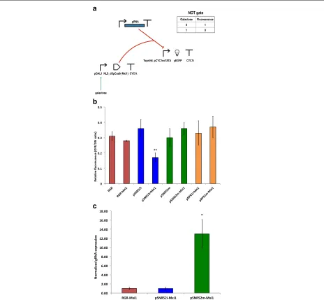
Each of these NOT gates contained a transcrip-tion unit that expressed the guide RNA constitutively. Three variants of this transcription unit were con-structed: pADH1–RGR–ADH1t (always into an inte-grative plasmid), pRPR1–gRNA–RPR1t, and pSNR52– gRNA–SUP4t. The two units based on RNA polymerase III-dependent promoters and terminators were placed into both multicopy and integrative plasmids. Synthesis of dSpCas9 or dSpCas–Mxi1 was induced by galactose. Finally, the circuit output (fluorescence) was expressed under Tsynth8_pCYC1noTATA (see Fig.1a).
With the exception of pRPR1–gRNA–RPR1t, which was used with dSpCas9–Mxi1 only, we tested the working of each transcription unit for gRNA expression together with both the bare dSpCas9 and that fused to the Mxi1 repression domain. As shown in Fig.1b, the strongest flu-orescence repression (83%) was observed in the presence of dSpCas9–Mxi1 and the RNA polymerase III-dependent transcription unit pSNR52–gRNA–SUP4t placed on an integrative plasmid (concisely, we refer to this configura-tion as pSNR52i–Mxi1). The other circuit configuraconfigura-tions showed repression varying from 63% (pRPR1m–Mxi1, i.e., pRPR1–gRNA–RPR1t on a multicopy plasmid and dSpCas9–Mxi1) to 72% (RGR–Mxi1, i.e., the RGR cassette
and dSpCas9–Mxi1). In statistical terms, pSNR52i–Mxi1 was significantly different from each of the other seven NOT gates (see Additional file 1: Figure S1). More-over, the only other statistically significant difference was between RGR–Mxi1 and pSNR52m–Mxi1. The latter cir-cuit employs pSNR52 on a multicopy plasmid for the synthesis of the gRNA.
As pointed out above, RNA polymerase III-dependent promoters are considered to be weak promoters. Hence, it was assumed that transcription units such as pSNR52–
gRNA–SUP4t or pRPR1–gRNA–RPR1t would not
express enough gRNA molecules upon integration into theS. cerevisiaegenome to result in a working CRISPR– (d)SpCas9 system. To the best of our knowledge, however, this assumption has never been proven. In fact, our results (Fig.1b) indicate that the quantity of gRNA is not a limiting factor, at least for certain kinds of synthetic gene circuits such as NOT gates.
For the construction of biosensors based on anti-CRISPR proteins we selected three configurations. First, we chose the design that outperformed all the others in terms of repression of fluorescence, i.e., pSNR52i– Mxi1. Then, we picked two other schemes that employed dSpCas9–Mxi1: RGR–Mxi1 (the second-best NOT gate in terms of mean relative fluorescence) and the lower-performing pSNR52m–Mxi1 (64% fluorescence repres-sion; see Fig.1b). We made this choice so as to examine the possible influence of the gRNA expression system (the integrative/multicopy plasmid carrying the pSNR52 promoter, or the RGR cassette) on the working of the anti-CRISPR-containing circuits. It should be noted that there were statistically significant differences in the relative flu-orescence of these three NOT gates.
We carried out real-time quantitative polymerase chain reaction (RT-qPCR) [6,31] to quantify the relative amount of gRNA expressed by each of these three NOT gates.
ACT1 was used a reference gene in these measurements. pSNR52m–Mxi1, the least efficient among these circuits, expressed almost 13-fold more gRNA than the other two gates (Fig.1c). Moreover, pSNR52i–Mxi1 and RGR– Mxi1 produced roughly the same quantities of gRNA. Nevertheless, the fluorescence repression achieved with pSNR52i–Mxi1 was clearly higher than that achieved with RGR–Mxi1. Taken together, these results confirmed that the working of our NOT gates was independent of the gRNA expression level.
In a biosensing device, the production (or the transloca-tion into the nucleus) of anti-CRISPR proteins is triggered by an input signal, whereas both gRNAs and dSpCas9– Mxi1 are constitutively expressed. For this reason, we tested the efficiency of three sub-circuits based on the chosen NOT gate configurations. These differed from the circuit shown in Fig. 1a in terms of the promoter
a
b
c
(pGPD) was used instead of pGAL1 (see Additional file1: Figure S2A). In each case, we compared the fluorescence level (low) of the whole three-gene circuit with that of a two-gene circuit lacking either dSpCas9–Mxi1 or the gRNA. With respect to the NOT gates, we did not detect any difference in the repression level associated with the three-gene circuit hosting a multicopy plasmid (64%). However, the configuration with the RGR cassette per-formed better than that with pSNR52 in an integrative plasmid: 87% of fluorescence repression versus 75% (see Additional file1: Figure S2B and Table S2). We attributed this discrepancy with respect to the NOT gates mainly to the fact that we did not use the same two-gene con-trol circuit for each of the three sub-circuits. In particular, the control circuit for the RGR cassette did not contain the transcription unit for the synthesis of dSpCas9–Mxi1 under the strongGPD promoter. As a consequence, the fluorescence level of the RGR control was higher than that of the other two-gene devices, which lacked the gRNA expression plasmid. RT-qPCR experiments confirmed the results obtained for the NOT gates, since the circuits asso-ciated with the highest fluorescence repression had the lowest gRNA expression (Additional file1: Figure S2C). This further underlines that the gRNA amount is not a limiting factor in achieving a dSpCas9:gRNA complex concentration sufficient to repress transcription from the synthetic promoter Tsynth8_pCYC1noTATA.
Biosensors based on anti-CRISPR proteins
In order to construct biosensors that exploit the inhibi-tion of the dSpCas9–Mxi1:gRNA system by anti-CRISPR proteins, we extended the three sub-circuits described above with a further transcription unit encoding one of LmAcrIIA2, LmAcrIIA4 [30], and StAcrIIA5 [19]. LmAcrIIA2 and LmAcrIIA4 were shown to inhibit both SpCas9 and its nuclease-deficient version, the former in eukaryotic cells and the latter in bacteria. StAcrIIA5 was proven to suppress DNA cleavage from SpCas9 into bac-terial cells. However, the way StAcrIIA5 interacts with SpCas9 is unknown. Notably, some anti-CRISPR proteins, such as AcrIIC1, hinder Cas9 DNA cleavage not by pre-venting Cas9 from binding to the target DNA sequence (i.e., by occluding the Cas9 PAM recognition site) but by inactivating Cas9 nuclease activity upon binding to the Cas9 HNH nuclease domain [18]. In other words, AcrIIC1 turns Cas9 into a dCas9. Therefore, the fact that an anti-CRISPR protein, such as StAcrIIA5, prevents SpCas9 from cutting the DNA does not necessarily imply that the same protein impedes dSpCas9 from binding to the DNA.
We placed the anti-CRISPR coding sequence down-stream of the GAL1 promoter. Hence, the presence of galactose in the cell-growth solution should induce an increase in the fluorescence expressed by our circuits (buffer, or YES, gates; see Fig.2a) to a level significantly
different, in statistical terms, from that expressed in the presence of glucose.
We started with LmAcrIIA4 because this protein is known to be a strong SpCas9 inhibitor in human cells [30]. Circuit performance was assessed by calculating the ratio between the fluorescence levels in the pres-ence and abspres-ence of galactose (see Fig.2band Additional file1: Table S3). LmAcrIIA4 performed well only when paired with the RGR cassette for gRNA expression. The circuit hosting the original sequence of LmAcrIIA4 together with the RGR (A4-RGR) showed an almost 3.2-fold gain in fluorescence upon induction with galactose. We obtained a statistically equivalent result by using an LmAcrIIA4 sequence codon optimized for expres-sion in S. cerevisiae (yo_LmAcrIIA4. YES gate: yo_A4-RGR). When the RGR cassette was replaced with the transcription unit pSNR52–gRNA–SUP4t (on both the integrative and the multicopy plasmid), only one of the three circuit variants functioned correctly, i.e., showed a statistically significant difference between fluorescence levels with and without galactose. This configuration made use of yo_LmAcrIIA4 and pSNR52 on an inte-grative plasmid for gRNA synthesis (yo_A4-pSNR52i). The corresponding gain in fluorescence, however, was modest (1.41-fold).
The reason for the malfunctioning of the galactose biosensors hosting LmAcrIIA4 and a gRNA expression
system based on theSNR52promoter (A4-pSNR52i and
a
b
c
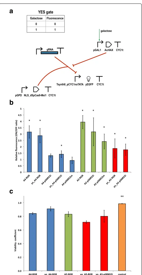
Fig. 2Galactose biosensors based on anti-CRISPR proteins.aUpon induction with galactose, an anti-CRISPR protein of the AcrIIA family is produced. An increase in fluorescence is detected if the anti-CRISPR is able to prevent the dSpCas9-Mxi1:gRNA system from binding the Tsynth8_pCYC1noTATA synthetic promoter.bGalactose biosensor performance. Main values of the ON/OFF ratio were calculated over at least 3 independent experiments for each circuit. The symbol “*” indicates a working biosensor i.e. there is a statistically significant difference between the mean fluorescence of the ON and OFF state (two-sided Welch’s t-test,p-value<0.05). A detailed statistical comparison of the relative fluorescence of these ten anti-CRIPSR-based constructs is given in Additional file1: Figure S3.cViability test. The viability coefficients of five YES gates presenting slow growth rate underline the presence of only moderate toxic effects. The control strain (A5-RGR grown in glucose-supplied synthetic medium) is associated with a viability coefficient, equal to 0.98, significantly higher in statistical terms than all the other viability coefficients calculated in this test (as denoted by the symbol “**”). A further statistical analysis on these results is reported in Additional file1: Figure S8
A faster growth rate (though only after about 11 h) with respect to these five strains was achieved by mod-ifying byMM234 with the insertion of LmAcrIIA4 and dSpCas9–Mxi1 only, i.e., without any transcription unit for gRNA synthesis. In this configuration (termed A4-dCas9; Fig.3a), LmAcrIIA4 could not bind to dSpCas9– Mxi1. We obtained an almost identical growth curve by expressing a “scrambled” gRNA – via the RGR cas-sette – in the presence of LmAcrIIA4 and dSpCas9–Mxi1 (A4-RGR(scrambled)). The scrambled gRNA contained a 20-nt spacer that targeted the LacI bacterial repressor and had no match in theS. cerevisiaegenome. Control exper-iments on the three-gene sub-circuits lacking LmAcrIIA4 confirmed that the RGR cassetteper sehad no negative influence on yeast cell growth but provoked a slowdown of the growth rate only when producing gRNA molecules able to bind to their target on the DNA (Additional file1: Figure S5). However, it is not clear why this effect was less evident when the gRNA was expressed by theSNR52 pro-moter placed on a multicopy plasmid and almost absent when an integrative plasmid was used instead.
We next constructed galactose-sensing devices based on LmAcrIIA2 in order to see whether we could observe any of the features present in the LmAcrIIA4-based circuits.
LmAcrIIA2 has been reported to have lower efficiency than LmAcrIIA4, in both bacteria and human cells. In our experiments, we did not observe a clear gap between these proteins; thus, we can assert that, inS. cerevisiae, LmAcrIIA2 and LmAcrIIA4 have comparable perfor-mance on dSpCas9:gRNA. Each of the three galactose sensors based on LmAcrIIA2 returned an average gain in fluorescence greater than two-fold upon induction with galactose. However, the uncertainty on the relative flu-orescence values were also rather large, especially when the gRNA was expressed via pSNR52 on an integrative plasmid (A2-pSNR52i; see Fig.2b and Additional file1: Table S3). Similar to the circuits realized with LmAcrIIA4, the best performance was observed with the biosensor hosting the RGR cassette (A2-RGR), a 3.94-fold average gain. This circuit was significantly different from all the other galactose-biosensing networks we constructed, with the exception of A4-RGR and A2-pSNR52i (it should be noted, though, that A2-pSNR52i was statistically indis-tinguishable from any of our working biosensors; see Additional file1: Figure S3). In contrast to LmAcrIIA4, we managed to build a working biosensor based on LmAcrIIA2 and the gRNAs expressed via pSNR52 on a multicopy plasmid, A2-pSNR52m, that gave a 2.41-fold average gain in fluorescence.
a
b
c
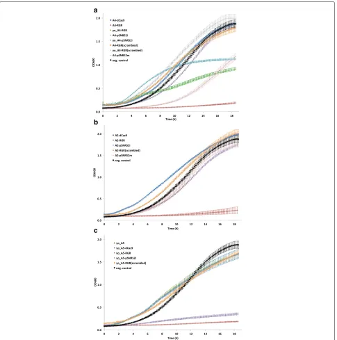
Fig. 3Growth curves in synthetic medium supplied with 2% galactose.aLmAcrIIA4-hosting circuits. The three working biosensors (A4-RGR, yo_A4-RGR, and yo_A4-pSNR52i) grew more slowly than byMM234, the negative control that expresses only yEGFP. By removing the gRNA from the circuit (dCas9-A4) or substituting it with a scrambled one with no match along the yeast genome (A4-RGR(scrambled) and yo_A4-RGR(scrambled)) a quick growth was re-established.bBiosensors based on LmAcrIIA2 confirmed that the RGR cassette is responsible for a slow growth when gRNAs targeting Tsynth8_pCYC1noTATA are expressed. In the presence of a scrambled gRNA (A2-RGR(scrambled)) as well as in the absence of any gRNA molecules (A2-dCas9) cell growth rate appeared even faster than that of the negative control. pSNR52 both on integrative (A2-pSNR52i) and multicopy (A2-pSNR52m) plasmid did not provoke drastic changes in cell growth either.cBoth biosensors containing yo_StAcrIIA5 (yo_A5-RGR and yo_A5-pSNR52i) grew extremely slowly. However, the protein yo_StAcrIIA5 on its own has no negative effects on yeast cell growth as witnessed by the fast growth rate of the three control circuits (yo_A5, yo_A5-dCas9, and yo_A5-RGR(scrambled). Every average OD600 value was calculated on three replicates (single experiment)
Tsynth8_pCYC1noTATA (A2-RGR). However, in con-trast to the LmAcrII4-based biosensors, the expression of gRNAs from multicopy plasmids containing pSNR52
pSNR52 on an integrative plasmid (A2-pSNR52i) induced only a small slowdown in cell growth rate. These two circuits also had little effect on cell growth in glucose-containing solution (Additional file1: Figure S4B).
Finally, we constructed two biosensors by integrat-ing into the genome of S. cerevisiae a yeast codon-optimized version of the anti-CRISPR protein StAcrIIA5 (yo_StActIIA5) together with either the RGR cassette (yo_A5-RGR) or the pSNR52–gRNA–SUP4 transcription unit (yo_A5-pSNR52i) for gRNA expression. For both circuits, the main values of fluorescence expressed in the presence of galactose were statistically significantly different from those obtained in the absence of galac-tose, and the fluorescence gain was close to two-fold (Fig. 2b); however, the cell populations did not show well-defined shapes in the fluorescence activated cell sort-ing (FACS) dot plots (Additional file 1: Figure S6). Fur-thermore, both yo_A5-RGR and yo_A5-pSNR52i showed remarkably slow growth rates in synthetic medium con-taining galactose, as shown in Fig. 3c (in the pres-ence of glucose, only the growth curve of yo_A5-RGR deviated considerably from that of the negative con-trol; see Additional file 1: Figure S4C). To determine whether yo_StActIIA5 was less tolerated byS. cerevisiae
than (yo_)LmAcrIIA4 and LmAcrIIA2, we built three control circuits in which byMM234 was modified with the expression of (1) the sole yo_StActIIA5 (yo_A5), (2) yo_StActIIA5 plus dSpCas9–Mxi1 (yo_A5-dCas9), or (3) yo_StActIIA5 together with dSpCas9–Mxi1 and an RGR cassette synthesizing the scrambled gRNA described above (yo_A5-RGR(scrambled)). In galactose-containing solution, these three circuits showed almost indistin-guishable growth curves, with shapes similar to that of the negative control (Fig. 3c). These results, which are consistent with our measurements on analogous circuits involving (yo)_LmAcrIIA4 and LmAcrIIA2, indicate that the anti-CRISPR protein yo_StActIIA5 is not toxic per se to yeast cells. However, mild toxicity effects might be present when the system yo_AcrIIA5–dSpCas9:gRNA interacts with the DNA and causes the irregular yeast population shapes that emerged during the FACS exper-iments. To verify this hypothesis, we ran a viability test on the strains yo_A5-RGR and yo_A5-pSNR52i. For com-parison with the other circuits that had growth curves significantly lower than that of byMM234 in galactose-containing medium (Additional file1: Figure S7), we also calculated the viability coefficients for A4-RGR, yo_A4-RGR, and A2-RGR. As a negative control, we took yo_A5-RGR grown in glucose-containing solution (Fig. 2c); yo_A5-RGR gave the lowest mean value of the viabil-ity coefficient (0.71), a statistically significant difference compared with all the other circuits except for yo_A5-pSNR52i (Additional file1: Figure S8 and Table S4). The high relative error (11.25%) on the viability coefficient of
yo_A5-pSNR52i caused this measurement to be statisti-cally indistinguishable from all the others (apart from the negative control). This result was consistent with the con-siderable variability in cell size highlighted by the FACS dot plot on yo_A5-pSNR52i. A4-RGR and A2-RGR gave comparable results (0.84 and 0.83, respectively), signifi-cantly higher than the value of yo_A5-RGR and lower than that of yo_A4-RGR (0.91), the circuit with the highest growth rate in galactose-containing medium. Overall, the viability test showed that only moderate toxicity effects, which did not preclude the working of our YES gates, were induced to different extents by the interactions of diverse anti-CRISPR proteins with the same dSpCas9:gRNA com-plex and target DNA sequence.
In summary, LmAcrIIA2 and LmAcrIIA4 have simi-lar performance in S. cerevisiae as inhibitors of tran-scriptional repression by dSpCas9–Mxi1:gRNA. In our galactose-sensing devices, where the presence of galac-tose stimulates the production of anti-CRISPR proteins, the highest gain in fluorescence was obtained by express-ing the gRNA via an RGR cassette. This configuration provoked a drastic decrease in the growth of yeast cells, without relevant toxicity effects. The use of a yeast codon-optimized version of LmAcrIIA4 together with the RGR cassette speeded up the growth of biosensor-carrying cells, increased (above 0.90) the value of the viability coef-ficient, and returned an ON/OFF ratio comparable with that of the A4-RGR circuit.
LmAcrIIA2 performed well not only in the presence of the RGR but also when paired with the RNA polymerase III-type SNR52 promoter on integrative and multicopy plasmids. In both cases, the decrease in cell growth rate was almost negligible.
StAcrIIA5-based biosensors, by contrast, showed lower ON/OFF ratios and a slowdown in the growth curves of cells containing both the RGR and pSNR52–gRNA– SUP4t transcription unit on an integrative plasmid. High variability in cell size was also apparent in the dot plots from FACS experiments. Moreover, the viability coeffi-cient calculated on the yo_A5-RGR circuit was signifi-cantly lower than those associated with the LmAcrIIA2-and (yo_)LmAcrIIA4-hosting biosensors.
in fluorescence, or by using an yeast codon-optimized version of the anti-CRISPR protein, as in the case of LmAcrIIA4. yo_StAcrIIA5, by contrast, showed more sig-nificant negative side effects on cell growth, and thus requires further characterization before being employed inside synthetic gene networks inS. cerevisiae.
Biosensors responding toβ-estradiol
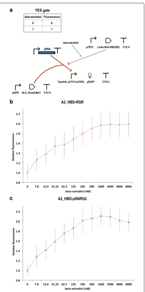
As LmAcrIIA2 performed well with every gRNA expres-sion system, we chose this anti-CRISPR protein to build a further biosensor that responds toβ-estradiol. We fused LmAcrIIA2 to the HBD(ER) [20], which is known to inter-act with and be bound by the heat-shock protein Hsp90. As a result, a chimeric protein containing HBD(ER), such as our LmAcrIIA2–HBD(ER), would be sequestered into the cytoplasm by Hsp90. Upon binding to β-estradiol, HBD(ER) undergoes a conformational change that pre-vents any further interactions with Hsp90. Hence, the chimeric protein can migrate into the nucleus. In our case, the presence ofβ-estradiol induced the interaction of LmAcrIIA2–HBD(ER) with dSpCas9–Mxi1:gRNA.
So far, in S. cerevisiae, HBD(ER) has been used
mainly as a means to engineer synthetic transcription factors [20,21,26,28].
We constructed two circuits, one expressing the gRNAs via the RGR cassette (A2_HBD-RGR), the other through pSNR52–gRNA–SUP4t on an integrative
plasmid (A2_HBD-pSNR52i). As depicted in Fig. 4a,
the chimeric protein LmAcrIIA2–HBD(ER) was placed downstream of the yeastTEF2promoter (47% as strong as pGPD [35]).
We tested the two circuits at different concentrations of β-estradiol, from 7.81nMto 8μM. At each concentration, we calculated the fluorescence expressed by each circuit relative to the fluorescence level detected in the absence of the hormone (Fig.4b-c). The two circuits had compara-ble performance. Their steady state, which corresponded to an approximately two-fold gain in fluorescence expres-sion, was reached at 250nM β-estradiol (2.01 ± 0.26) by A2_HBD–pSNR52i and at 1μM β-estradiol (1.96 ± 0.25) by A2_HBD–RGR. In a later attempt to improve the circuit performance, we tried to express LmAcrIIA2– HBD(ER) under pGPD; however, this resulted in the syn-thesis of too much LmAcrIIA2–HBD(ER), such that a substantial amount of this chimeric protein could enter the nucleus and inhibit dSpCas9–Mxi1:gRNA even in the absence ofβ-estradiol, preventing correct functioning of our circuit (Additional file1: Figure S9).
Notably, A2_HBD-RGR resulted in a slower growth rate with respect to A2_HBD–pSNR52i, both in the presence and absence ofβ-estradiol, confirming the trend observed with A2-RGR and A2-pSNR52i. However, the slowdown in the growth of A2_HBD–RGR was much less remark-able than that corresponding to A2-RGR. Moreover, the
a
b
c
viability coefficients calculated for the two β-estradiol sensors were proximal to 1 both in the presence and absence ofβ-estradiol (Additional file1: Figure S10 and Table S5).
Conclusions
In this work, we have described a new class of synthetic biosensors in the yeastS. cerevisiae, based on the inter-actions between anti-CRISPR proteins and the dSpCas9– Mxi1:gRNA system. First, we assessed how different dSp-Cas9:gRNA configurations could repress the synthesis of a reporter protein from a synthetic promoter built in our laboratory. The 20-mer spacer sequence of the gRNA was chosen both to bind in the proximity of the promoter TATA-box (to enhance transcriptional repression) and to avoid off-target matches within the yeast genome. Con-trary to the belief that RNA polymerase III-dependent promoters are much weaker than promoters bound by RNA polymerase II and need to be present in a high copy number to assure a reasonable gRNA expression, we achieved the strongest fluorescence repression by insert-ing the transcription unit pSNR52–gRNA–SUP4t into an integrative plasmid. dSpCas9 was fused to the Mxi1 repression domain and placed downstream of theGAL1
promoter. Uninduced systems, where dSpCas9–Mxi1 was produced by pGPD, showed slightly better performance when the gRNAs were synthesized via an RGR cassette under the ADH1 promoter. However, the transcription unit pSNR52–gRNA–SUP4t was again more effective if integrated into the yeast genome rather than placed on a multicopy plasmid.
We chose three anti-CRISPR proteins for the con-struction of galactose-biosensing devices. LmAcrIIA2 and LmAcrIIA4 were previously shown to be efficient inhibitors of SpCas9 (and dSpCas9) in bacterial and human cells [30] and were recently tested also on S. cerevisiae [1] . Once bound to the gRNA, they act on SpCas9 by occluding its PAM binding site. StAcrIIA5 was proven to inhibit SpCas9 in bacterial cells [19]. However, the mechanism through which this anti-CRISPR works is unknown. According to previous reports, LmAcrIIA4 performs better than LmAcrIIA2 in human and bacterial cells. We have shown that, in S. cerevisiae, the perfor-mance of LmAcrIIA2 is comparable to that of LmAcrIIA4. Moreover, LmAcrIIA2 allowed the construction of work-ing biosensors with every gRNA expression system con-sidered here.
The growth curves of yeast cells re-engineered with anti-CRISPR-based biosensors highlighted how the best-performing devices (those where the gRNAs were pro-duced via the RGR cassette) grew much more slowly than the circuits containing an RNA polymerase III-dependent transcription unit for gRNA synthesis. This growth slowdown, however, was not associated with
any drastic reduction in cell viability. In the case of LmAcrIIA4, the growth of the cells hosting the RGR cassette was increased – although it was still not com-parable to that of the control strains – by using a yeast codon-optimized version of the anti-CRISPR protein. No substantial changes in circuit performance were detected between A4-RGR and yo_A4-RGR circuits.
StAcrIIA5 was also employed in a yeast codon-optimized version. However, even though the corre-sponding biosensors worked properly, as evidenced by the fluorescence levels observed, more serious concerns arose based on the growth curves, viability test, and dot plots. Hence, this anti-CRISPR protein does not seem to be a reliable component of synthetic circuits in
S. cerevisiaecells.
Finally, we built β-estradiol-sensing devices by fus-ing LmAcrIIA2 to the hormone bindfus-ing domain of the human estrogen receptor. The two biosensors we constructed (one with the RGR cassette, the other with pSNR52–gRNA–SUP4t on an integrative plasmid) worked effectively and reached an approximately
two-fold gain when induced with 1μM(A2_HBD-RGR) and
250nM(A2_HBD-pSNR52i)β-estradiol.
Overall, our results indicate that anti-CRISPR proteins can work efficiently inS. cerevisiaecells. We have shown how they can be used to construct biosensing systems responding to a single chemical. Anti-CRISPR represent a useful means to control the activity of both SpCas9 and its nuclease-deficient variants. Hence, a deep understanding of their mechanisms may lead to these proteins becom-ing widely-used components in future yeast synthetic regulatory networks and DNA-editing circuits.
Methods
Plasmid construction
All integrative and multicopy plasmids realized in this work (a complete list is provided in Additional file1: Table S6) are based on the pRSII4XX yeast shuttle-vector col-lection (available at Addgene–their codes are reported below–a gift from Steven Haase) [4].
The vector targeted by the CRISPR-dSpCas9
sys-tem (pMM260) contains the transcription unit
Tsynth8_pCYC1noTATA-yEGFP-CYC1t. The assembly of this plasmid is described in our previous work [35].
As a template for gRNA expression via the RPR1
promoter and terminator we used the plasmid
In order to assemble a gRNA expression cassette via the
SNR52promoter and SUP4 terminator we constructed, via isothermal assembly, the gRNA-spacer acceptor vector pMM465 that contains the transcription unit pSNR52-BpiI(GATC)-place_for_spacer-BpiI(GTTT)-SpCas9_repeat-SUP4t. Backbone for pMM465 was pRSII404. The spacer sequence was extended with BpiI sites compatible with those present on pMM465. Primers containing the extended spacer sequence (synthesized, annealead, and purified by COMATE, Harbin, China) were inserted into pMM465 via Golden Gate method [9]– see below for details. The resulted plasmid was named pMM524. The transcription unit pSNR52-gRNA-SUP4t from pMM524 was inserted into the multicopy shuttle vector pRSII424 via digestion with KpnI (NEB-R0142S) and SacI (NEB-R0156S), and successive ligation with T4 DNA ligase (NEB-M0202S).
The RGR sequence (HH_ribozyme-spacer-SpCas9_ repeat-HDV_ribozyme) followed by theADH1terminator was synthesized by GENEWIZ Inc., Suzhou (China), and then assembled, with the Gibson method, into pRSII404 together with theADH1promoter. The new plasmid was named pMM554.
NLS-dSpCas9 was extracted from the plasmid
“pTPGI_dCas9_VP64” (Addgene-49013, a gift from Tim-othy Lu) via digestion with XbaI (NEB-R0145S) and SalI (NEB-R0138S).
In order to express NLS-dSpCas9 under the GPD
promoter we constructed the acceptor vector pMM551.
pGPD and CYC1t were extended via PCR–and
then assembled, via Gibson method, into pRSII406 (Addgene-35442)–to have the sequence: pGPD-ATG-XbaI-space_for_NLS-dSpCas9-SalI-GG-STOP-CYC1t, where G stands for glycine. This plasmid served as a template to build pMM559, another acceptor vec-tor for NLS-dSpCas9, where pGAL1 replaced pGPD. NLS-dSpCas9 was inserted into both plasmids after digestion with XbaI and SalI followed by ligation with T4 DNA ligase.
pMM551 and pMM559 were used to construct pMM573 and pMM574, respectively. They are the NLS-dSpCas9 acceptor vectors that contain the Mxi1 repression domain. For instance, pMM573 includes the
sequence:
pGPD-ATG-XbaI-space_for_NLS-dSpCas9-SalI-GG-GS-Mxi1-STOP-CYC1t. Both pMM573 and pMM574 were assembled, with the Gibson method, into pRSII406. NLS-dSpCas9 was inserted into them via digestion with XbaI and SalI, and ligation with T4 DNA ligase.
Finally, all plasmids expressing anti-CRISPR proteins were build into the pRSII403 backbone (Addgene-35436) via isothermal assembly. LmAcrIIA2, LmAcrIIA4, yo_LmAcrIIA4, and yo_StAcrIIA4 were synthesized by GENEWIZ Inc., Suzhou, China. The hormone binding
domain of the human estrogen receptor was extracted, via PCR, from the plasmid "pHCA/GAL4(1-93).ER.VP16" [20] (courtesy of Didier Picard, University of Geneva, Switzerland).
Touchdown PCR was employed to extract DNA sequences from the above cited plasmids (the sequences of all parts used in this work–promoters, coding regions, and terminators–are provided in the Additional file 1). DNA elution from agarose gel was carried out with the “AxyPrep DNA extraction kit” (Axigen–AP-GX-250). Isothermal assembly required always one hour at 50◦C. As for the Golden Gate assembly, the insert (spacer) and the acceptor vector (pMM465) were combined in a 8:1 molar ratio and mixed with a master mix (1μlBpiI 10 units/μl, Thermo Scientific-ER1011; 2μl G buffer, Thermo Sci-entific; 1μlT4 DNA ligase 400 units/μl, NEB-M0202S; 2μl 10mMATP, Sigma-Aldrich-A7699) to a final 15μl
volume. The thermocycler program was set to: 3 cycles of 10 min at 37◦C and other 10 min at 16◦C. These cycles were followed by 10 min at 37◦C, 20 min at at 65◦C, and the final temperature was set to 4◦C.
E. coli competent cells (strain DH5α, Life Technol-ogy 18263-012) transformed with our plasmids (30-s heat(30-shock at 42◦C) have been grown overnight at 37◦C in LB broth or plates (Bacto-tryptone 10%, Yeast extract 5%, NaCl 10%, Agar 15% for the plates) supplied with ampicillin. Plasmid extraction from bacterial cells was carried out with standard methods [16]. All plas-mids built via isothermal assembly have been sequenced (Sanger method) to check the correctness of their insert sequences.
Yeast strain construction
Our integrative plasmids were placed into the genome of the yeast S. cerevisiae strain CEN.PK2-1C (MATa; his31; leu2-3_112; ura3-52; trp1-289; MAL2-8c; SUC2), Euroscarf (Johann Wolfgang Goethe University, Frank-furt, Germany). Genomic integration was carried out as described in [14]. About 5μgof plasmidic DNA were lin-earized at the corresponding auxotrophic marker with a proper restriction enzyme. Transformed cells were grown on plates containing synthetic selective medium (2% glu-cose, 2% agar) from 2 up to 4 days at 30◦C. A similar procedure (without plasmid linearization) was followed to transform yeast cells with multicopy plasmids. A list of all synthetic yeast strains realized in this work is given in Additional file1: Table S7.
Flow cytometry
Yeast cells were grown overnight in synthetic complete medium (SDC containing either 2% glucose or 2% galac-tose) at 30◦C. They were diluted, in the morning, approx-imately 1 : 100 and let grow in SDC up to five more
and 2.0 (exponential phase). When β-estradiol (Sigma-Aldrich–E8875) was used to induce the translocation of the anti-CRISPR proteins into the nucleus, yeast cells were grown overnight in synthetic complete medium (SDC with 2% glucose) at 30◦C. They were diluted, in the morn-ing, approximately 1 : 100 and let grow in SDC up to five more hours. Cells were then diluted into SDC sup-plied with β-estradiol (concentrations up to 8μM) and grown overnight. In the morning, cells were diluted, again, roughly 1:100 in SDC plusβ-estradiol. FACS experiments were run after four-five hours upon reaching the exponen-tial phase.
Fluorescence measurements were performed with a BD FACScalibur machine (488nm laser, 530/30 filter). The FACS machine set-up was reproduced at each experiment by using fluorescent beads (AlignFlown Life Technologies-A16500). We placed their peak (mean value) as close as possible to 400 AU. The measurement was repeated at the end of each experiment to assure that the machine conditions did not change considerably over the whole experiment. We considered as reliable only the measurements where the relative difference between the initial and the final value of the peaks of the beads was lower than 5%. Data were analyzed with the flow-core R-Bioconductor package [17]. As referenced in the main text, statistical analysis was conducted via two-sided Welch’s t-test (p-value <0.05). Fluorescence levels were estimated as the mean values of at least three independent experiments (i.e. carried out in different days–each time 30000 samples were recorded). Standard deviations were calculated on these mean values.
RT-qPCR
Purified RNA was extracted from yeast cells with the YeaStar RNA kit (Zymo Research-R1002). In order to synthesize cDNA, an RNA-oligo solution was first pre-pared by mixing 2μg of purified RNA together with 2μl of 9-nt-long random primers (Takara-3802), and nuclease-free water up to an overall volume of 5μl. The RNA-oligo solution was heated at 70◦C for 5 min, then kept on ice for 5 more min, and finally centrifuged for 10 s. At this point, the whole 5μl of RNA-oligo solu-tion were added, into a PCR tube, to a 15μl “RT mix” (1μl GoScript Reverse Transcriptase–Promega, A5003; 4μl GoScript 5X RT buffer; 1.5μl MgCl2; 4μl 0.5mM
dNTPmix; nuclease-free water up to the overall
vol-ume of 15μl). The PCR tube containing the
RNA-oligo solution together with the RT mix was placed in a thermal shaker and the following program was run: 5 min at 25◦C; 1 h at 42◦C; 15 min at 70◦C. The obtained cDNA was used as a template for qPCR. The guide RNA targeting the Tsynth8_pCYC1noTATA synthe tic promoter was amplified with forward primer (oMM9 40) 5’-TAAACTCATTTACTTATGTGTTTTAGAG-3’ and
reverse primer (oMM941) 5’-GACTCGGTGCCACTTTT T-3’.ACT1 was chosen as reference gene and amplified with forward primer (oMM919) 5’-CAGGTATTGCCG AAAGAA-3’ and reverse primer (oMM920) 5’-CCACA TTTGTTGGAAGGTA-3’. A variable quantity of cDNA (up to 50ng) was mixed with: 5μlSYBR premix (Takara-RR820A); 0.5μl of 10μMforward and reverse primers; 0.2μlROX(II), and water up to an overall volume of 10μl. qPCR was run on an Applied Biosystems ViiA 7 machine with the following protocol: 1) Hold stage: 2 min at 50◦C
followed by 10 min at 95◦C; 2) PCR stage: 15 s at 95◦C fol-lowed by 34 s at 58◦C. The PCR stage was cycled 45 times. qPCR permitted the estimation of the threshold cycle for ACT1 gene and the guide RNA. Each sample was present in three replicates.
We estimated the efficiency of amplification of both pairs of primers used in our experiments by calculating their standard curve (see Additional file 1: Figure S11). The ratio between the amount of gRNA and mRNA corre-sponding to theACT1gene was calculated with the Pfaffl formula [29].
Growth curves
Yeast cells were grown overnight (either in SDC or SD-TRP supplied with 2% glucose or 2% galactose) at 30◦C. They were diluted, in the morning, approximately 1 : 100 and grown in the corresponding medium up to six more hours. Cells were then diluted to roughlyOD600=
0.2 into 2 ml of the corresponding growth medium and poured into 24-well plates–in triplicates. Finally, yeast cells were grown for 18 h inside a TECAN NanoQuant Infinite M200 machine (orbital shaking: amplitude 1
mm, frequency: 87.5 RPM). OD600 was measured
every 10 min.
Viability test
A 0.08% solution of trypan blue (Beyotime–ST798) was prepared by dissolving trypan blue powder in water. Yeast cells were grown overnight in synthetic medium sup-plied with either 2% galactose or 2% glucose (with 1μM β-estradiol for the two hormone-sensing circuits) then diluted to OD600between 0.2 and 0.4. Yeast cells were then stained with trypan blue (1 : 1 ratio, 200μlfor each solution) and, after 5 ÷ 10 min, loaded on a hemocy-tomer (MC Qiujing–02270113). The viability coefficient, defined as the number of alive cells divided by the total number of cells, was evaluated into 4 quadrants of the hemocytometer. Mean value and corresponding standard deviation were finally calculated.
Additional file
Abbreviations
ADH1t:ADH1terminator; CRISPR: Clustered regularly interspaced short palindromic repeats; CYC1t:CYC1terminator; dSpCas9: Nuclease-deficient SpCas9; FACS: Fluorescence activated cell sorting; gRNA: Guide RNA; HBD(ER): Hormone binding domain of the human estrogen receptor; HDV: Hepatitis delta virus (ribozyme); HH: Hammerhead (ribozyme); Hsp90: Heat shock protein 90; LB: Luria-Bertani (medium); LmAcrIIA2/A4:L. monocytogenes
anti-CRISPR type II-A number 2/4 (protein)–type IIA refers to the inhibited CRISPR-Cas system; NLS: Nuclear localization sequence; pADH1:ADH1
promoter; PAM: protospacer adjacent motif; pCYC1noTATA: MinimalCYC1
promoter without TATA boxes; pGAL1:GAL1promoter; pGPD:GPDpromoter; pRPR1:RPR1promoter; pRPR1i: pRPR1-gRNA-RPR1t on integrative plasmids; pRPR1m: pRPR1-gRNA-RPR1t on multicopy plasmids; pSNR52:SNR52promoter; pSNR52i: gRNA-SUP4t on integrative plasmids; pSNR52m: pSNR52-gRNA-SUP4t on multicopy plasmids; RGR: ribozyme-gRNA-ribozyme; RPR1t:
RPR1terminator; RT-qPCR: Reverse transcription quantitative polymerase chain reaction; SDC: Synthetic complete medium; SpCas9:S. pyogenes
CRISPR-associated 9 (protein); StAcrIIA5:S. thermophilusanti-CRISPR type II-A number 5 (protein); SUP4t:SUP4terminator; yEGFP: Yeast enhanced green fluorescent protein; yo_LmAcrIIA4/A5: yeast codon-optimized LmAcrIIA4/A5
Acknowledgements
We want to thank for their kind help all the students of the Synthetic Biology lab (Harbin Institute of Technology). We also want to express our gratitude, for their help and support, to Prof Yu Li, Prof Huan Nie, Prof Zhiwei Huang, Mr Qingyong Yu, and Prof Ying Hu together with her students.
Funding
We acknowledge financial support by the National Natural Science Foundation of China, grant number 31571373.
Availability of data and materials
A complete list of the plasmids and the yeast strains we constructed, together with the DNA sequences of each genetic part (promoters, coding sequences, and terminators) used in this work and further experimental data are provided in the Additional file1.
Authors’ contributions
Main project idea: MAM. Plasmid construction and integration into yeast cells: JL, ZX, and AC. Growth curves: JL and AC. RT-qPCR: JL. FACS experiments: JL, ZX, and AC. Viability test: AC. Data analysis: JL, ZX, AC, and MAM. Manuscript writing: MAM. All authors read and approved the final manuscript.
Ethics approval and consent to participate Not applicable.
Competing interests
The authors declare they have no competing interests.
Publisher’s Note
Springer Nature remains neutral with regard to jurisdictional claims in published maps and institutional affiliations.
Received: 8 January 2018 Accepted: 4 May 2018
References
1. Basgall EM, Goetting SC, Goeckel ME, Giersch RM, Roggenkamp E, Schrock MN, Halloran M, Finnigan GC. Gene drive inhibition by the anti-CRISPR proteins AcrIIA2 and AcrIIA4 in Saccharomyces cerevisiae. Microbiology. 2018;164(4):464–74.
2. Benenson Y. Biomolecular computing systems: principles, progress and potential. Nat Rev Genet. 2012;13(7):455–68.
3. Bondy-Denomy J, Garcia B, Strum S, Du M, Rollins MF, Hidalgo-Reyes Y, et al. Multiple mechanisms for CRISPR-Cas inhibition by anti-CRISPR proteins. Nat Publ Group. 2015;526(7571):136–139.http://doi.org/10. 1038/nature15254.
4. Chee MK, Haase SB. New and Redesigned pRS Plasmid Shuttle Vectors for Genetic Manipulation of Saccharomycescerevisiae. G3 Genes Genomes Genet. 2012;2(5):515–26.
5. Curran KA, Morse NJ, Markham KA, Wagman AM, Gupta A, Alper HS. Short Synthetic Terminators for Improved Heterologous Gene Expression in Yeast. ACS Synth Biol. 2015;4(7):824–32.
6. Deaner M, Mejia J, Alper HS. Enabling Graded and Large-Scale Multiplex of Desired Genes Using a Dual-Mode dCas9 Activator in Saccharomyces cerevisiae. ACS Synth Biol. 2017;6(10):1931–43.
7. DiCarlo JE, Norville JE, Mali P, Rios X, Aach J, Church GM. Genome engineering in Saccharomyces cerevisiae using CRISPR-Cas systems. Nucleic Acids Res. 2013;41(7):4336–43.
8. Dong D, Guo M, Wang S, Zhu Y, Wang S, Xiong Z, et al. Structural basis of CRISPR-SpyCas9 inhibition by an anti-CRISPR protein. Nat Publ Group. 2017;546(7658):436–439.http://doi.org/10.1038/nature22377.
9. Engler C, Kandzia R, Marillonnet S. A one pot, one step, precision cloning method with high throughput capability. PLoS ONE. 2008;3(11):e3647. 10. Farzadfard F, Perli SD, Lu TK. Tunable and multifunctional eukaryotic
transcription factors based on CRISPR/Cas. ACS Synth Biol. 2013;2(10): 604–13.
11. Gander MW, Vrana JD, Voje WE, Carothers JM, Klavins E. Digital logic circuits in yeast with CRISPR-dCas9 NOR gates. Nat Commun. 2017;8: 15459.
12. Gao Y, Zhao Y. Self-processing of ribozyme-flanked RNAs into guide RNAs in vitro and in vivo for CRISPR-mediated genome editing. J Integr Plant Biol. 2014;56(4):343–9.
13. Gibson DG. Synthesis of DNA fragments in yeast by one-step assembly of overlapping oligonucleotides. Nucleic Acids Res. 2009;37(20):6984–90. 14. Gietz RD, Woods RA. Transformation of yeast by lithium
acetate/single-stranded carrier DNA/polyethylene glycol method. Methods Enzymol. 2002;350:87–96.
15. Gilbert LA, Horlbeck MA, Adamson B, Villalta JE, Chen Y, Whitehead EH, Guimaraes C, Panning B, Ploegh HL, Bassik MC, Qi LS, Kampmann M, Weissman JS. Genome-Scale CRISPR-Mediated Control of Gene Repression and Activation. Cell. 2014;159(3):647–61.
16. Green MR, Sambrook J, (eds). Molecular Cloning, fourth edition. New York: Cold Spring Harbor Laboratory Press; 2012.
17. Hahne F LeMeur N, Brinkman RR, Ellis B, Haaland P, Sarkar D, Spidlen J, Strain E, Gentleman R. flowCore: a Bioconductor package for high throughput flow cytometry. BMC Bioinformatics. 2009;10(1):106–8. 18. Harrington LB, Doxzen KW, Ma E, Liu J-J, Knott GJ, Edraki A, Garcia B,
Amrani N, Chen JS, Cofsky JC, Kranzusch PJ, Sontheimer EJ, Davidson AR, Maxwell KL, Doudna JA. A Broad-Spectrum Inhibitor of CRISPR-Cas9. Cell. 2017;170(6):1224–1233.e15.
19. Hynes AP, Rousseau GM, Lemay M-L, Horvath P, Romero DA, Fremaux C, Moineau S. An anti-CRISPR from a virulent streptococcal phage inhibits Streptococcus pyogenes Cas9. Nat Microbiol. 2017;2(10):1374–80. 20. Louvion JF, Havaux-Copf B, Picard D. Fusion of GAL4-VP16 to a
steroid-binding domain provides a tool for gratuitous induction of galactose-responsive genes in yeast. Gene. 1993;131(1):129–34. 21. Marchisio MA. In silico design and in vivo implementation of yeast gene
Boolean gates. J Biol Eng. 2014;8(1):6.
22. Marchisio MA, Huang Z. CRISPR-Cas type II-based Synthetic Biology applications in eukaryotic cells. RNA Biol. 2017;265(24):1–8.
23. Marchisio MA, Rudolf F. Synthetic biosensing systems. Int J Biochem Cell Biol. 2011;43(3):310–9.
24. Marchisio MA, Stelling J. Automatic design of digital synthetic gene circuits. PLoS Comput Biol. 2011;7(2):e1001083.
25. Maxwell KL. The Anti-CRISPR Story: A Battle for Survival. Mol Cell. 2017;68(1):8–14.
26. McIsaac RS, Gibney PA, Chandran SS, Benjamin KR, Botstein D. Synthetic biology tools for programming gene expression without nutritional perturbations in Saccharomyces cerevisiae. Nucleic Acids Res. 2014;42(6): e48–e48.
27. Naito Y, Hino K, Bono H, Ui-Tei K. CRISPRdirect: software for designing CRISPR/Cas guide RNA with reduced off-target sites. Bioinformatics. 2015;31(7):1120–3.
28. Ottoz DSM, Rudolf F, Stelling J. Inducible, tightly regulated and growth condition-independent transcription factor in Saccharomyces cerevisiae. Nucleic Acids Res. 2014;42(17):e130–e130.
29. Pfaffl MW. A new mathematical model for relative quantification in real-time RT-PCR. Nucleic Acids Res. 2001;29(9):e45.
31. Schwartz CM, Hussain MS, Blenner M, Wheeldon I. Synthetic RNA Polymerase III Promoters Facilitate High-Efficiency CRISPR-Cas9-Mediated Genome Editing in Yarrowia lipolytica. ACS Synth Biol. 2016;5(4):356–9. 32. Sheff MA, Thorn KS. Optimized cassettes for fluorescent protein tagging
in Saccharomyces cerevisiae. Yeast. 2004;21(8):661–70. 33. Shin J, Jiang F, Liu J-J, Bray NL, Rauch BJ, Baik SH, Nogales E,
Bondy-Denomy J, Corn JE, Doudna JA. Disabling Cas9 by an anti-CRISPR DNA mimic. Science Adv. 2017;3(7):e1701620.
34. Smith JD, Suresh S, Schlecht U, Wu M, Wagih O, Peltz G, et al. Quantitative CRISPR interference screens in yeast identify chemical-genetic interactions and new rules for guide RNA design. Genome Biol. 2016;17(1):45.http://doi.org/10.1186/s13059-016-0900-9. 35. Song W, Li J, Liang Q, Marchisio MA. Can terminators be used as
insulators into yeast synthetic gene circuits?. J Biol Eng. 2016;10(1):19. 36. Yang H, Patel DJ. Inhibition Mechanism of an Anti-CRISPR Suppressor