R E S E A R C H A R T I C L E
Open Access
A study on paracetamol cardiotoxicity
Udaya Ralapanawa
1*, Kushalee Poornima Jayawickreme
1, Ekanayake Mudiyanselage Madhushanka Ekanayake
1and A. M. S. Dhammika Menike Dissanayake
2Abstract
Background:Sri Lanka has a high suicide rate, with more than 40 % of poisoning admissions due to overdose of drugs with Paracetamol being the commonest. Data regarding cardiotoxicity to paracetamol is very minimal though hepatotoxicity following poisoning is well studied. Paracetamol cardiotoxicity has rarely been clinically significant and may have well been overlooked. The possibility that paracetamol overdose might be directly cardiotoxic has been the subject of a few reports. Unexplained deaths and electrocardiographic changes associated with paracetamol poisoning have also been reported in which cardiac origin cannot be clearly ruled out. Although some studies state that paracetamol poisoning has no direct cardiotoxic effect, electrocardiographic changes due to metabolic derangement of hepatotoxicity have been shown in certain studies. Thus, we conducted this study to assess in detail the cardiotoxic effect of
paracetamol poisoning.
Methods:This is a cross sectional descriptive study done on those with confirmed paracetamol poisoning. Serum paracetamol levels, Electrocardiogram, Echocardiogram, troponin I, and other basic investigations were done. Results:Paracetamol ingestion is more common among teenagers and the young population in Sri Lanka. Although several cases of paracetamol poisoning induced cardiotoxicity has been described in the past, this study demonstrated no electrocardiographic, echocardiographic or cardiac biomarkers changes of myocardial toxicity.
Conclusion:Though literature review support cardiotoxicity following paracetamol poisoning, our study does not provide enough evidence for this. Continuous cardiac monitoring, serial troponin and echocaediogram assessment would be voluble adjunct in its management. Further experiments and research in this subject would be useful with a larger number of samples to further evaluate this important problem.
Background
Sri Lanka has a very high suicide rate, mainly due to acute poisoning with pesticides. Since 1995 the suicide rate in Sri Lanka has declined to 23 per 100,000 popula-tion in 2006, and is attributed to a drop in case fatality due to the reduction in toxicity of accessible pesticides, and the implication of regulations to restrict the accessi-bility of toxic pesticides [1]. Ingestion of pesticides as a method of self-harm has become less frequent in recent years in Sri Lanka. In urban districts in Sri Lanka, more than 40 % of poisoning admissions were due to the over-dose of drugs, medicaments and biological substances but this trend is not restricted to urban areas alone. However, though there is a shift of substances used in self-poisoning from pesticides to medicinal drugs, there is a steep rise in rates of self-poisoning in most districts,
which is a serious issue [2]. Over the past few years paracetamol poisoning has shown a marked increase. It is the most widely used over-the -counter analgesic/anti-pyretic. The main reason for paracetamol to be the com-monest drug responsible for self-poisoning is that it is readily available [3–6]. It was the commonest drug responsible for deliberate self-harm in the National Hospital Sri Lanka in 2003 [4, 5]. It was also the com-monest drug responsible for self-poisoning in the United Kingdom, with 25 000 admissions from paracetamol be-ing recorded in 2001 alone [7].
Paracetamol is predominantly metabolised to glucuro-nide and sulphate conjugates, which are excreted in the urine. Hepatotoxicity is related to the conversion of a small proportion of the ingested dose to N-acetyl-p-benzo-quinoneimine. In therapeutic doses, N-acetyl-p-benzoqui-noneimine is detoxified by conjugation with glutathione in the liver, but hepatic and renal damage may ensue once the protective intracellular glutathione stores are depleted. * Correspondence:udayapralapanawa@yahoo.com
1Department of Medicine, University of Peradeniya, Peradeniya, Sri Lanka
Full list of author information is available at the end of the article
N-acetylcysteine and methionine replenish glutathione stores in the liver and kidney. Methionine is given orally whereas N-acetylcysteine is available in oral and intraven-ous form. There is also some evidence that N- acetylcys-teine reduces mortality in paracetamol induced liver failure, although the drug acts via a different mechanism from glutathione repletion [8–13].
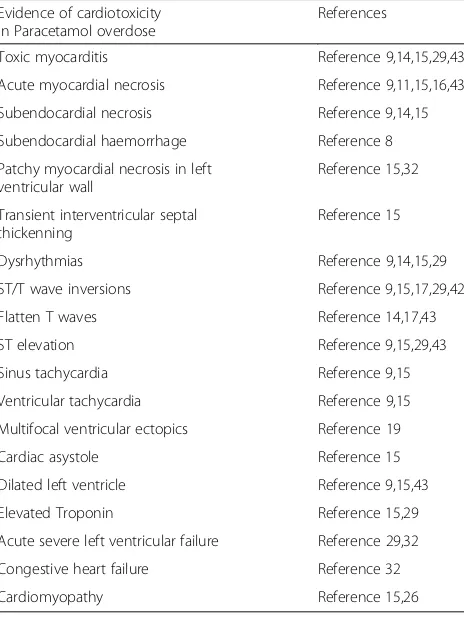
Though hepatotoxicity following paracetamol poison-ing is well studied data regardpoison-ing cardiotoxicity is very minimal. While general experience suggests that para-cetamol induced cardiotoxicity has rarely been clinically significant, it may have been overlooked. The possibility that an overdose of paracetamol might be directly cardi-otoxic has been the subject of a few reports [14–19]. (Table 1) However the true incidence is unknown as ser-ial electrocardiograph (ECG) monitoring is not part of routine clinical management. Moreover, anxieties have been expressed regarding unexplained deaths associated with paracetamol poisoning, with some occurring within 24 h of ingestion [13]. It cannot be ruled out that a number of these may have been cardiac in origin [9]. Dysrhythmias and other ECG abnormalities, especially of the ST segment or T wave, frequently occur in coma-tose encephalopathic patients [4, 12], but ST/T wave changes in non-encephalopathic subjects have been
reported [4, 9, 12–14]. Sudden unexplained death due to paracetamol overdose may occur early before acute hep-atic failure is established, and even in the complete ab-sence of histological evidence of hepatic necrosis [9, 14]. A direct toxic myocarditis has been postulated from aut-opsy evidence of myocardial necrosis being present par-ticularly in the sub endocardium [9, 14, 15]. Cases of Toxic myocarditis, acute myocardial necrosis following paracetamol poisoning have been reported [9, 11, 14, 15]. The cause of the ECG abnormalities in encephalopathic patients is probably multifactorial and at least partly re-lated to severe metabolic changes. However, these cannot be implicated in patients with less hepatic damage or in sudden death early in the course of paracetamol poisoning [4, 9]. Because of thisArmouret al. suggested an ECG to be taken on admission and checked daily if the overdose is significant [9] They also further stated that if ST/T wave abnormalities or a dysrhythmia are present, the treatment with a standard acetylcysteine infusion should be consid-ered, probably irrespective of the plasma concentration of paracetamol or time laps from ingestion [9]. Thum and Borlak showed the presence of metabolic enzyme(Cyto-chrome p450s) in the heart and suggested the possibility of metabolic activation of paracetamol [16]. Also literature review showed a very minimal amount of research had been carried out to determine paracetamol cardio toxicity which seems to have been overlooked.
The above evidence would justify paracetamol cardi-otoxicity being studied in detail amongst our patients. We carried out a study on patients presenting to the Toxicology Unit of the Teaching Hospital Peradeniya. We selected this unit of the Teaching Hospital Peradeniya as all acute poisoning patients are admitted here.
Methods
The objective of our study was to assess the demo-graphic and clinical patterns of paracetamol poisoning induced cardiotoxicity in the Sri Lankan population. The study population included all patients with a history of Paracetamol poisoning presenting to the toxicology unit of the Teaching Hospital Peradeniya over a period of one year. All patients who were clinically diagnosed to have paracetamol poisoning after detailed history at the time of presentation were included in this study. Those patients with multiple drug poisoning and those who re-fused to participate were excluded from the study. Ethical clearance was obtained from the Ethical Review Committee of the Faculty of Medicine, University of Peradeniya, and informed written consent was obtained from all subjects prior to data collection.
All patients presenting with paracetamol poisoning to the Toxicology Unit of the Teaching Hospital Peradeniya were assessed by detailed history and examination by in-vestigators recruited to the study. The patients who were
Table 1Evidence of cardiotoxicity in paracetamol overdose
Evidence of cardiotoxicity in Paracetamol overdose
References
Toxic myocarditis Reference 9,14,15,29,43
Acute myocardial necrosis Reference 9,11,15,16,43
Subendocardial necrosis Reference 9,14,15
Subendocardial haemorrhage Reference 8
Patchy myocardial necrosis in left ventricular wall
Reference 15,32
Transient interventricular septal thickenning
Reference 15
Dysrhythmias Reference 9,14,15,29
ST/T wave inversions Reference 9,15,17,29,42
Flatten T waves Reference 14,17,43
ST elevation Reference 9,15,29,43
Sinus tachycardia Reference 9,15
Ventricular tachycardia Reference 9,15
Multifocal ventricular ectopics Reference 19
Cardiac asystole Reference 15
Dilated left ventricle Reference 9,15,43
Elevated Troponin Reference 15,29
Acute severe left ventricular failure Reference 29,32
Congestive heart failure Reference 32
Cardiomyopathy Reference 15,26
confirmed to have a paracetamol poisoning were se-lected for the study. The toxic dose of paracetamol for adults is considered to be either a single dose of 7.5– 10 g, which is equivalent to 20 tablets of 500 mg each, or repeated sub-therapeutic doses exceeding 100 mg/kg/ day. As for healthy children aged between 1 and 6 years the toxic dose is considered to be between 150 and 200 mg/kg. This toxic dose is defined by the occurrence of hepatotoxicity which is the main toxic effect of para-cetamol poisoning, and the cardiotoxic dose has not yet been determined. All the subjects of this study were above the age of 15, therefore all subjects who had con-sumed more than 20 paracetamol tablets were consid-ered to have paracetamol poisoning.
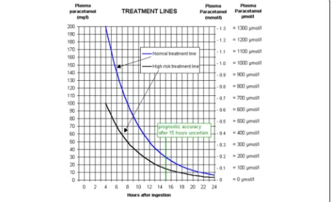
All the study subjects underwent routine hematological and biochemical investigations on admission, followed by 4 h blood paracetamol level. In case of unknown time of ingestion random blood paracetamol level was measured. Blood paracetamol concentration level against the time since ingestion was plotted on the Rumak-Mathew nor-mogram (Fig. 1) to assess paracetamol toxicity.
Liver functions and renal functions were assessed daily over the next few days. Daily 12 leads ECG was taken, and was interpreted by a Consultant Physician special-ized in Cardiology. Serum Troponin I level was mea-sured at 4 h after ingestion of paracetamol in patients who were found to have detectable serum paracetamol
levels. In addition basic demographic data was also ob-tained. Data was collected using a comprehensive data collection sheet and was analyzed using the SPSS (Statis-tical package for social sciences) 20.0 software.
Results
Basic socio-demographic data
Studied sample included 68 patients who are ingested 20 or more Paracetamol tablets, with a mean age of 21.38 +/-5.11 (range 15–36). This included 26(38.2 %) male patients and 42(61.8 %) females (Table 2) and reason for Paracetamol ingestion is given in Table 3. Clinical findings on admission were recorded and is shown in Table 4.
Out of 68 patients, 58(85.3 %) were Sinhalese, 9(13.2 %) were Muslims and 1(1.5 %) patient was Christian; 16(23.5 %) patients were regular smokers and 18(26.5 %) were regular alcohol users.
Among the Studied population, 44(64.7 %) only com-pleted their primary education (Ordinary level), 21(30.9 %) educated up to Advanced level and only 3(4.4 %) completed tertiary education.
Out of 68, 47(69.12 %) answered the source of para-cetamol as Home and 21(30.88 %) patients were taken Paracetamol over the counter.
All the patients had ingested paracetamol tablets and the mean number of paracetamol tablets was 37+/- 18.36 (Range20–96) (19 g); 2(2.9 %) of them ingested alcohol
with paracetamol and 3(4.4 %) ingested cholrophenira-mine with paracetamol.
Of the 68 subject, 27 (39.70 %) had detectable paracta-mol levels in serum, and 21 (30.88 %) had Alanine Transaminase levels (ALT)≥1000 IU/L.
Out of studied sample (68), 9 (13.2 %) had a past his-tory of Depression, 11(16.2 %) had a chronic illness, and only 10(14.7 %) had a history of previous suicidal at-tempts. Eight(11.8 %) had family history of suicide and 6(9 %) had family history of depression.
ECG and Echocardiography were done in all patients. Approximately, 20.59 % had bradycardia while 2.94 % had tachycardia. Majority(76.47 %) had normal heart rate. Fourteen patients(20.59 %) had first degree heart block. The duration of QRS was normal in all the patients. Small Q waves were detected in 13.24 % patients and they were not pathological. ST segment depression was observed in 2.94 % patients and was less than 1 mm and non signifi-cant.10.29 % patients had T wave inversions in inferior leads and were considered normal (Table 5). Standard treatment was given to all patients on admission and dur-ing the stay in the hospital (Table 6).
Echocardiogram was done on all the patients and did not reveal any significant abnormality. All of them had normal ejection fraction and there were no any regional wall motion abnormalities. There were no hypokinetic or akinetic segments.
Cardiac Biomarker Troponin I was done only in patients who had detectable serum paracetamol levels (27 Patients). Surprisingly Troponin I was negative in all of them.
The average time taken for hospital admission follow-ing paracetamol follow-ingestion was 3.25 ± 2.27 h.
Discussion
Paracetamol is the most commonly encountered drug leading to toxicity due to overdose and can lead to severe complications and even result in death. The commonest organ system involved in toxicity of paracetamol is the liver.(H,I) A few previous studies have reported cases of
paracetamol toxicity affecting organs other than the liver, including the heart, which was assessed in detail by this study [10, 15, 17–19].
Currently there are a few hypothesized mechanisms for the occurrence of the previous reported cases of car-diotoxicity due to paracetamol overdose, which could ei-ther be a direct or indirect effect. A few studies have shown the Acetaminophen moiety to have a direct toxic effect on the heart [9, 14, 20–23]. One hypothesis for the direct toxic effect on the heart suggests that myocar-dial injury occurs due to a similar mechanism which causes hepatic damage, with paracetamol partly con-verted to a toxic metabolite, N-acetyl-p-benzoquinoni-mine, which is normally inactivated by reduction with glutathione and acts as a direct toxin on the myocar-dium [9]. Paracetamol has shown to bind covalently to proteins in both the liver and cardiac tissue which re-sults in the alteration of protein structure and function, potentially precipitating cytokine release and tissue in-jury [22, 23]. Another hypothesis is one resulting in is-chemia due to paracetamol, which can be due to the depletion of sulfydryl groups. Acetaminophen can de-plete sulfydryl groups, which interferes with nitric oxide production, which results in coronary ischaemia [9, 20]. This sulfydryl deficit also may interfere with endothe-lium derived vascular relaxing factor, which leads to functional coronary ischemia [9, 20].
The most commonly postulated hypothesis for the mechanism for cardiotoxicty in paracetamol poisoning is metabolic derangements such as hyperkalemia, meta-bolic acidosis and increase in serum fatty acid levels, resulting by hepatic failure, which could lead to arryth-mias [9, 12]. Previously reported cases of paracetamol poisoning with cardiac tissue damage has almost always been associated with hepatotoxicity. Lesna et al. stated that there have been no cases of cardiac arrest so far in the absence of hepatotoxicity, which further justifies the hypothesis which states that cardiac tissue damage in paracetamol poisoning occurs following metabolic de-rangements due to hepatotoxicity [18, 24–27]. Similarly, in this study 30.88 % had ALT≥1000 IU/L indicating hepatic tissue injury, but all had negative Troponin I,
Table 2Age and sex Distribution
Age Group Number Total = 68 Sex
Male(n 26) Female(n 42)
<18 22(32.4 %) 8 14
18-24 31(45.6 %) 11 20
>24 15(22.0 %) 7 8
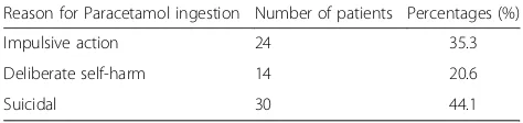
Table 3Reason for Paracetamol ingestion
Reason for Paracetamol ingestion Number of patients Percentages (%)
Impulsive action 24 35.3
Deliberate self-harm 14 20.6
Suicidal 30 44.1
Table 4Clinical data on admission
Clinical findings Number of patients Percentages %
Nausea 55 80.9
Vomiting 48 70.6
Chest Pain 20 29.4
RUQ pain 15 22.1
Jaundice 2 2.9
Confusion 1 1.5
Easy Bleeding 0 0
Table 5Electrocardiogram findings of the sample
Heart Rate PR interval(Sec) QRS duration(Sec) Q wave ST depresion T inversion
<60 60-100 >100 <0.12 0.12-0.20 >0.2 <0.12 >0.12 Present Absent Present Absent Present Absent
Number 14(20.59 %) 52(76.47 %) 2(2.94 %) 0 54(79.41 %) 14(20.59 %) 68(100 %) 0 9(13.24 %) 59(86.76 %) 2(2.94 %) 66(97.09 %) 7(10.29 %) 61(89.71 %)
et
al.
BMC
Pharmaco
logy
and
Toxicology
(2016) 17:30
Page
5
of
echocardiogram and ECG, indicating the absence of car-diac tissue injury in any of the subjects. A case of myo-cardial damage following acute paracetamol poisoning by Ohtani et al. reported heart failure to occur 2 weeks following hepatic injury, and had return of hepatic func-tion tests to normal and had no elevated levels of serum paracetamol by that time, which shows that though there may be no features of cardiac tissue injury or im-pairment at the time there is hepatic damage, but car-diac toxicity could occur at a later stage even in the absence of features of hepatic tissue injury at that stage [28–32]. Mi Jin et al. in their experiment have demon-strated that paracetamol induced cytotoxicity and altered gene expression in cultures cardiomyocytes of H9C2 cells [23]. This study was plan to identify the alterations in gene expression in cultured cardiomyocytes;H9C2 cells, which are rat cardiomyocytes, were treated with paracetamol, to evaluate the possible toxic mechanism in the heart. During this study authors have demon-strated some of the DNA damage and upregulation of genes including those associated with oxidative stress, DNA damage and apoptosis [23]. During this study to examine toxic effects of paracetamol,H9C2 rat cardio-myocytes were incubated with different concentratins of paracetamol and viability was determined at different time intervals and showed H9C2 cells viability was de-creased by treatment with paracetamol in a time- and dose-dependant manner [16]. Another laboratory study done byInoue et al. to study cardiotoxicity and hepato-toxicity of different compounds using neonatal rat heart cells and rat hepatocytes confirmed that paracetamol was cardiotoxic in significant overdose [33].
Previous studies have shown that cardiotoxicity follow-ing paracetamol poisonfollow-ing may be either immediate or delayed [20, 21], similar to the case reported byOhtani et al. which showed a delay in onset of cardiac involve-ment [28]. The average time taken for hospital admis-sion following paracetamol ingestion in this study was 3.25 ± 2.27 h. Probably if these patients were followed up weeks later, some of them may have developed cardiac toxicity and showed cardiac tissue damage by elevated troponin I, or positive echocardiogram or ECG changes. Since the cardiotoxicity due to paracetamol poisoning is believed to be either immediate or delayed, it is of paramount importance to administer N-acetylcysteine
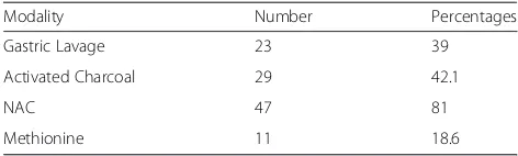
(NAC) as early as possible because it may be beneficial in reducing the functional coronary ischaemia as well as the direct toxic effects, irrespective of the time of ingestion [34–40]. Since a majority of 81 % of subjects were treated with NAC, and since the time taken for hospital admission following paracetamol ingestion was short, effective treat-ment with NAC would’ve been protective against the devel-opment of cardiotoxicity in this sample. Gastric aspiration or lavage should be carried out, or emesis induced within 4 h of ingestion of 100 mg/kg of paracetamol to reduce absorption, and this has been carried out in 39 % of patients. Activated charcoal and cholestyramine reduce absorption of paracetamol if given within 1 h of ingestion [24–26]. 42.1 % of the patients underwent treatment with charcoal, but since it was done after 1 h of ingestion, the protective effect from this measure would have been min-imal or absent. However, other effective measures for the treatment of paracetamol poisoning may be the reason for cardiotoxicity not to develop in these patients.
In contrast to the believed toxic potential of paraceta-mol on the heart, literature has also demonstrated para-cetamol to have a cardioprotective potential in ischaemia reperfusion syndrome. At concentrations of 45–50 mg/ml, well below the clinically toxic dose of 300 mg/ml, para-cetamol can reduce tissue injury by preventing the open-ing of the mitochondrial permeability transition pore and reducing apoptosis [27, 28]. This dose dependent cardio-protective effect has a different mechanism from that of the cardiotoxic effect [41, 42]. However, the mean amount of paracetamol ingested by this study sample was 19 g, ac-counting to about 315 mg/kg, indicating that this cardio-protective effect of paracetamol is quite unlikely in this sample according to the facts stated in literature.
In patients with severe paracetamol poisoning, the ECG often reveals minor nonspecific ST changes and T wave flattening with U waves during the first 48 h, which is likely to be related to hyperkalaemia, but not due to direct cardio-toxicity [29]. Nonspecific ECG changes and elevated serum creatine kinase has been reported without clinical evidence of toxic myocarditis [30, 31]. A post mortem analysis of 20 fatal cases of paracetamol poisoning showed that there was no evidence of cardiomyopathy. Some degree of focal nec-rotizing myopathy has been found to occur in patients with fatal massive hepatic necrosis of diverse etiology [32, 33]. Serious cardiovascular complications in paracetamol over-dose are not heard of in the absence of hepatic failure, and cardiac abnormalities reported in certain fatal cases are due to the profound metabolic disturbances of acute liver failure rather than paracetamol itself.
In contrast, there have been a few very rare cases of paracetamol overdose reported with abnormal ECG pat-terns in the absence of hepatotoxicity, metabolic de-rangement or encephalopathy [32, 43]. A similar case of a 29 year old man with significant paracetamol overdose
Table 6Treatment modalities
Modality Number Percentages
Gastric Lavage 23 39
Activated Charcoal 29 42.1
NAC 47 81
Methionine 11 18.6
was found to have an abnormal ECG in the absence of hepatic encephalopathy [9].
Approximately 35.3 % ingested paracetamol overdose by impulsive action, 20.6 % for deliberate self-harm, and 44.1 % for suicide. 11.8 % had a family history of suicide and 9 % had a family history of depression. Considering these facts, there is also a possibility that a certain num-ber of patients may have mentioned an exaggerated number of paracetamol tablets ingested, and some may also not be very sure of the time and details of ingestion of the overdose. There is also the possibility that para-cetamol tablets may have a more diluted concentration of the drug in its composition with a hindrance of qual-ity, resulting in a lower level of serum paracetamol than expected compared to the number of tablets ingested.
Conclusion
Cardiotoxicity is not a common presentation in para-cetamol poisoning. It further justifies certain previous studies which state that if at all any cardiac tissue dam-age only occurs due to metabolic derangements follow-ing hepatotoxicity.
Abbreviations
ALT, Alanine Transaminase levels; ECG, Electrocardiograph; NAC, N-acetylcysteine
Acknowledgements
We all express our gratitude to all participants who consented to take part in this study.
Funding
Faculty of Medicine, University of Peradeniya has made this study possible through Earned Funds Research Grant (No: RG/EF/2013/12) offered to UR.
Availability of data and materials
The raw datasets supporting this article can be made available by emailing the corresponding author.
Authors’contributions
UR conceived the research idea and guided it. Data collection, analysis and interpretation of data and literature review were done by UR, KPJ, and EMME.UR and AMSDMD guided the other authors in data analysis, interpretation and corrected the final manuscript. All authors were involved in the study and read and approved the final manuscript.
Authors’information
UR [MBBS, MD, MRCP (UK), FRCP (London)] is a Senior Lecturer and Consultant Physician, Department of Medicine, University of Peradeniya. KPJ [MBBS] and EMME [MBBS] are Temporary Lecturers and Research Assistants, Department of Medicine, University of Peradeniya, Sri Lanka. AMSDMD [MBBS, D.Path (Colombo), MD-Heamatology (Colombo), PhD (London)] is a Professor, Department of Pathology, Faculty of Medicine, University of Peradeniya.
Competing interests
The authors declare that they have no competing interests.
Consent for publication
Not applicable.
Ethics approval and consent to participate
Ethical approval for the research work has been obtained from the Ethical review committee, Faculty of Medicine, University of Peradeniya, Sri Lanka. All the data were collected from the participants after obtaining written consent.
Author details
1Department of Medicine, University of Peradeniya, Peradeniya, Sri Lanka. 2
Department of Pathology, Faculty of Medicine, University of Peradeniya, Peradeniya, Sri Lanka.
Received: 17 November 2015 Accepted: 1 July 2016
References
1. Gunnell D, Fernando R, Hewagama M, Priyangika WDD, Konradsen F, Eddleston M. The impact of pesticide regulations on suicide in Sri Lanka. Int J Epidemiol. 2007;36(6):1235–42.
2. Hanwella R, Senanayake SM, de Silva VA. Geographical variation in admissions due to poisoning in Sri Lanka: a time series analysis. Ceylon Med J. 2012;57:152–8.
3. Hawton K, Ware C, Mistry H, et al. Why patients choose paracetamol for self poisoning and their knowledge of its dangers. BMJ : British Medical Journal. 1995;310(6973):164.
4. Galappatthy P, Dawson A, Fernando R. Management of paracetamolovedose. Sri Lanka Prescriber. 2006;14(4):1–4.
5. Rajapakse T, Griffiths KM, Christensen H. Characteristics of non-fatal self-poisoning in Sri Lanka:a systematic review. BMC Public Health. 2013;13:331. pages 1-15.
6. Eddleston M, Karunaratne A, Weerakoon M, Kumarasinghe S, Rezvi Sheriff MH, Gunnell D. Choce of poison for intentional self-poisoning in rural Sri Lanka. Clin Toxicol(Phila). 2006;44(3):283–6.
7. Morgan O, Griffiths C, Majeed A. Impact of paracetamol pack size restrictions on poisoning from paracetamol in England and Wales: An observational study. J Public Health. 2005;27:19–24.
8. Routledge P, Allister Vale J, Nicholas Bateman D, et al. BMJ. 1998;317(7173): 1609–10.
9. Armour A, Slater SD. Paracetamolcardiotoxicity. Postgrad Med J. 1993;69:52–4. 10. Isbister GK, Downes F, Whyte M. Regular medication and paracetamol
overdose. Aliment Pharmac of Ther. 2003;17(4):609–10.
11. Tenenbein M. Acetamenophen:the 150mg/kg myth. A Toxicol Clin Toxicol. 2004;42(2):145–48.
12. Anderson BD, Shepherd JG, Klein-Schwartz W. Outcome of acetaminophen overdose. J Pediatr. 1998;132(6):1080.
13. Hoffman C, Gibel L. Acetomenophen overdose. Nursing. 2005;35(1):88. 14. Will EJ, Tomkins AM. Acute myocardial necrosis in paracetamol poisoning.
Br Med J. 1971;4:430–1. letter.
15. Wakeel RA, Davies HT, Williams JD. Toxic myocarditis in paracetamol poisoning. Br Med J. 1987;295:1097.
16. Weston MJ, Talbot IC, Howorth PJN, Mant AK, Capildeo R, Williams R. Frequency of arrhythmias and other cardiac abnormalities in fulminant hepatic failure. Br Heart J. 1976;38:1179–88.
17. Mehrpour O, Afshari R, Delshard P, Jalazaden M, Khodashenas M. Cardiotoxicity due to paracetamol overdose-A case study and review of the literature. Indian Journal of Forensic Medicine and Toxicology. 2011;5(2):31. 18. Smilkstein MJ. APAP-induced heart injury?may be yes.may be no.next
question? J Toxicol, Clin Toxicol. 1996;34(2):155–6.
19. Brent J. New ways of looking at an old molecule. J Toxicol, Clin Toxicol. 1996;34(2):149–53.
20. Lim AY, Segarra I, Chakravarthi S, Akram S, Judson JP. Histology and biochemistry analysis of the interaction between sunitinib and paracetamol in mice. BMC Pharmacol. 2010;10:14. doi:10.1186/1471-2219-10-14. 21. Jacob S, Cherian P, Preusz C, Kovacs R. Heartbreaking case of
Acetaminophen poisoning. Circulation. 2008;118:e514–23.
22. Sanerkin NG. Acute myocardial necrosis in par-acetamol poisoning. Br Med J. 1971;iii:478.
23. Jin SM, Park K. Acetaminophen induced cytotoxicity and altered gene expression in cultured cardiomyocytes of H9C2 cells. Environmental Health and Toxicology. 2012;27:e2012011.
25. Hamwi I, Picksak G, Stichtenoth DO. Accidental acetaminophen overdose. Med Monatsschr Pharm. 2008;31(6):224–5.
26. Pimstone BL, Uys CJ. Liver necrosis and myocardiopathy following paracetamoloverdosage. S Afr Med J. 1968;42(11):259–62.
27. Lesna M, Watson AJ, Douglas A, Hamlyn AN, James O, Wardle EN, Chenery R, Fisher C, McLean AEM. Toxicity of paracetamol. The Lancet 307(7952):191 28. Ohtani N, Matsuzaki M, Anno Y, Ogawa H, Matsuda Y, Kusukawa R. A case of
myocardial damage following acute paracetamol poisoning. Japanese Ciculation Journal. 1989;53:278–82.
29. Jones AL, Prescott LF. Unusual complications of paracetamol poisoning. Q J Med. 1997;90:161–8.
30. Lip GYH, Vale JA. Does acetaminophen damage the heart? Clin Toxicol. 1996;34:145–7.
31. Lacour S, Gautier J-C, Pallardy M, Roberts R. Cytokines as potential biomarkers of liver toxicity. Cancer Biomark. 2005;1:2939.
32. Weston MJ, Williams R. Letter:Paracetamol and the heart. Lancet. 1976; 1(7958):536.
33. Inoue T, Tanaka K, Mishima M, Watanabe K. Predictivein vitrocardiotoxicity and hepatotoxicity screening system using neonatal rat heart cells and rat hepatocytes. AATEX. 2008;14:457–62.
34. Dordoni B, Willson RA, Thompson RPH, Williams R. Reduction of absorption of paracetamol by activated charcoal and cholestyramine: a possible therapeutic measure. Br Med J. 1973;3:86–7.
35. Levy G, Houston JB. Effect of activated charcoal on acetaminophen absorption. Pediatrics. 1976;58:432–5.
36. Prescott LF, Critchley JAJH, Balali-Mood M, Pentland B. Effects of microsomal enzyme induction on paracetamol metabolism in man. Br J Clin Pharmacol. 1981;12:149–53.
37. Hamlyn AN, Douglas AP, James O. The spectrum of paracetamol (acetaminophen) overdose: clinical and epidemiological studies. Postgrad Med J. 1978;54:400–4.
38. Maclean D, Peters TJ, Brown RAG, McCathie M, Baines GF, Robertson PGE. Treatment of acute paracetamol poisoning. Lancet. 1968;2:849–52. 39. Dixon MF. Paracetamol hepatotoxicity. Lancet. 1976;I:35.
40. Ojeda VJ, Shilkin KB, Wright EA, Williams R. Massive hepatic necrosis and focal necrotising myopathy. Lancet. 1982;I:172–3.
41. Hadzimichalis NM, Baliga SS, Golfetti R, Jaques KM, Firestein BL, Merrill GF. Acetaminophen-mediated cardioprotection via inhibition of the mitochondrial permeability transition poreinduced apoptotic pathway. Am J Physiol Heart Circ Physiol. 2007;293:H3348–55.
42. Merrill G, McConnell P, Vandyke K, Powell S. Coronary and myocardial effects of acetaminophen: Protection during ischemiareperfusion. Am J Physiol Heart Circ Physiol. 2001;280:H2631–8.
43. Contractor H, Gauge V, Nabi S, Titu H, Arya S, Naqvi N. ST segment elevation secondary to paracetamol overdose. Ther Adv Cardiovasc Dis. 2011;5(1):8385.
• We accept pre-submission inquiries
• Our selector tool helps you to find the most relevant journal
• We provide round the clock customer support
• Convenient online submission
• Thorough peer review
• Inclusion in PubMed and all major indexing services
• Maximum visibility for your research
Submit your manuscript at www.biomedcentral.com/submit