O R I G I N A L A R T I C L E
Open Access
Effect of simultaneous exposure to
extremely short pulses of blue and green
light on human pupillary constriction
Soomin Lee
1*, Shougo Ishibashi
2,3, Yoshihiro Shimomura
2and Tetsuo Katsuura
2Keywords:Blue light, LED-pulsed light, Pupillary constriction, Subadditive response
Abbreviations:NIF, Non-image-forming; ipRGC, Intrinsically photosensitive retinal ganglion cell; LED, Light-emitting diode; KSS, Kwansei Gakuin Sleepiness Scale; VAS, Visual analog scale; PD, Pupil diameter; ANOVA, Analysis of variance
Background
Light has various influences on all species, including humans. In natural environments, the only light source is sunlight. Humans have been evolving and adapting under such natural light environments. In modern soci-ety, illumination in the workplace has a great influence on work efficiency and the health of workers [1]. The ef-fects of illumination are classified as visual efef-fects and non-visual or non-image-forming (NIF) effects. Recently, a number of studies in the field of physiological anthro-pology have focused on the NIF effects of illumination on humans [2–8].
In 2002, melanopsin-containing intrinsically photo-sensitive retinal ganglion cells (ipRGCs), a novel type of photoreceptor cells, were found in the mammalian retina [9, 10]. It was confirmed that ipRGCs respond to short-wavelength (blue) light of around 480 nm [9, 11, 12]. The ipRGCs in the retina of the eye affect the interlamellar nuclei of the lateral geniculate nucleus, suprachiasmatic nucleus, intergeniculate leaflet, olivary pretectal nucleus, and ventrolateral preoptic nucleus [10, 13–15] and act as the primary photoreceptors for NIF functions such as melatonin suppression [3, 6, 14, 16–18] and pupillary constriction [5, 7, 8, 14, 18–28].
Recently, it was pointed that the input from cones and rods could potentially affect the ipRGC response [11, 14, 18, 21, 29]. Most vertebrates, including fishes,
amphibians, reptiles, and birds, have three or four types of cones and trichromatic or tetrachromatic color vision. However, in the history of evolution, mammals lost a portion of these cones and have di-chromatic color vision. Some primates (catarrhines) acquired a third cone and have trichromatic color vision. Humans have three types of cones (S-cones, M-cones, and L-cones) and have trichromatic color vi-sion [30, 31], which is rare in mammals. Figueiro et al. [29] studied the effects of blue (450 nm, 7.7 μW/cm2) and green (525 nm, 21.1μW/cm2) light on melatonin suppression at night. They found that simultaneous exposure to blue and green light resulted in less mela-tonin suppression than monochromatic exposure to blue or green light. This effect is called the subadditive response to light [29]. Figueiro et al. [32, 33] and Revell et al. [34] also identified the subadditive effects of monochromatic and polychromatic light on mela-tonin suppression, suggesting that cones affected the ipRGC response. However, it remains unclear whether the subadditive response affects pupillary constriction.
The response of mouse ipRGCs to a single photon was examined, and it became clear that ipRGCs have an exceptionally large and prolonged response in com-parison with rods and cones [12]. However, ipRGCs are far less sensitive than rods and cones to light intensity [19, 21, 23, 35], so we hypothesized that exposure to high irradiance pulsed light might produce higher NIF function. Therefore, in the present study, we examined the effects of separate and simultaneous exposure to ex-tremely short pulses of blue and green light at different
* Correspondence:[email protected]
1Center for Environment, Health and Field Sciences, Chiba University, 6-2-1,
Kashiwanoha, Kashiwa 277-0882, Japan
Full list of author information is available at the end of the article
irradiance levels on pupillary constriction and sought to confirm the subadditive response to light.
Methods
Eleven healthy young Japanese males (mean ± standard deviation age 23 ± 0.9 years, body height 172.7 ± 6.7 cm, body mass 66.2 ± 9.7 kg) with dark eyes participated in this study. They were screened for normal color vision using the Farnsworth-Munsell 100 hue color vision test. Each subject gave his informed consent to participate in the study. The Ethics Committee of the Graduate School of Engineering at Chiba University approved the proto-col for the study (#24-25).
The experiment was conducted in a lighting laboratory controlled at a temperature of 26 ± 0.5 °C and relative humidity of 50 ± 5 %. Each subject sat in a chair with his head facing a diffusion panel, which was located in front of an integrating sphere. Light-emitting diodes (LEDs) were arrayed in the integrating sphere. The spectral ir-radiance of blue and green LEDs was measured at each subject's eye level using a spectroradiometer (HSR-8100, MAKI Manufacturing, Co. Ltd., Hamamatsu, Japan). The peak wavelength of blue light was 470 nm and that of green light was 532 nm (Fig. 1). Each subject was ex-posed to nine different light conditions, i.e., a pulse of blue and/or green light of 10, 15, and 20 μW/cm2, simultaneously or separately (Table 1). The melanopsin-stimulating irradiance and photon density at the sub-ject’s retinal level were estimated for each light condition [35] based on the spectral absorption of the crystalline lens [36] and a template [37] indicating the spectral ab-sorption characteristics of photopigment with a peak wavelength of 484 nm [9].
After 45 min of dark adaptation (<0.5 lx), the subject was exposed to three pulses of lights with a 1-ms-pulse width in a square waveform every 1 min in each of the
nine light conditions. Each subject took a 10-min rest between exposure to each light condition (Fig. 2). The experiments were carried out at 9 a.m. to midday or at 1 p.m. to 4 p.m.. The order of the nine light conditions was counterbalanced among the subjects.
We measured the pupil diameter (EMR-8B, NAC Image Technology Inc., Tokyo, Japan) in the subject’s left eye and used the Kwansei Gakuin Sleepiness Scale (KSS) and the visual analog scale (VAS) for subjective evaluation of “sleepiness.” Pupil diameter measure-ments during 10 s before and 10 s after exposure to three pulses of light were averaged for each subject and under each light condition. The mean value for the averaged pupil diameter in the 10 s before expos-ure to the pulses of light was defined as the baseline value. From the averaged pupil diameter (PD) meas-urement, we calculated the percent pupil constriction and recovery time (Fig. 3) as follows:
Percent pupil constriction ¼ ½ðbaseline PD–minimum PD after exposure to pulsed lightÞ= baseline PDÞ 100
Recovery time: time (s) until recovery to 15 % pupil constriction after exposure to pulsed light.
KSS and VAS for “sleepiness” were evaluated by the change from the value before exposure to each pulse of light to the value after exposure.
Two-way repeated measures analysis of variance (ANOVA) was applied to clarify the effects of irradiance intensity (10, 15, and 20μW/cm2) and wavelength (blue, green, and simultaneous blue and green). In the case of a significant interaction, one-way repeated measures ANOVA was applied to evaluate the effects of wave-length under each irradiance condition on these mea-surements. When any significant main effect was found, multiple comparisons of the light condition were per-formed using the Bonferroni procedure. The data were analyzed using SPSS version 23.0 software (IBM Corp., Armonk, NY, USA). The level of statistical significance was set at 0.05.
Results
Percent pupil constriction
Pupillary constriction was observed during exposure to pulsed light under all light conditions. Table 2 shows the results for percent pupil constriction during the nine light conditions. Two-way repeated measures ANOVA revealed that the main effects of irradiance and wave-length on percent pupil constriction were significant (F(2, 20) = 14.78, p= 0.000 and F(2, 20) = 10.79, p= 0.001, respectively). However, the interaction effect of ir-radiance and wavelength on percent pupil constriction was not significant (F(4, 40) = 2.19,p= 0.087).
Fig. 1Spectral irradiance of light. Spectral irradiance of blue and green light-emitting diodes and blue plus green light of 15μW/ cm2. The light of 10 and 20μW/cm2irradiance had the same
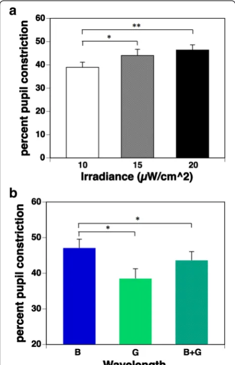
Figure 4a shows the percent pupil constriction under three irradiance conditions. Multiple comparisons using the Bonferroni procedure found that 15 and 20μW/cm2 irradiance conditions resulted in significantly more pro-nounced pupillary constriction than the 10 μW/cm2 condition (p= 0.014 and 0.005, respectively). However, there was no significant difference between 15 and 20μW/cm2irradiance conditions (p= 0.106).
The percent pupil constriction under the three wave-length conditions is shown in Fig. 4b. Multiple compari-sons found that the percent pupil constriction during exposure to a pulse of blue light (B) was significantly (p= 0.010) more pronounced than during exposure to a pulse of green light (G). Interestingly, the percent pupil constriction during simultaneous exposure to blue and green (B + G) light was significantly (p= 0.031) more inhibited than during exposure to a pulse of blue light (B), despite the double irradiance intensity and the 1.5× melanopsin-stimulating photon density, as shown in Table 1.
Recovery time
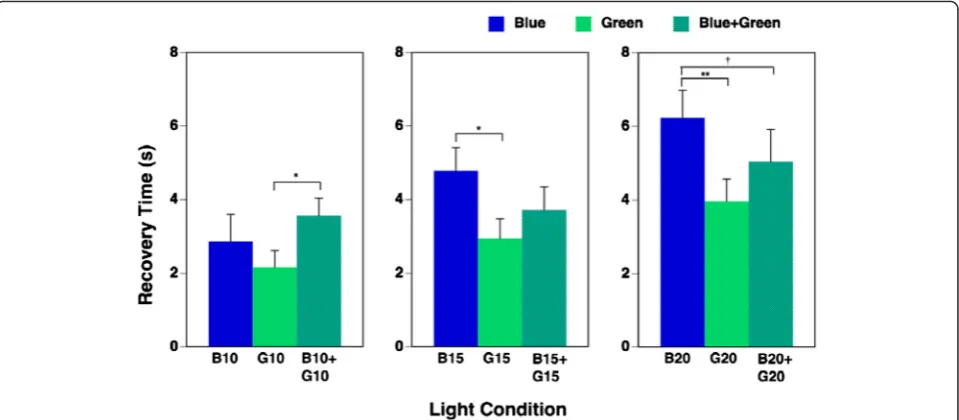
The results for time taken for pupillary constriction to recover upon exposure to the nine light conditions are shown in Table 3. Two-way repeated measures ANOVA determined that the main effect of irradiance on time taken to recovery was significant (F(2, 20) = 19.08, p= 0.000). Higher irradiance resulted in a longer recovery
time. The main effect of wavelength was also significant (F(2, 20) = 8.42, p= 0.002). The interaction of irradiance and wavelength was significant (F(4, 40) = 3.59, p= 0.014), so one-way repeated measures ANOVA was applied to evaluate the effects of wavelength under each irradiance condition on the recovery time. We found that the main effects of wavelength on recovery time under each 10, 15, and 20μW/cm2irradiance condition were all significant (10 μW/cm2: F(2, 20) = 3.81, p= 0.040; 15 μW/cm2: F(2, 20) = 5.46, p= 0.013; 20 μW/ cm2: F(2, 20) = 10.72, p= 0.001, respectively). Multiple comparisons showed that recovery of pupillary constric-tion upon exposure to the highest irradiance of simul-taneous exposure to blue and green (B20 + G20) had a tendency (p= 0.073) to be longer on exposure to blue light (B20) despite the double irradiance intensity of the combination, as shown in Fig. 5.
There was no significant main effect of light condition on subjective evaluations using KSS and VAS scores for “sleepiness.”
Discussion
We found pupillary constriction under all light condi-tions used in the present study. It has been suggested that pupillary constriction is controlled mainly by rods under lower irradiance light exposure and by ipRGCs under higher irradiance light exposure [14, 21, 23, 35]. The ipRGCs have been reported to contribute to the
Fig. 2Experimental procedure
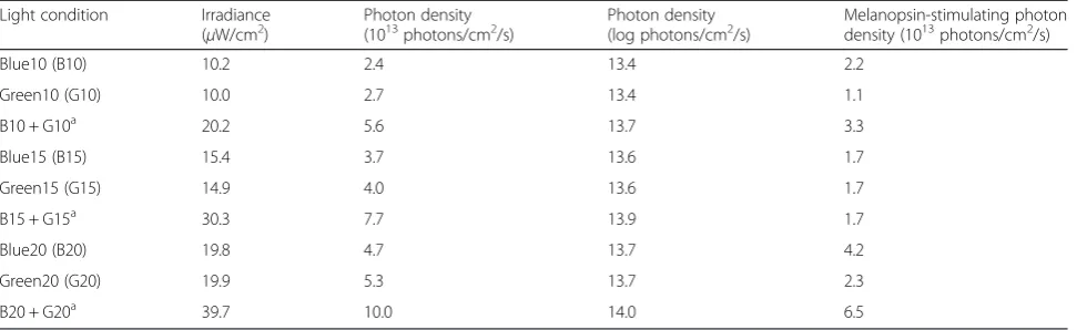
Table 1Characteristics of the light conditions
Light condition Irradiance
(μW/cm2) Photon density(1013photons/cm2/s) Photon density(log photons/cm2/s) Melanopsin-stimulating photondensity (1013photons/cm2/s)
Blue10 (B10) 10.2 2.4 13.4 2.2 Green10 (G10) 10.0 2.7 13.4 1.1 B10 + G10a 20.2 5.6 13.7 3.3
Blue15 (B15) 15.4 3.7 13.6 1.7 Green15 (G15) 14.9 4.0 13.6 1.7 B15 + G15a 30.3 7.7 13.9 1.7
Blue20 (B20) 19.8 4.7 13.7 4.2 Green20 (G20) 19.9 5.3 13.7 2.3 B20 + G20a 39.7 10.0 14.0 6.5 B + Ga
pupillary response to light in mice at an irradiance level greater than about 13 log photons/cm2/s of 470 nm light at the eye level [23]. It was also reported that the thresh-old retinal irradiance for depolarization of ipRGCs in rats was about 12.7 log photons/cm2/s of 500 nm light [9]. In the present study, the lowest irradiance intensity of blue (B10) was 13.4 log photons/cm2/s, which was higher than the threshold intensity for the activation of ipRGC. The lowest irradiance intensity of green (G10) was 13.4 log photons/cm2/s; however, the melanopsin-stimulating photon density of G10 was 1.1 photons/cm2/ s, which was half the value of B10. From the standpoint of the melanopsin-stimulating effect, G10 corresponded to about 5 μW/cm2or 13.1 log photons/cm2/s of blue light. This value was still higher than the threshold intensity of ipRGC activation.
In the present study, we used extremely short pulses of monochromatic light with a pulse width of 1 ms. There has been very little research on NIF function using such short pulses of light. We previously con-ducted an experiment with pulsed blue light (irradiance 11.2 μW/cm2, pulse width 100μs) and continuous blue light (irradiance 1.4 μW/cm2), which had the same multiplication value for irradiance and duration, and found that pupillary constriction was significantly
greater under the extremely short pulsed light condition than under the continuous light condition [5]. Recently, Vartanian et al. [25] studied pupillary constriction using flickering light stimuli under combined conditions with seven flicker frequencies (0.1, 0.25, 0.5, 1, 2, 4, and 7 Hz), three total photons (13.7, 14.7, and 15.7 log pho-tons/cm2), and three duty cycles (12, 47, and 93 %). They found the greatest pupillary constriction was evoked by the stimuli of flickering at 2 Hz with a 12 % duty cycle and 13.7 log photons/cm2 conditions, which was 71 % greater than that evoked by equal-intensity (12.3 log pho-tons/cm2/s) continuous light. This frequency and duty cycle were also optimal for 14.7 log photons/cm2stimuli, which was 38 % greater than that evoked by equal-intensity constant light. The pulse width of the stimuli at 2 Hz with a 12 % duty cycle is 60 ms. Although the pulse width in the study by Vartanian et al. [25] was much longer than that used in the present study, their results suggest that pulsed light has a greater influence on pupillary constriction.
We also found that pupillary constriction during ex-posure to pulsed blue light was significantly greater than during exposure to pulsed green light. These results are in accordance with those of a previous study [27] com-paring pupillary constriction during 45 s of exposure to
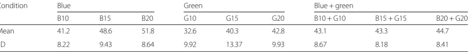
Table 2Percent pupil constriction during the nine light conditions
Condition Blue Green Blue + green
B10 B15 B20 G10 G15 G20 B10 + G10 B15 + G15 B20 + G20 Mean 41.2 48.6 51.8 32.6 40.3 42.8 43.1 43.3 44.7 SD 8.22 9.43 8.64 9.92 13.37 9.93 8.67 8.18 8.41
Percent pupil constriction was calculated by the following equation: [(baseline PD−minimum PD after exposure to pulsed light) / baseline PD] × 100 SDstandard deviation
continuous blue and green monochromatic light at three irradiance intensities after 15 min of dark adaptation. The greater pupillary constriction during exposure to blue light might be involved in the higher melanopsin-stimulating photon density of blue light.
In the present study, the most important finding was that pupillary constriction during simultaneous exposure to blue and green light was significantly decreased when compared with separate exposure to blue light, despite the double irradiance intensity and a 1.5× melanopsin-stimulating photon density. Recovery of pupillary con-striction during 20μW/cm2of simultaneous exposure to blue and green light (B20 + G20) also had a tendency to be more rapid than during separate exposure to blue light (B20). These findings indicate that the effect of blue light on ipRGCs is inhibited by simultaneous exposure to green light.
It has been reported that ipRGCs receive synaptic in-put from rods and cones [11, 14, 18, 21, 29]. Information on light wavelength relayed by the rods and the three types of cones is processed by bipolar cells, horizontal cells, and amacrine cells in the retina [38]. It is well known that the human visual system segregates cone responses into color information processed by two chan-nels [32], one class of bipolar cells forms the red versus green (r/g) channel with opposing input from L-cones and M-cones, and the other class forms the blue ver-sus yellow (b/y) channel from S-cones opposed to the combined input from the L-cones and M-cones and brightness information in the ganglion cells; and this information is sent to the visual cortex via the optic nerve [29]. In humans, spectral opponent blue versus yellow (b/y) bipolar cells have been hypothesized to provide direct input to the ipRGCs [29]. In fact, the ipRGCs in the in vitro primate retina show an unusual “color-opponent” receptive field in which an S-cone-mediated OFF response is antagonistic to an (L + M)-cone-mediated ON response on electrophysiological recordings [11], and an S-cone-mediated ON response is opposed to an (L + M)-cone-mediated OFF response in a similar way [39]. Thus, a b/y pathway originates in the small bistratified RGCs and associated interneurons that combine excitation from S-cones and inhibition from (L + M)-cones [39]. Therefore, the responses of ipRGCs activated by S-cones might be reduced by inhibition from (L + M)-cones on simultaneous exposure to blue and green light, and NIF functions might show subadditivity to some types of polychromatic light [32–34] and two Fig. 4aPercent pupil constriction under three irradiance conditions
(mean + standard error of the mean). Percent pupil constriction was calculated by the equation [(baseline PD−minimum PD after exposure to pulsed light) / baseline PD] × 100. The main effect of irradiance on percent pupil constriction was significant (p< 0.01). Multiple comparisons by the Bonferroni procedure found that irradiance conditions under 15 and 20μW/cm2resulted in significantly pronounced
pupillary constriction when compared with conditions under 10μW/ cm2; however, there was no significant difference in those under 15
and 20μW/cm2irradiance conditions.bPercent pupil constriction
under three wavelength conditions (mean + standard error of the mean). The main effect of wavelength on percent pupil constriction was significant (p< 0.01). Multiple comparisons found that the percent pupil constriction during exposure to a pulse of blue light (B) was significantly more pronounced than during exposure to a pulse of green light (G). Interestingly, the percent pupil constriction during simultaneous exposure to blue and green (B + G) light was significantly inhibited than during exposure to a pulse of blue light (B). *p< 0.05 **p<0.01
Table 3Recovery time of pupillary constriction during the nine light conditions
Condition Blue Green Blue + green B10 B15 B20 G10 G15 G20 B10 +
G10 B15 + G15
B20 + G20 Mean 2.94 4.79 6.24 2.17 2.95 3.97 3.57 3.72 5.04 SD 1.66 2.06 2.47 1.47 1.77 2.01 1.56 2.09 2.90
Recovery time denotes the time (s) until recovery to 15 % pupil constriction after exposure to pulsed light
simultaneous exposures to monochromatic light [29], as in the present study.
This study confirms for the first time that the subaddi-tive response affects pupillary constriction during expos-ure to extremely short pulses of blue and green light. This response might be involved in the activation of cones, which provide input to the ipRGCs.
Conclusion
We examined pupillary constriction during separate and simultaneous exposure to extremely short pulses of blue and green light of three irradiance intensities. We found that higher irradiance resulted in more pronounced pupillary constriction, with pupil constriction during ex-posure to a pulse of blue light being significantly greater than during exposure to a pulse of green light in all ir-radiance conditions. Interestingly, pupillary constriction during the simultaneous exposure to pulses of blue and green light was smaller than during exposure to a pulse of blue light despite the double irradiance intensity of the combination. This indicates that the effect of blue light on ipRGCs may be inhibited by simultaneous exposure to green light and shows the subadditive response in terms of pupillary constriction.
Acknowledgements
We would like to thank all the subjects who willingly participated in this study. This work was supported by JSPS KAKENHI grant numbers 23370104, 15K14617, and 26291098.
Funding
This work was supported by JSPS KAKENHI grant numbers 23370104, 15K14617 and 26291098.
Authors’contributions
SL, SI, YS, and TK conceived and designed the experiments. SI performed the experiments. SL, SI, YS, and TK analyzed the data and drafted the manuscript. SL wrote this manuscript and revised the manuscript. All authors have read and approved the final draft of the manuscript.
Competing interests
The authors declare that they have no competing interests.
Ethics approval and consent to participate
Our experiment received institutional ethics approval from the Ethics Committee of the Graduate School of Engineering at Chiba University (#24-25), and written informed consent was obtained from each participant.
Author details
1Center for Environment, Health and Field Sciences, Chiba University, 6-2-1,
Kashiwanoha, Kashiwa 277-0882, Japan.2Graduate School of Engineering,
Chiba University, Chiba, Japan.3Present address: East Japan Railway Company, Tokyo, Japan.
Received: 14 June 2016 Accepted: 19 August 2016
References
1. Münch M, Bromundt V. Light and chronobiology: implications for health and disease. Dialogues Clin Neurosci. 2012;14(4):448–53.
2. Fukuda Y, Higuchi S, Yasukouchi A, Morita T. Distinct responses of cones and melanopsin-expressing retinal ganglion cells in the human electroretinogram. J Physiol Anthropol. 2012;31:20.
3. Fukushige H, Fukuda Y, Tanaka M, Inami K, Wada K, Tsumura Y, Kondo M, Harada T, Wakamura T, Morita T. Effects of tryptophan-rich breakfast and light exposure during the daytime on melatonin secretion at night. J Physiol Anthropol. 2014;33:33.
Fig. 5Recovery time during each of nine light conditions (mean + standard error of mean). Recovery time denotes the time (s) until recovery to 15 % pupil constriction after exposure to pulsed light. The main effects of irradiance and wavelength on recovery time was significant (p< 0.01), and the interaction of irradiance and wavelength was also significant (p< 0.05); therefore, one-way repeated measures ANOVA was applied. The main effects of wavelength on recovery time under each 10, 15, and 20μW/cm2irradiance condition were all significant (
4. Kakitsuba N, Mekjavic IB, Katsuura T. The core interthreshold zone during exposure to red and blue light. J Physiol Anthropol. 2013;32:6. 5. Katsuura T, Ochiai Y, Senoo T, Lee S, Takahashi Y, Shimomura Y. Effects of
blue pulsed light on human physiological functions and subjective evaluation. J Physiol Anthropol. 2012;31:23.
6. Kozaki T, Kubokawa A, Taketomi R, Hatae K. Effects of day-time exposure to different light intensities on light-induced melatonin suppression at night. J Physiol Anthropol. 2015;34:27.
7. Lee SI, Hida A, Tsujimura S, Morita T, Mishima K, Higuchi S. Association between melanopsin gene polymorphism (I394T) and pupillary light reflex is dependent on light wavelength. J Physiol Anthropol. 2013;32:16. 8. Daneault V, Dumont M, Masse E, Vandewalle G, Carrier J. Light-sensitive
brain pathways and aging. J Physiol Anthropol. 2016;35:9.
9. Berson DM, Dunn FA, Takao M. Phototransduction by retinal ganglion cells that set the circadian clock. Science. 2002;295(5557):1070–3.
10. Hattar S, Liao HW, Takao M, Berson DM, Yau KW. Melanopsin-containing retinal ganglion cells: architecture, projections, and intrinsic photosensitivity. Science. 2002;295(5557):1065–70.
11. Dacey DM, Liao HW, Peterson BB, Robinson FR, Smith VC, Pokorny J, Yau KW, Gamlin PD. Melanopsin-expressing ganglion cells in primate retina signal colour and irradiance and project to the LGN. Nature. 2005;433(7027):749–54. 12. Do MT, Yau KW. Intrinsically photosensitive retinal ganglion cells. Physiol
Rev. 2010;90(4):1547–81.
13. Hattar S, Kumar M, Park A, Tong P, Tung J, Yau KW, Berson DM. Central projections of melanopsin-expressing retinal ganglion cells in the mouse. J Comp Neurol. 2006;497(3):326–49.
14. Güler AD, Ecker JL, Lall GS, Haq S, Altimus CM, Liao HW, Barnard AR, Cahill H, Badea TC, Zhao H, et al. Melanopsin cells are the principal conduits for rod-cone input to non-image-forming vision. Nature. 2008;453(7191):102–5. 15. Pickard GE, Sollars PJ. Intrinsically photosensitive retinal ganglion cells. Rev
Physiol Biochem Pharmacol. 2012;162:59–90.
16. Brainard GC, Sliney D, Hanifin JP, Glickman G, Byrne B, Greeson JM, Jasser S, Gerner E, Rollag MD. Sensitivity of the human circadian system to short-wavelength (420-nm) light. J Biol Rhythms. 2008;23(5):379–86.
17. Cajochen C, Munch M, Kobialka S, Krauchi K, Steiner R, Oelhafen P, Orgul S, Wirz-Justice A. High sensitivity of human melatonin, alertness, thermoregulation, and heart rate to short wavelength light. J Clin Endocrinol Metab. 2005; 90(3):1311–6.
18. Lall GS, Revell VL, Momiji H, Al Enezi J, Altimus CM, Guler AD, Aguilar C, Cameron MA, Allender S, Hankins MW, et al. Distinct contributions of rod, cone, and melanopsin photoreceptors to encoding irradiance. Neuron. 2010;66(3):417–28.
19. Herbst K, Sander B, Lund-Andersen H, Broendsted AE, Kessel L, Hansen MS, Kawasaki A. Intrinsically photosensitive retinal ganglion cell function in relation to age: a pupillometric study in humans with special reference to the age-related optic properties of the lens. BMC Ophthalmol. 2012;12:4. 20. Kardon R, Anderson SC, Damarjian TG, Grace EM, Stone E, Kawasaki A.
Chromatic pupil responses: preferential activation of the melanopsin-mediated versus outer photoreceptor-melanopsin-mediated pupil light reflex. Ophthalmology. 2009;116(8):1564–73.
21. Lucas RJ, Hattar S, Takao M, Berson DM, Foster RG, Yau KW. Diminished pupillary light reflex at high irradiances in melanopsin-knockout mice. Science. 2003;299(5604):245–7.
22. Mure LS, Rieux C, Hattar S, Cooper HM. Melanopsin-dependent nonvisual responses: evidence for photopigment bistability in vivo. J Biol Rhythms. 2007;22(5):411–24.
23. Panda S, Provencio I, Tu DC, Pires SS, Rollag MD, Castrucci AM, Pletcher MT, Sato TK, Wiltshire T, Andahazy M, et al. Melanopsin is required for non-image-forming photic responses in blind mice. Science. 2003;301(5632):525–7. 24. Tsujimura S, Ukai K, Ohama D, Nuruki A, Yunokuchi K. Contribution of
human melanopsin retinal ganglion cells to steady-state pupil responses. Proc Biol Sci. 2010;277(1693):2485–92.
25. Vartanian GV, Zhao X, Wong KY. Using flickering light to enhance nonimage-forming visual stimulation in humans. Invest Ophthalmol Vis Sci. 2015;56(8): 4680–8.
26. Young RS, Kimura E. Pupillary correlates of light-evoked melanopsin activity in humans. Vision Res. 2008;48(7):862–71.
27. Daneault V, Vandewalle G, Hebert M, Teikari P, Mure LS, Doyon J, Gronfier C, Cooper HM, Dumont M, Carrier J. Does pupil constriction under blue and green monochromatic light exposure change with age? J Biol Rhythms. 2012;27(3):257–64.
28. Gamlin PD, McDougal DH, Pokorny J, Smith VC, Yau KW, Dacey DM. Human and macaque pupil responses driven by melanopsin-containing retinal ganglion cells. Vision Res. 2007;47(7):946–54.
29. Figueiro MG, Bierman A, Rea MS. Retinal mechanisms determine the subadditive response to polychromatic light by the human circadian system. Neurosci Lett. 2008;438(2):242–5.
30. Jacobs GH. The evolution of vertebrate color vision. In: López-Larrea C, editor. Sensing in nature. New York: Landes Bioscience and Springer Science+Business Media; 2012. p. 156–72.
31. Jacobs GH. Losses of functional opsin genes, short-wavelength cone photopigments, and color vision—a significant trend in the evolution of mammalian vision. Vis Neurosci. 2013;30:39–53.
32. Figueiro MG, Bullough JD, Parsons RH, Rea MS. Preliminary evidence for spetral opponency in the suppression of melatonin by light in humans. Neuroreport. 2004;15:313–6.
33. Figueiro MG, Bullough JD, Parsons RH, Rea MS. Preliminary evidence for a change in spectral sensitivity of the circadian system at night. J Circadian Rhythms. 2005;3:14.
34. Revell VL, Barrett DC, Schlangen LJ, Skene DJ. Predicting human nocturnal nonvisual responses to monochromatic and polychromatic light with a melanopsin photosensitivity function. Chronobiol Int. 2010;27(9-10):1762–77. 35. Takahashi Y, Katsuura T, Shimomura Y, Iwanaga K. Prediction model of
light-induced melatonin suppression. J Light Vis Env. 2011;35:123–35. 36. Stockman A, Sharpe LT, Fach C. The spectral sensitivity of the human
short-wavelength sensitive cones derived from thresholds and color matches. Vision Res. 1999;39(17):2901–27.
37. Lamb T. Photoreceptor spectral sensitivities: common shape in the long-wavelength region. Vision Res. 1995;35:3083–91.
38. Kolb H. How the retina works. Am Sci. 2003;91:28–35.
39. Dacey DM. Parallel pathways for spectral coding in primate retina. Annu Rev Neurosci. 2000;23:743–75.
• We accept pre-submission inquiries
• Our selector tool helps you to find the most relevant journal • We provide round the clock customer support
• Convenient online submission • Thorough peer review
• Inclusion in PubMed and all major indexing services • Maximum visibility for your research
Submit your manuscript at www.biomedcentral.com/submit