R E V I E W
Open Access
The roles of miRNAs in human breast
cancer and canine mammary tumor
Richard Ming Chuan Yu
1and Yoke Kqueen Cheah
1,2,3*Abstract:MicroRNAs have become a hot topic in cancer research nowadays due to their important role not only
on cancer development, progression, invasion but also on repression of cancer related genes. With advanced technologies, these microRNAs can easily be detected from biopsy samples and blood for early diagnosis,
prognosis and treatment. Due to increasing demand of research in exploring expression profile of microRNAs with respect to different subtypes of breast cancer, this review aimed to provide an update on microRNA database available resources, canine breast cancer models, the role of microRNA as oncomir or oncosupressor, detection of microRNAs and potential of miRNAs for breast cancer treatment.
Keywords:Breast cancer, MicroRNAs, Oncomir, Oncosupressor, Targets of miRNA, Breast cancer model, miRNA
profiles, miRNA database
Background
MicroRNA (miRNA) was discovered in 1993 in Caenor-habditis elegans [1]. These non-coding short RNAs known as miRNAs from eukaryotes such as in plants and animals are highly important for regulation of gene expression at post-transcriptional level. Further, miRNAs are also responsible for cellular growth, differentiation, proliferation and apoptosis [2]. Predominant expression of a particular miRNA varies according to the type of animal tissue. Changes of normal miRNA expression levels indicate abnormal or disease conditions of tissues or body systems. Researchers were convinced to focus more about miRNAs after they found the first miRNA lin-4 ofC. elegansthat inhibited the expression of lin-14 gene [1]. Since then researchers introduced more and more worthy new findings related to several miRNAs and their roles in different diseases and cancers includ-ing breast cancers that potentially led clinicians to be able use circulating miRNAs as affordable, non-invasive detectable biomarkers for earlier diagnosis, prognosis and treatments of breast cancers.
Processing of microRNAs in the body
MicroRNAs (miRNAs, miRs) are 20 to 23 nucleotide RNAs and modulate gene expressions [3]. MicroRNAs processing in our body can be observed two parts as they started working in the nucleus of cells and continue its further processing in the cytoplasm [4]. The process in the nucleus initiates with the transcription of non-coding miRNAs genes with their own promoters. These primary transcriptions are carried out mainly by RNA polymerase II and the products are known as pri-miRNAs [5]. The stem-loop structure of pri-pri-miRNAs are further processed by enzyme Drosha and dsRNA-binding protein DGCR8 [6–8]. The cleavage of pri-miRNAs produces 70 nucleotide long pre-pri-miRNAs [9]. These pre-miRNAs are then exported into the cytoplasm by exportin 5 for further processing [10]. In the cyto-plasm, pre-miRNAs are cleaved by RNase III enzyme, Dicer 1 that works together with transactivation respon-sive RNA binding protein 2 (TRBP) and AGO2 to gener-ate double stranded miRNA-miRNA* duplex [11–13]. From the two miRNA strands, the guide strands or ma-ture miRNA strands are incorporated into RISC (RNA-induced silencing complex) in order to bind its comple-mentary target mRNA sequences of the target genes to block translation process. The passenger strands miRNA* are degraded [14, 15]. However, it was also re-ported that some miRNAs; e.g. miRNA 145 are proc-essed without Dicer, alternative precursors of miRNAs called mirtrons are processed without Drosha. Some * Correspondence:ykcheah@upm.edu.my
1Department of Biomedical Science, Faculty of Medicine and Health Sciences, University Putra Malaysia, 43400 Serdang, Selangor Darul Ehsan, Malaysia
2Centre for Diagnostic Nuclear Imaging, University Putra Malaysia, 43400 Serdang, Selangor Darul Ehsan, Malaysia
Full list of author information is available at the end of the article
miRNAs are processed along the pathway of tRNA [16– 19]. Up to now, scientists have found miRNAs 2619 in human [20].
The process of miRNA begins in the nucleus of cells and continue in the cytoplasm of cells. Primary miRNA (pri-miRNA) transcription is done mainly by RNA poly-merase II. Drosha and DGCR8 trim 100 nt to 120 nt long pri-miRNA to become a shorter 70 nt pre-miRNA and it is exported into cytoplasm by exportin 5 for fur-ther processes. Pre-miRNA in the cytoplasm is cleaved by RNase III, Dicer I and Argonaute 2 (AGO2) to be-come a 20 nt to 30 nt long duplex miRNA consists of a guide strand and a passenger strand. From the duplex miRNA, the strand, 3′to 5′miRNA known as passenger miRNA is degraded due to PIWI domain in RISC. The guide strand 5′ to 3′ miRNA which is engaged with PAZ domain in RISC binds to its target mRNAs and causes translational repression and cleavage of target mRNAs (Fig. 1).
Mechanisms of miRNA action
Single stranded mature microRNAs bind 3′ UTR (un-translated region) of target mRNAs as a post transcrip-tional regulation of gene expressions that inhibits translation of target mRNAs for proteins or cleavage of the target mRNAs by Argonaut ribonuclease of RISC (RNA-induced silencing complex) [21]. MicroRNA does not need to bind its whole length of complete nucleotide sequences to target mRNA but miRNA can perform gene expression regulation as long as minimum 2 to 8 base pairs are perfectly complementary between 3′ re-gion of target mRNA and 5′region of miRNA (miRNA seed sequence) in human and animals [22]. In human and animals, seed region of miRNA is usually 100% complementary to target mRNA but the rest sequences of miRNA may include mismatches to target mRNA that causes distended appearance due to limited base paring [23]. Two silencing mechanisms; slicer dependent that shows targeted mRNA cleavage irreversibly by Ago2 and slicer independent way that causes reversible translation repressions [24].
One miRNA can regulate concurrently the expression of many genes and thus it is involved in multiple cellular signaling pathway [3]. Furthermore, in recent studies, miRNAs was found to be involved in increasing the translation of a target mRNA directly and they were also be able to indirectly increase target mRNA levels by interacting repressor proteins which deter the translation of target mRNA [25]. MicroRNAs (miRNAs), either be as oncogenes or tumor suppressor genes play crucial role in cell differentiation, development, apoptosis and cell cycle. MicroRNAs are known to be expressed differ-entially in cancers but they show unique expression sig-nature for a specific tumor type. It was reported that
miRNAs influence on metastasis and resistance to differ-ent types of therapies [26]. Furthermore, researchers have found pro and anti-metastatic miRNA [27].
MicroRNAs for diagnosis, prognosis and therapeutics
Detecting circulating miRNA has also become a promis-ing diagnostic tool. MicroRNA profiles are useful not only for diagnosis and prognosis but also for treatments in terms of choosing effective chemotherapeutic drug in cancers. This is why nowadays detecting miRNA profiles from breast cancer become more and more common in research field and also this happens to clinicians. In a study with zebra fish, researcher found that certain miR-NAs dominantly expressed in specific tissues and they became noticeably reduced expression levels when the tissues are in tumors state [28]. Commonly used breast cancers diagnostic methods such as mammography, ultrasound, X-rays and MRI still exist limitations of its use in patients and also required skills to read the im-ages for proper interpretations. Although detecting cir-culating miRNA is not a substitute for other conventional diagnostic methods, it simply becomes es-sential for most cancer diseases due to its less invasive than tissue biopsy which has to be done for genetic tests such as specific mRNA expression. It has been reported several miRNAs have been proved to be deregulated. Up-regulated miRNAs that may be used for diagnosis, prognosis and therapeutic are miR21, miR155, miR221/ 222, miR9, miR10b, miR29a, miR96, miR146a, miR181, miR373, miR375, miR520c and miR589 [29, 30]. Some of these up-regulated miRNAs in tumors play major role in controlling and working together with multiple tar-gets genes and led them to be invasive types. Down-regulated miRNAs are those with tumor suppressor properties. Recent study reported that let7c is possible biomarker which can discriminate breast cancer samples grade 1 to grade 3 [3]. Other consistently down-regulated miRNAs are miR30a, miR31, miR34, miR92a, miR93, miR125, miR126, miR146a, miR195, miR200, miR205, miR206, miR503 and let7 family.
found to be upregulated. Anti-miRNAs to those upregu-lated oncomirs not only inhibited cancer cells growth but also increased apoptosis and decreased cell prolifera-tion. Oncosupressors such as miR204, miR34a,b,c and let-7 family of microRNAs are found to be down-regulated in breast cancers. In one study, let-7 mimics showed effectiveness in the treatment of lungs cancer in mouse model when it was administered I.V. in the form of neutral lipid emulsion. MetastamiRs are recently rec-ognized and introduced to those small non-coding miR-NAs that regulates migration and invasion of cancer cells [33, 34]. These metastamiRs plays as major role in epithelial-mesenchymal transition (EMT), apoptosis and angiogenesis. It was reported that miRNAs 10b expres-sion is 50% higher in invasive type of cancer cell line MDA-MB-231 compare to non-invasive type MCF-7
able to target multiple mRNAs. In vivo study further provided an evidence that silencing of overexpression miR10b with systemic antagomirs suppressed breast cancer metastasis in mice [35]. Another study reported that miR373 was up-regulated in patients with breast cancer metastasized to lymph nodes. Like other circulat-ing miRNAs, metastamiRs also become a popular mo-lecular marker for breast cancers. Circulating miRNAs biomarkers offered affordable and non-invasive method of early detection, differentiating subtypes of breast can-cers which allows clinicians to predict prognosis and fur-ther adjust their treatment regime as required. Table 1 miRNAs expression level, their targets, functional roles and potentials as biomarker in human breast cancer (female).
Methods to detect miRNAs in breast cancer
Micro-RNAs are quite a smaller size and impossible to detect them with the methods used for mRNA. However, since miRNAs roles become important and also found to be significant biomarkers of diseases including can-cers, scientists invested enormous efforts in order to de-tect them. Nowadays, about 30 methods in total are available to detect miRNA expression profiles. Micro-RNA expression profiles in certain diseases, give critical useful information about diagnosis and prognosis. Among these miRNA detection methods, commonly ap-plied to detect miRNA are Northern blotting, Micro-array, Bead based flow cytometry, qRT-PCR, In sithu hybridization and Next-Generation Sequencing [36]. Among them, the most standardized method for miRNA analysis is Northern blotting although it is low-throughput [37].
Being a high throughput, miRNA microarrays can be applied several miRNAs in a large number of samples. However, miRNA microarrays is low specificity and low sensitivity due to their extremely short sequence which allows only very little room to fine-tune the hybridization conditions of miRNAs [38]. Another high throughput, high specificity and high sensitivity method for assessment of miRNA is qRT-PCR and this method can also be used for validation of data obtained from other detection platforms [39]. Bead-based flow cytome-try and in situ hybridization detection methods are lim-ited to known miRNA structures. However, the latest detection method which gives high throughput, high specificity and high sensitivity is Next-generation se-quencing and this method allows to discovery of new miRNA as well as to confirm known miRNA [40].
In breast cancers, miR10b, miR125b and miR145 are consistently found to be down-regulated and miR21, miR155 are consistently up-regulated [41, 42]. It was also reported that miRNAs can still be detected from formalin fixed and paraffin embedded tissue samples,
collected blood and serum too. MicroRNAs are stable due to their smaller size that showed tolerance to ribo-nuclease degradation.
Breast cancer model
All previous studies have identified that canine mam-mary tumor models are great to study the molecular pathogenesis of human breast cancer due to several similarities such as epidemiological factors and histo-pathological aspects [43–46]. In addition, it is very com-mon that dogs and humans are exposed to equivalent carcinogens as frequently they are living in the same en-vironment. Risk factors and molecular changes in af-fected mammary tissues for both species are comparable. Most of canine mammary tumors and hu-man breast cancers are also originated from epithelial tissues [47]. In fact, different types of human cancers can be found in dogs too. For example, lymphoma, breast cancer, bone cancer and etc. Furthermore, studies have pointed out specifically that the age of onset and peak incidence among humans and dogs are about the same if their age is calculated proportionately [43]. Spontaneous mammary tumors incidence rate in female dogs is about 25% (1 in 4) while breast cancer incidence rate in women is about 12% (1 in 8). Readily availability of spontaneous canine mammary tumors is one of the factors that makes them a good model to study breast cancer as an alternative to experimental animal models. In particular, similarities including expression of hor-mone receptors, tumor growth markers, epidermal growth factor receptor, and P53 mutations also strengthen the canine mammary tumor model. More-over, overexpression of cyclooxygenase-2 (COX-2), a molecule directly involved with mammary carcinogen-esis, and matrix metalloproteinase-2 (MMP-2) which plays a role in invasion and metastasis process was de-tected in both species too [48, 49].
Table 1miRNAs expression level, their targets, functional roles and potentials as biomarker in human breast cancer (female) miRNA
Human (Female)
Sample Source Expression Level
Target Functional Role, Biomarker as Diagnostic, Prognostic, Therapeutic
Authors, year of publication
Let 7 family
Cell line, Tissue, Blood, Serum
Down RAS, HMGA2, H-RAS, KRAS, MYC, CCND2, PBX3, LIN28, PEBP1, BMI-1
Proliferation, differentiation, self-renewal, EMT, Tamoxifen response, diagnostic and prognostic biomarker
Kurozumi et al. 2016 [57], Bertoli et al. 2015 [3]
Schooneveld et al. 2015 [36], Kaboli et al. 2015 [58], Takahashi et al. 2015 [59], Zhang et al. 2014 [60], Schwarzenbacher et al. 2013 [61], Fu et al. 2011 [62], O′Day et al. 2010 [2]
7 Tissue Down FAK, IGFR, EGFR, REGγ Regulating gene expression, cell growth and survival control, Tamoxifen response, Docetaxel and Cisplatin sensitive, diagnostic biomarker
Bertoli et al., 2015 [3], Kaboli et al. 2015 [58]
9 ER+ tissue, Cell line
Up CCND1, E-CAD, CDH1 Regulate CDKs and control cell cycle progression G1 to S, cellular adhesion and metastasis, diagnostic and prognostic biomarker
Bertoli et al., 2015 [3], Kaboli et al. 2015 [58],
Schooneveld et al. 2015 [36], Zhang et al. 2014 [60]
10b Cell line, Tissue
Up HOXD 10, SYNDECAN-1, TIAM1, E-CAD, RHOC
Promote cell migration, invasion, metastasis, EMT, stemness of BCSC, Tamoxifen resistance, diagnostic biomarker
Bertoli et al. 2015 [3], Schooneveld et al. 2015 [36]
Kaboli et al. 2015 [58], Takahashi et al. 2015 [59],
Shafi et al. 2014 [32], Zhang et al. 2014 [60], Fu et al. 2011 [62], O′Day et al. 2010 [2]
16 Plasma, Tissue, Cell line, Serum
Down WIP1, BCL2, E2F, CDK6, CCND1
Regulate cell proliferation and death, diagnostic and prognostic, Doxorubicin and Docetaxel response
Bertoli et al. 2015 [3], Kaboli et al. 2015 [58],
Zhang et al. 2014 [60], Schwarzenbacher et al. 2013 [61],
17-5p Tissue Down E2F1, CCND1, AIB1 Regulate CDKs and control cell cycle progression G1 to S, cancer cell proliferation, diagnostic biomarker
Kurozumi et al. 2016 [57], Bertoli et al. 2015 [3],
Fu et al. 2011 [62], O′Day et al. 2010 [2]
17/92 Cell line Up E2Fs, ERα,C-MYC, AIB1, CYCLIN D1, MEKK2
Control cell cycle, proliferation, tumorigenesis, proapoptosis, metastasis
Kaboli et al. 2015 [58], Schooneveld et al. 2015 [36], Xiang et al. 2010 [63], Bonauer et al. 2009 [64]
21 Plasma/Tissue/ Cell line, Serum, Blood
Up PTEN, BCL-2, TPM1, TIMP3, HER, PDCD4, MASPIN, CDC25, PTEN, BCL2, RHOB, MMPs, HIF1A
Promote cell migration, invasion, metastasis, EMT, diagnostic and prognostic biomarkers, resistant to Cisplatin, Doxorubicin, Topotecan
Kurozumi et al. 2016 [57], Bertoli et al. 2015 [3],
Schoonevel et al. 2015 [36], Kodahl et al.2014 [65], Shafi et al. 2014 [32], Zhang et al. 2014 [60]
22 Cell line Down CDK6. SIRT1, SP1, TET1– 3, TIP60
Regulator of cellular senescence, inhibit tumor growth and metastasis, regulate EMT genes by repressing TIP60, prognostic biomarker (TIP60/miR-22)
Kaboli et al. 2015 [58], Takahashi et al. 2015 [59], Schwarzenbacher et al. 2013 [61]
27a Tissue Up FOXO1, ZBTB10/RINZF,
MYT-1, ZBTB10
Cell cycle progression G2 to M check point regulation, tumor development, invasion and metastasis, diagnostic and prognostic biomarker
Kurozumi et al. 2016 [57], Bertoli et al. 2015 [3], Zhang et al. 2014 [60], Tang et al. 2012 [66], Fu et al. 2011 [62], Mertens-Talcott et al. 2007 [67]
27b TNBC, Cell line and tissue
Up CYP1B1, ARFGEF1, FOXO1, PPARγ, ST14, NISCH
Regulate cell cycle progression, proliferation, metastasis, angiogenesis, drug resistance, generation of breast cancer stem cells (BCSCs), prognostic biomarker
Ding et al. 2017 [68], Kurozumi et al. 2016 [57], Bertoli et al. 2015 [3], Zhang et al. 2014 [60], Schoonevel et al. 2015 [36], Fu et al. 2011 [62]
29b Tissue Down ITGB1, MMP2, TIAM1,
VEGFA, ANGPTL4, LOX
Inhibit proliferation, angiogenesis and metastasis. Diagnostic biomarker
Kurozumi et al. 2016 [57], Bertoli et al. 2015 [3]
30 TNBC, Cell line and BCSCs
Down UBC9, ITGB3, AVEN Regulate self-renewal and antiapoptotic, in-vitro mammosphere formation.
Bertoli et al. 2015 [3], Kaboli et al. 2015 [58]
Schwarzenbacher et al. 2013 [61]
30c Tissue Down TWF1, IL-11, VIM Tamoxifen response, suppresses interleukin 11 expression and inhibit resistance to paclitaxel and doxorubicin
Kurozumi et al. 2016 [57], Bertoli et al. 2015 [3],
Table 1miRNAs expression level, their targets, functional roles and potentials as biomarker in human breast cancer (female) (Continued)
31 BC and TNBC
cell line
Down RHOA, RDX, ITGA5, FZD3, M-RIP, MMP16, WAVE3, PKCepilon
Inhibits several steps of the invasion-metastasis cascade in breast cancer
Kurozumi et al. 2016 [57], Bertoli et al. 2015 [3],
Kaboli et al. 2015 [58], Schooneveld et al. 2015 [36],
Zhang et al. 2014 [60], Fu et al. 2011 [62], O′Day et al. 2010 [2]
34a, b, c BC and TNBC cell line, Tissue
Down NOTCH4, NOTCH 1, CCND1, AXL, WIP1 C-MYC, FRA1, CDK4, CDK6, SIRT1, E2F3
Cell Cycle control, invasion and metastasis, EMT, self-renewal and EMT, diagnostic biomarker, radiotherapy sensitive
Bertoli et al. 2015 [3], Zhang et al. 2014 [60],
Schwarzenbacher et al. 2013 [61] Kaboli et al. 2015 [58], Fu et al. 2011 [62] O′Day et al. 2010 [2]
125a-5p Cell line, Tissue
Down HDAC4, HDAC5, HER3, HUR
Inhibit cell proliferation and differentiation, induce apoptosis, Docetaxel sensitive, diagnostic biomarker
Hsieh et al. 2015 [69], Bertoli et al. 2015 [3], Kaboli et al. 2015 [58], Zhang et al. 2014 [60]
125b Cell line, Tissue
Down HER2, EST1, E2F3, EPO, EPOR, ENPEP, CK2-α, CCNJ, MEGF9, ERBB2, HUR, BAK
Inhibit cell proliferation and
differentiation, diagnostic biomarker, FEC chemotherapy resistant, Taxol resistant, Trastuzumab sensitive.
Kurozumi et al. 2016 [57], Bertoli et al., 2015 [3],
Schooneveld et al. 2015 [36], Zhang et al. 2014 [60], Feliciano et al. 2013 [70], Wang et al. 2012 [71]
126 BC and TNBC cell line, Tissue
Down IGFBP2, PITPNC1, MERTK, VEGF, IRS-1, PIK3R2
Cell cycle progression from G1/G0 to S, reduces metastasis and angiogenesis, diagnostic biomarker
Kurozumi et al. 2016 [57], Bertoli et al. 2015 [3],
Zhang et al.2014 [60], Fu et al. 2011 [62]
128 Cell line and BCSCs
Down BMI-1, ABCC5 Regulate cell cycle, inhibit tumor growth and angiogenesis, Doxorubicin sensitive
Bertoli et al. 2015 [3], Kaboli et al. 2015 [58],
Schwarzenbacher et al. 2013 [61]
143 Serum Down HER3 Inhibit cell invasion and metastasis,
diagnostic biomarker
Schooneveld et al. 2015 [36], Kaboli et al. 2015 [58], Kodahl et al. 2014 [65]
145 Serum/
Plasma/Tissue, Cell line
Down EGF, C-MYC, VEGF, N-CADHERIN, HIF-2α, MUCIN1, HER3, IRS1, RTKN
Inhibit cell invasion and metastasis, diagnostic biomarker
Bertoli et al. 2015 [3], Schooneveld et al. 2015 [36], Zhang et al. 2014 [60], Kodahl et al. 2014 [65]
Fu et al. 2011 [62]
146 BC and TNBC cell line, Tissue
Down BRCA1, NFκB, TRAF6, IRAK1, ROCK1, CXCR4, EGFR
Proliferation, antiapoptotic, diagnostic biomarker
Bertoli et al. 2015 [3], Shafi et al. 2014 [32]
146a BC and TNBC cell line, Tissue
Down ICAM1, VHRF1, NF-Κb, EGFR
Antimetastasis, symmetric and asymmetric division of CSCs
Kurozumi et al. 2016 [57], Bertoli et al. 2015 [3], Kaboli et al. 2015 [58]
146b BC and TNBC cell line, Tissue
Down ICAM1, VHRF1, NF-κB, STAT3, EGFR
Anti-metastasis Kurozumi et al. 2016 [57], Schooneveld et al. 2015 [36], Kaboli et al. 2015 [58]
155 Serum,
Heterogenous BC, Tissue
Up CXCR4, FOXO3, TRF1, SHIP, TP53INPI, RHOA, SOCS1
Cell growth, proliferation, metastasis, telomere synthesis, TGF-βSignaling, diagnostic and prognostic biomarker, Taxane response
Bertoli et al. 2015 [3], Schooneveld et al. 2015 [36],
Kodahl et al.2014 [65], Zhang et al. 2014 [60], Kaboli et al. 2015 [58], Fu et al. 2011 [62], O′Day et al. 2010 [2]
181 Cell line and BCSCs, Serum
Up ATM Regulate in vitro mammosphere
formation, anti-apoptotic
Bertoli et al. 2015 [3], Schwarzenbacher et al. 2013 [61], Kaboli et al. 2015 [58]
181b Tissue, Blood, Cell line
Up SMAD3, BIM, CDK8 Promotes cell proliferation, migration and metastasis, associated with the resistance to Doxorubicin, diagnostic biomarker
Zheng et al. 2015 [72], Kaboli et al. 2015 [58]
182 Cell line and tissue
Up BRCA1, FOXO1 Proliferation, antiapoptotic, Tamoxifen response
Kurozumi et al. 2016 [57], Bertoli et al. 2015 [3]
Kaboli et al. 2015 [58], Zhang et al. 2014 [60]
194 Tissue Up TLN2, CDH2, RAC1,
THBS1, ITGA9
Antimetastastic, inhibit cell migration, enhance chemosensitivity, associate with trastuzumab (Herceptin) response, diagnostic biomarker.
Bertoli et al., 2015 [3], Kaboli et al. 2015 [58], Calura et al. 2014 [73], Le et al. 2012 [74]
Table 1miRNAs expression level, their targets, functional roles and potentials as biomarker in human breast cancer (female) (Continued)
Tissue, Cell line
Regulate proliferation, invasion and metastasis, prognostic biomarker
200 Tissue and Cell line
Down BMI-1, SUZ12, ZEB1, ZEB2, PLCG1, TGFβ2, FAP-1, SNAI-1, SNAI-2, CTNNB1
Reduces tumor growth, anti-metastatic, stemness and EMT.
Bertoli et al. 2015 [3], Kaboli et al. 2015 [58],
Shafi et al. 2014 [32], Hilmarsdottir et al. 2014 [77], Schwarzenbacher et al. 2013 [61], O′Day et al. 2010 [2]
200a Tissue and Cell line
Down SLUG, BMI1, ZEB1, ZEB2, EPHA2
Reduces tumor growth, anti-metastatic Tsouko et al. 2015 [78], Bertoli et al. 2015 [3], Kaboli et al. 2015 [58]
200b Tissue and Cell line
Down SP1, RAB21, RAB23, RAB18, RAB3B
Control cell proliferation and apoptosis, Prognostic biomarker
Yao et al. 2015 [79], Ye et al. 2014 [80]
200c Tissue and cell line
Down BMI1, ZEB1, ZEB2, FHOD1, PPM1F
Reduces tumor growth, antimetastatic, inhibit clonogenicity of BCSCs, induce differentiation, sensitize breast cancer cells to doxorubicin, therapeutic biomarker
Bertoli et al. 2015 [3], Kaboli et al. 2015 [58],
Zhang et al. 2014 [60], Jurmeister et al. 2012 [81], Fu et al. 2011 [62], Shimono et al. 2009 [82]
204 Tissue and cell line
Down JAK2, ZEB2, FOXA1, BDNF, IL-11, PDEF, SIX1, SAM68
Inhibit cell growth, invasion and metastasis, induce cell apoptosis, suppress BCSCs, diagnostic and prognostic biomarker, Tamoxifen response
Shen et al. 2017 [83], Li et al. 2016 [84], Flores-Pérez et al. 2016 [85], Bertoli et al. 2015 [3]
205 Ductal BC,BC and TNBC cell line
Down HER3, E2F, P53, ZEB-1, HMGB3, VEGF-A, BRBB3
Inhibit proliferation, invasion and EMT, prognostic biomarker
Bertoli et al. 2015 [3], Schooneveld et al. 2015 [36],
Takahashi et al. 2015 [59], Zhang et al.2014 [60], Fu et al. 2011 [62]
206 BC and TNBC cell line
Down ERα, CCND2, ESR1, NOTCH3, SRC-1, SRC-3, GATA-3, ERα, Cx43
Reduces migration, invasion and anti-metastatic
Bertoli et al. 2015 [3], Schooneveld et al. 2015 [36],
Kaboli et al. 2015 [58], Shafi et al. 2014 [32], Zhang et al. 2014 [60], Fu et al. 2011 [62], O′Day et al. 2010 [2]
210 IDC, Cell line, Tissue, Plasma
Up E2F3, NPTX1, RAD52, ACVR1B, MNT, CASP8AP2, FGFRL1, HOXA-1, HOXA-9
Prognostic and diagnostic biomarker, Herceptin resistance
Bertoli et al. 2015 [3], Schooneveld et al. 2015 [36], Devlin et al. 2011 [86], Huang et al. 2010 [87]
216b Tissue and cell line
Down SDCBP, P2X7 Suppress breast cancer growth and metastasis, proapoptoic, therapeutic biomarker
Jana et al. 2016 [88], Kaboli et al. 2015 [58], Zheng et al. 2014 [60]
222 Serum, Tissue Up ABCG2, MMP1, SOD2, TIMP3, GNAI3
Inhibit migration and invasion, enhance breast cancer cells to cisplatin responsiveness, diagnostic and prognostic biomarker
Bertoli et al. 2015 [3], Schooneveld et al. 2015 [36], Zhao et al. 2015 [89], Kodahl et al. 2014 [65]
221/222 Cell line Up ERα, P27kip1, KIT, P57, PTEN
Tamoxifen resistant luminal type breast cancer and Fulvestrant resistant
Bertoli et al. 2015 [3], Takahashi et al. 2015 [59]
Piva et al. 2013 [30], Fu et al. 2011 [62]
224 Tissue and Cell line
Up CXCR4, CDC42, RKIP, FZD5, FZD4,
Inhibited cell proliferation and migration Liu et al. 2014 [90], Zhang et al.2014 [60]
335 Cell line, Tissue
Down SOX4, SPL, BCL-W, SOX4, TNC, PTPRN2, MERTK, RSP1, IGF1, ID4, ERα
Suppress metastasis and migration, proapoptotic, diagnostic and prognostic biomarkers
Bertoli et al. 2015 [3], Schooneveld et al. 2015 [36]
Kaboli et al. 2015 [58], Shafi et al. 2014 [32], Zhang et al. 2014 [60]
339-5p Tissue Down BCL6 Proapoptotic, Tamoxifen response Bertoli et al. 2015 [3], Kaboli et al. 2015 [58]
342-5p Heterogenous BC, Tissue
Down EGRF, HER2, AKT, PKC, ESR1, ERN2, PELP1, SRC
Regulate cell cycle, antiproliferative, diagnostic and prognostic biomarker, regulate Tamoxifen response
Bertoli et al. 2015 [3], Schooneveld et al. 2015 [36]
Kaboli et al. 2015 [58], Leivonen et al. 2014 [91], Romero-Cordoba et al. 2012 [92]
cancer, upregulated miRNA210 was reported as a diag-nostic and progdiag-nostic marker [3, 36].
A study reported that protein expression profiles were comparable between canine mammary tumors and hu-man breast cancers. However, 12 other differentially expressed proteins in metastatic canine mammary tu-mors have not been studied yet and it remains to be established whether they are associated or not with me-tastasis of human breast cancers [54]. It is noted that re-searchers are able to study myoepithelial cell proliferation in canine breast cancer model rather than in human breast cancer due to their incidence rate > 20% and <0.1% respectively [55].
Besides the aforementioned similarities between ca-nine and human breast cancers, other aspects that make canine tumors a good model for human breast cancer is faster aging (7 times more) compared to human due to their shorter life span and additionally the average shorter overall survival rate of dogs diagnosed with can-cers [56].
Resources of miRNAs database
MicroRNA database including targets of miRNAs can be retrieved from the following web-based online programs.
1. miRBase:http://www.mirbase.org/
This site was founded by Sam Griffths-Jones and cur-rently governed by Griffiths-Jones lab of University of Manchester, UK. The version 1 of miRNAs database was introduced in 2002 with 218 entries. Total entries be-came 28, 645 in their latest version 21 that was released in 2014. These entries can be viewed and downloaded by names, keywords, references and annotations of
respective miRNAs. This site is also available for naming of newly found miRNAs in order to avoid overlaps.
2. miRwalk:http://zmf.umm.uni-heidelberg.de/apps/
zmf/mirwalk2/
Current version is miRNA walk2 and the site handle 15 different species’ information on genes, mRNA and miRNA. This website is controlled by University Heidel-berg, Faculty of medicine, Germany. Total 11,748 miRNA and 308,700 genes are documents in this version and generate predicted and validated miRNA-target interactions.
3. miRTarBase:http://mirtarbase.mbc.nctu.edu.tw/
This site was established by Department of Biological Science and Technology, institute of Bioiformatics Na-tional Chiao Tung University of Taiwan. This site is mainly used for identifying not only miRNA targets but also to understand interaction networks such as logical functions of miRNAs against their targets in bio-logical pathways (KEGG pathways).
4. miRNAmap:http://mirnamap.mbc.nctu.edu.tw/
Established from the same organization mentioned above in (3). This site facilitates to search experimental verified microRNAs as well as experimental verified miRNA target genes in human, mouse, rat, and other metazoan genomes. Data recorded in this site includes expression profiles of 224 human miRNAs in 18 major normal tissues.
5. miRGator:http://mirgator.kobic.re.kr/
Table 1miRNAs expression level, their targets, functional roles and potentials as biomarker in human breast cancer (female)
(Continued)
Bertoli et al. 2015 [3], Schooneveld et al. 2015 [36], Shafi et al. 2014 [32], Fu et al. 2011 [62]
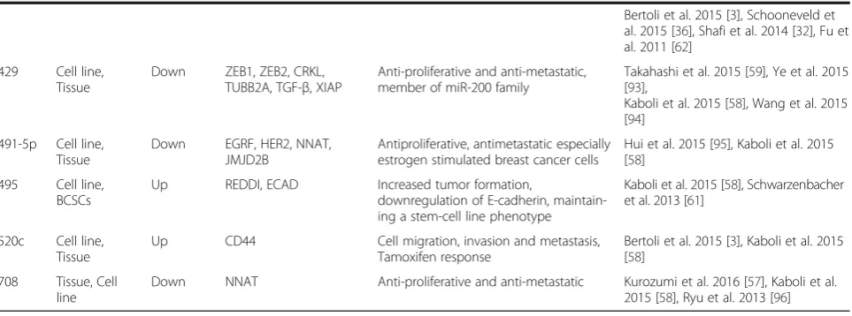
429 Cell line, Tissue
Down ZEB1, ZEB2, CRKL, TUBB2A, TGF-β, XIAP
Anti-proliferative and anti-metastatic, member of miR-200 family
Takahashi et al. 2015 [59], Ye et al. 2015 [93],
Kaboli et al. 2015 [58], Wang et al. 2015 [94]
491-5p Cell line, Tissue
Down EGRF, HER2, NNAT, JMJD2B
Antiproliferative, antimetastatic especially estrogen stimulated breast cancer cells
Hui et al. 2015 [95], Kaboli et al. 2015 [58]
495 Cell line, BCSCs
Up REDDI, ECAD Increased tumor formation,
downregulation of E-cadherin, maintain-ing a stem-cell line phenotype
Kaboli et al. 2015 [58], Schwarzenbacher et al. 2013 [61]
520c Cell line, Tissue
Up CD44 Cell migration, invasion and metastasis,
Tamoxifen response
Bertoli et al. 2015 [3], Kaboli et al. 2015 [58]
708 Tissue, Cell line
miRGator is based in Seoul, Korea. Current version 3 provides deep sequencing data of miRNAs that facilitate users to comprehend precursor of miRNAs, its se-quences, their final products. Furthermore, miRGator has another two features such as miRNA catalogues and Expression profiles, and miRNA-mRNA target relations and expression correlations.
6. miRDB:http://mirdb.org/miRDB/
This data based was created by the department of radi-ation oncology, Washington university school of medi-cine in St. Louis. This web page provides predicted microRNA targets in human, mouse, rat, dog and chicken. This miRDB search engine allows users to search targets of miRNAs by miRNA name directly or gene accession number from GenBank, Gene Symbol and NCBI Gene ID.
7. PhenomiR:http://mips.helmholtz-muenchen.de/
phenomir/
Established by German Research Center for Environ-mental Health. Differentially regulated miRNA expres-sion in diseases and other biological processes can be divulged from this site.
8. miRecords:http://c1.accurascience.com/miRecords/
The Predicted Targets component of miRecords gener-ates integrated results from 11 established miRNA target prediction programs such as DIANA-microT, MicroIn-spector, miRanda, MirTarget2, miTarget, NBmiRTar, Pic-Tar, PITA, RNA22, RNAhybrid, and TargetScan/ TargertScanS.
9. miRGen:http://carolina.imis.athena-innovation.gr/ diana_tools/web/
miRGen, developed at the University of Pennsylvania, generates miRNA gene transcription start sites (TSSs), coupled with genome wide maps of transcription factor (TF) binding sites in order to understand mechanisms of miRNA transcription regulation.
Other useful online miRNA related sites were micro-rna.org, targetscan.org, ChIPBase, TarBase, starBase, ebi.ac.uk, PmmR (Putative microRNA-microRNA Regu-lations) and pictar.mdc-berlin.de.
Conclusion
MicroRNAs can be easily detected with advanced tech-nologies and the continuous investigations supply miRNA as a novel clue for diagnosis and prognosis marker to therapeutic targets. Furthermore, research on
delivering miRNAs mimics and/or inhibitors directly into breast cancer tissue to regulate the balance of miR-NAs is gaining attention in the field. The miRNA re-sources mentioned in this paper are useful to generate predicted targeted mRNAs for interested miRNAs candi-date. Aberrant miRNA expression in our body such as oncomirs (upregulated miRNAs) and oncosupressors (downregulated miRNAs) could be the sign of diseases and they can be clinically detected by RT-qPCR, digital PCR, microarrays, and next generation sequencing tech-nologies. Signature miRNA profile can be used to distin-guish the stages of breast cancer progression and help in early diagnosis, prognosis, and effective treatment for human breast cancer and canine mammary tumor.
Abbreviations
AGRO2:Protein argonaute-2; DGCR8: Digeorge syndrome chromosomal region 8; KEGG: Kyoto encyclopedia genes and genomes; miR/ miRNA: microRNA; MRI: Magnetic resonance imaging; mRNA: messenger RNA; PAZ: Protein PAZ; PIWI: Protein PIWI; pre-miRNA: precursor microRNA; pri-miRNA: primary microRNA; RISC: RNA-induced silencing complex; TRBP: TAR RNA binding protein; tRNA: transfer RNA
Acknowledgements Not applicable.
Funding
The authors acknowledge the research funding from Trans Disciplinary Research Grant Scheme (TRGS) (5535000) for the financial support.
Availability of data and materials Not applicable.
Authors’contributions
RMCY wrote and reviewed the manuscripts. CYK constructed and reviewed the manuscript. Both authors read and approved the final manuscript.
Authors’information
CYK: Associate professor, Department of Biomedical Science, Molecular Biology laboratory, Faculty of medicine and health sciences, University Putra Malaysia. Founder and president of BioMedKL. Technical advisor for molecular diagnostic at Ramsay Sime Darby Medical Centre, Malaysia. RMCY: PhD candidate, Department of Biomedical Science, Molecular Biology laboratory, Faculty of medicine and health sciences, University Putra Malaysia.
Ethics approval and consent to participate Not applicable.
Consent for publication Not applicable.
Competing interests
The authors declare that they have no competing interests.
Publisher’s Note
Springer Nature remains neutral with regard to jurisdictional claims in published maps and institutional affiliations.
Author details
Received: 12 June 2017 Accepted: 29 August 2017
References
1. Lee R, Feinbaum R, Ambrost V. The C. Elegans Heterochronic gene lin-4 encodes small RNAs with antisense Complementarity to & II-14. Cell. 1993; 75:843–54.
2. O’Day E, Lal A. MicroRNAs and their target gene networks in breast cancer. Breast Cancer Res. 2010;12:201.
3. Bertoli G, Cava C, Castiglioni I. T h e r a n o s t i c s MicroRNAs : new biomarkers for diagnosis, prognosis, therapy prediction and therapeutic tools for breast cancer; 2015. p. 5.
4. Grosshans H, Filipowicz W. Molecular biologyThe expanding world of small RNAs. Nature. 2008;451:414–6. https://doi.org/10.1016/j.devcel.2014.01.009. 5. Lee Y, Kim M, Han JJ, Yeom KH, Lee S, Baek SH, et al. MicroRNA genes are
transcribed by RNA polymerase II. EMBO J. 2004;23:4051–60.
6. Filippov V, Solovyev V, Filippova M, Gill SS. A novel type of RNase III family proteins in eukaryotes. Gene. 2000;245:213–21.
7. Gregory RI, Yan K-P, Amuthan G, Chendrimada T, Doratotaj B, Cooch N, et al. The microprocessor complex mediates the genesis of microRNAs. Nature. 2004;432:235–40.
8. Denli AM, Tops BBJ. Plasterk RH a, Ketting RF, Hannon GJ. Processing of primary microRNAs by the microprocessor complex. Nature. 2004;432:231–5. 9. Czech B, Hannon GJ. Small RNA sorting: matchmaking for Argonautes. Nat
Rev Genet. 2011;12:19–31. https://doi.org/10.1038/nrg2916.
10. Lund E, Gu S. Nuclear Export of MicroRNA. Science (80- ). 2004;303 January:95–8. 11. Bernstein E. Caudy a a, Hammond SM, Hannon GJ. Role for a bidentate
ribonuclease in the initiation step of RNA interference. Nature. 2001;409:363–6. 12. Chendrimada TP, Gregory RI, Kumaraswamy E, Cooch N, Nishikura K,
Shiekhattar R. NIH Public Access. 2010;436:740–4.
13. Lau P-W, Guiley KZ, De N, Potter CS, Carragher B, MacRae IJ. The molecular architecture of human dicer. Nat Struct Mol Biol. 2012;19:436–40. https://doi. org/10.1038/nsmb.2268.
14. Bartel DP. MicroRNAs: genomics, biogenesis, mechanism, and function. Cell. 2004;116:281–97.
15. Ghildiyal M, Xu J, Seitz H, Weng Z, Zamore PD. Sorting of drosophila small silencing RNAs partitions microRNA* strands into the RNA interference pathway. RNA. 2010;16:43–56.
16. Cheng G. Circulating miRNAs: roles in cancer diagnosis, prognosis and therapy. Adv Drug Deliv Rev. 2015;81:75–93. https://doi.org/10.1016/j.addr. 2014.09.001.
17. Okamura K, Hagen JW, Duan H, Tyler DM, Lai EC. NIH Public Access. 2009; 130:89–100.
18. Cheloufi S, Dos Santos CO, Chong M. Hannon G. Ago Catalysis. 2010;465:584–9. 19. Yang J-S, Maurin T, Robine N, Rasmussen KD, Jeffrey KL, Chandwani R, et al.
Conserved vertebrate mir-451 provides a platform for dicer-independent, Ago2-mediated microRNA biogenesis. Proc Natl Acad Sci U S A. 2010;107:15163–8. 20. Chou C-H, Chang N-W, Shrestha S, Hsu S-D, Lin Y-L, Lee W-H, et al.
miRTarBase 2016: updates to the experimentally validated miRNA-target interactions database. Nucleic Acids Res. 2015;44 November 2015:gkv1258-. doi:https://doi.org/10.1093/nar/gkv1258.
21. He L, Hannon GJ. MicroRNAs: small RNAs with a big role in gene regulation. Nat Rev Genet. 2004;5:522–31.
22. Dalmay T. Mechanism of miRNA-mediated repression of mRNA translation. Essays Biochem. 2013;54:29–38.
23. Saxena S, Jónsson ZO, Dutta A. Small RNAs with imperfect match to endogenous mRNA repress translation. Implications for off-target activity of small inhibitory RNA in mammalian cells. J Biol Chem. 2003;278:44312–9. 24. MacFarlane L-A, Murphy PR. MicroRNA: biogenesis, function and role in
cancer. Curr Genomics. 2010;11:537–61. https://doi.org/10.2174/ 138920210793175895.
25. Eiring AM, Harb JG, Neviani P, Garton C, Oaks JJ, Spizzo R, et al. miR-328 functions as an RNA decoy to modulate hnRNP E2 regulation of mRNA translation in leukemic blasts. Cell. 2010;140:652–65. https://doi.org/10.1016/ j.cell.2010.01.007.
26. Tang J, Ahmad A, Sarkar FH. The role of MicroRNAs in breast cancer migration, invasion and metastasis. Int J Mol Sci. 2012;13:13414–37. https:// doi.org/10.3390/ijms131013414.
27. Baffa R, Fassan M, Volinia S, O’Hara B. MicroRNA expression profiling of human metastatic cancers identifies cancer gene targets†. J Pathol. 2009;219
28. Henry JC, Azevedo-Pouly ACP, Schmittgen TD. MicroRNA replacement therapy for cancer. Pharm Res. 2011;28:3030–42.
29. Christodoulatos GS, Dalamaga M. Micro-RNAs as clinical biomarkers and therapeutic targets in breast cancer: quo vadis? World J Clin Oncol. 2014;5: 71–81. https://doi.org/10.5306/wjco.v5.i2.71.
30. Piva R. Spandidos D a., Gambari R. From microRNA functions to microRNA therapeutics: novel targets and novel drugs in breast cancer research and treatment (review). Int J Oncol. 2013;43:985–94.
31. Blenkiron C, Goldstein LD, Thorne NP, Spiteri I, Chin S-F, Dunning MJ, et al. MicroRNA expression profiling of human breast cancer identifies new markers of tumor subtype. Genome Biol. 2007;8:R214.
32. Shafi G, Hasan TN, Syed NA, Paine A, Tegner J, Munshi A. Omics approaches in breast cancer; 2014. p. 171–82. https://doi.org/10.1007/978-81-322-0843-3. 33. Hurst DR, Edmonds MD, Welch DR. Metastamir: the field of
metastasis-regulatory microRNA is spreading. Cancer Res. 2009;69:7495–8.
34. Lopez-Camarillo C, Marchat LA, Arechaga-Ocampo E, Perez-Plasencia C, del Oral-Hernandez O, Castaneda-Ortiz EJ, et al. MetastamiRs: non-coding microRNAs driving cancer invasion and metastasis. Int J Mol Sci. 2012;13:1347–79. 35. Ma L, Reinhardt F, Pan E, Soutschek J, Bhat B, Teruya-feldstein J, et al.
Therapeutic silencing of miR-10b inhibits metastasis in a mouse mammary tumor model. Nat Biotechnol. 2010;28:341–7.
36. Schooneveld E, Wildiers H, Vergote I, Vermeulen PB, Dirix LY, Van Laere SJ. Dysregulation of microRNAs in breast cancer and their potential role as prognostic and predictive biomarkers in patient management. Breast Cancer Res. 2015;17:1–15. https://doi.org/10.1186/s13058-015-0526-y. 37. Várallyay E, Burgyán J, Havelda Z. MicroRNA detection by northern blotting
using locked nucleic acid probes. Nat Protoc. 2008;3:190–6. 38. Wang Z, Yang B. MicroRNA expression detection methods. 1st Editio.
Springer-Verlag: Berlin Heidelberg; 2010. doi:https://doi.org/10.1007/ 9783642049286.
39. Balcells I, Cirera S, Busk PK. Specific and sensitive quantitative RT-PCR of miRNAs with DNA primers. BMC Biotechnol. 2011;11:70. https://doi.org/10. 1186/1472-6750-11-70.
40. Creighton CJ, Reid JG, Gunaratne PH. Expression profiling of microRNAs by deep sequencing. Brief Bioinform. 2009;10:490–7.
41. Mattiske S, Suetani RJ, Neilsen PM, Callen DF. The oncogenic role of miR-155 in breast cancer. Cancer Epidemiol Biomark Prev. 2012;21:1236–43. 42. Han J-G, Jiang Y-D, Zhang C-H, Yang Y-M, Pang D, Song Y-N, et al. A novel
panel of serum miR-21/miR-155/miR-365 as a potential diagnostic biomarker for breast cancer. Ann Surg Treat Res. 2017;92:55–66. https://doi. org/10.4174/astr.2017.92.2.55.
43. Queiroga FL, Raposo T, Carvalho MI, Prada J, Pires I. Canine mammary tumours as a model to study human breast cancer: most recent findings. In Vivo. 2011;25:455–65. 44. Strandberg JD, Goodman DG. Animal model of human disease: canine
mammary neoplasia. Am J Pathol. 1974;75:225–8.
45. Schneider R. Comparison of age, sex, and incidence rates in human and canine breast cancer. Cancer. 1970;26:419–26.
46. Prier JE, Brodey RS. Canine Neoplasia: a prototype for human cancer study; 1959. p. 331–44.
47. Sorenmo KU. Canine mammary gland tumors. Vet Clin North Am Small Anim Pract. 2003;33:573–96. https://doi.org/10.1016/S0195-5616(03)00020-2. 48. Loukopoulos P, Mungall B a, Straw RC, Thornton JR, Robinson WF.
Matrix metalloproteinase-2 and -9 involvement in canine tumors. Vet Pathol. 2003;40:382–94.
49. Shah FD, Shukla SN, Shah PM, Shukla HK, Patel PS. Clinical significance of matrix metalloproteinase 2 and 9 in breast cancer. Indian J Cancer. 2013;46: 194–202. https://doi.org/10.4103/0019-509X.52953.
50. Wagner S, Willenbrock S, Nolte I, Murua EH. Comparison of non-coding RNAs in human and canine cancer. Front Genet. 2013:46. https://doi.org/10. 3389/fgene.2013.00046.
51. Boggs RM, Wright ZM, Stickney MJ, Porter WW, Murphy KE. MicroRNA expression in canine mammary cancer. Mamm Genome. 2008;19:561–9. 52. Zhou D, Li S, Wen J, Gong X, Xu L, Luo Y. Genome-wide computational
analyses of microRNAs and their targets from Canis Familiaris. Comput Biol Chem. 2008;32:60–5.
53. von Deetzen M-C, Schmeck BT, Gruber AD, Klopfleisch R. Malignancy associated MicroRNA expression changes in canine mammary cancer of different malignancies. ISRN Vet Sci. 2014;2014:148597. https://doi.org/10.1155/2014/148597. 54. Klopfleisch R, Klose P, Weise C, Bondzio A, Multhaup G, Einspanier R, et al.
55. Liu D, Xiong H, Ellis AE, Northrup NC, Rodriguez CO Jr, O’Regan RM, et al. Molecular homology and difference between spontaneous canine mammary cancer and human breast cancer. Cancer Res. 2014;74:5045–56. 56. Hawai SM, Ali MM, Niu Y, Alawad A, Aljofan M. Dogs : active role model for
cancer studies—a review. J Cancer Ther. 2013;2013 July:989–95. 57. Kurozumi S, Yamaguchi Y, Kurosumi M, Ohira M, Matsumoto H, Horiguchi J.
Recent trends in microRNA research into breast cancer with particular focus on the associations between microRNAs and intrinsic subtypes. J Hum Genet. 2016;62:15–24. https://doi.org/10.1038/jhg.2016.89.
58. Kaboli PJ, Rahmat A, Ismail P, Ling K-H. MicroRNA-based therapy and breast cancer: a comprehensive review of novel therapeutic strategies from diagnosis to treatment. Pharmacol Res. 2015;97:104–21. https://doi.org/10. 1016/j.phrs.2015.04.015.
59. Takahashi RU, Miyazaki H, Ochiya T. The roles of microRNAs in breast cancer. Cancers (Basel). 2015;7:598–616.
60. Zhang W-C, Liu J, Xu X, Wang G. The role of microRNAs in breast cancer progression. Med Oncol. 2014;30:675. https://doi.org/10.1007/s12032-013-0675-8. 61. Schwarzenbacher D, Balic M, Pichler M. The role of microRNAs in breast
cancer stem cells. Int J Mol Sci. 2013;14:14712–23.
62. Fu SW, Chen L, Man Y-G. miRNA biomarkers in breast cancer detection and management. J Cancer. 2011;2:116–22.
63. Xiang J, Wu J. Feud or friend? The role of the miR-17-92 cluster in tumorigenesis. Curr Genomics. 2010;11:129–35. https://doi.org/10.2174/ 138920210790886853.
64. Bonauer A, Dimmeler S. The microRNA-17∼92 cluster: still a miRacle? Cell Cycle. 2009;8:3866–73.
65. Kodahl AR, Lyng MB, Binder H, Cold S, Gravgaard K, Knoop AS, et al. Novel circulating microRNA signature as a potential non-invasive multi-marker test in ER-positive early-stage breast cancer: a case control study. Mol Oncol. 2014;8:874–83. https://doi.org/10.1016/j.molonc.2014.03.002.
66. Tang W, Zhu J, Su S, Wu W, Liu Q, Su F, et al. MiR-27 as a prognostic marker for breast cancer progression and patient survival. PLoS One. 2012;7:e51702. https://doi.org/10.1371/journal.pone.0051702.
67. Mertens-Talcott SU, Chintharlapalli S, Li X, Safe S. The oncogenic microRNA-27a targets genes that regulate specificity protein transcription factors and the G2-M checkpoint in MDA-MB-231 breast cancer cells. Cancer Res. 2007;67:11001–11. 68. Ding L, Ni J, Yang F, Huang L, Deng H, Wu Y, et al. Promising therapeutic
role of miR-27b in tumor. Tumor Biol. 2017;39:101042831769165. https://doi. org/10.1177/1010428317691657.
69. Hsieh T-H, Hsu C-Y, Tsai C-F, Long C-Y, Wu C-H, Wu D-C, et al. HDAC inhibitors target HDAC5, Upregulate MicroRNA-125a-5p, and induce apoptosis in breast cancer cells. Mol Ther. 2015;23:656–66. https://doi.org/ 10.1038/mt.2014.247.
70. Feliciano A, Castellvi J, Artero-Castro A, Leal JA, Romagosa C, Hernández-Losa J, et al. miR-125b acts as a tumor suppressor in breast tumorigenesis via its novel direct targets ENPEP, CK2-α, CCNJ, and MEGF9. PLoS One. 2013; 8:1–18.
71. Wang H, Tan G, Dong L, Cheng L, Li K, Wang Z, et al. Circulating mir-125b as a marker predicting chemoresistance in breast cancer. PLoS One. 2012;7: e34210. https://doi.org/10.1371/journal.pone.0034210.
72. Zheng Y, Lv X, Wang X, Wang B, Shao X, Huang Y. miR-181b promotes chemoresistance in breast cancer by regulating Bim expression; 2016. p. 683–90.
73. Calura E, Martini P, Sales G, Beltrame L, Chiorino G, D’Incalci M, et al. Wiring miRNAs to pathways: a topological approach to integrate miRNA and mRNA expression profiles. Nucleic Acids Res. 2014;42:e96. https://doi.org/10. 1093/nar/gku354.
74. Le XF, Almeida MI, Mao W, Spizzo R, Rossi S, Nicoloso MS, et al. Modulation of microrna-194 and cell migration by her2-targeting trastuzumab in breast cancer. PLoS One. 2012;7:1–14.
75. Fang C, Wang F-B, Li Y, Zeng X-T. Down-regulation of miR-199b-5p is correlated with poor prognosis for breast cancer patients. Biomed Pharmacother. 2016;84:1189–93. https://doi.org/10.1016/j.biopha.2016.10.006. 76. Fang C, Zhao Y, Guo B. MiR-199b-5p targets HER2 in breast cancer cells. J
Cell Biochem. 2013;114:1457–63.
77. Hilmarsdottir B, Briem E, Bergthorsson J, Magnusson M, Gudjonsson T. Functional role of the microRNA-200 family in breast morphogenesis and Neoplasia. Genes (Basel). 2014;5:804–20. https://doi.org/10.3390/ genes5030804.
78. Tsouko E, Wang J, Frigo DE, Aydogdu E, Williams C. miR-200a inhibits migration of triple-negative breast cancer cells through direct repression of
the EPHA2 oncogene. Carcinogenesis. 2015;36:1051–60. https://doi.org/10. 1093/carcin/bgv087.
79. Yao Y, Hu J, Shen Z, Yao R, Liu S, Li Y, et al. MiR-200b expression in breast cancer: a prognostic marker and act on cell proliferation and apoptosis by targeting Sp1. J Cell Mol Med. 2015;19:760–9.
80. Ye F, Tang H, Liu Q, Xie X, Wu M, Liu X, et al. MiR-200b as a prognostic factor targets multiple members of RAB family in glioma. Med Oncol. 2014; 31:1–10.
81. Jurmeister S, Baumann M, Balwierz A, Keklikoglou I, Ward A, Uhlmann S, et al. MicroRNA-200c represses migration and invasion of breast cancer cells by targeting Actin-regulatory proteins FHOD1 and PPM1F. Mol Cell Biol. 2012;32:633–51.
82. Shimono Y, Ugalde M, Cho R, Lobo N, Dalerba P, Qian D, et al. Down-regulation of miRNA-200c links breast cancer stem cells with normal stem cells. Cell. 2009;138:423–4.
83. Shen S-Q, Huang L-S, Xiao X-L, Zhu X-F, Xiong D-D, Cao X-M, et al. miR-204 regulates the biological behavior of breast cancer MCF-7 cells by directly targeting FOXA1. Oncol Rep. 2017:1–9. https://doi.org/10.3892/or.2017.5644. 84. Li T, Pan H, Li R. The dual regulatory role of miR-204 in cancer. Tumor Biol.
2016;37:11667–77. https://doi.org/10.1007/s13277-016-5144-5. 85. Flores-Pérez A, Marchat LA, Rodríguez-Cuevas S, Bautista-Piña V,
Hidalgo-Miranda A, Ocampo EA, et al. Dual targeting of ANGPT1 and TGFBR2 genes by miR-204 controls angiogenesis in breast cancer. Sci Rep. 2016;6:34504. https://doi.org/10.1038/srep34504.
86. Devlin C, Greco S, Martelli F, Ivan M. MiR-210: more than a silent player in hypoxia. IUBMB Life. 2011;63:94–100.
87. Huang X, Ding L, Bennewith K, Tong R, Ang KK, Le Q, et al. Hypoxia inducible mir-210 regulates normoxic gene expression involved in tumor initiation. 2010;35:856–867.
88. Jana S, Sengupta S, Biswas S, Chatterjee A, Roy H, Bhattacharyya A. miR-216b suppresses breast cancer growth and metastasis by targeting SDCBP. Biochem Biophys Res Commun. 2017;482:126–33. https://doi.org/10.1016/j. bbrc.2016.10.003.
89. Zhao L, Ren Y, Tang H, Wang W, He Q, Sun J, et al. Deregulation of the miR-222-ABCG2 regulatory module in tongue squamous cell carcinoma contributes to chemoresistance and enhanced migratory/invasive potential. Oncotarget. 2015;6:44538–50.
90. Liu F, Liu Y, Shen J, Zhang G, Han J. MicroRNA-224 inhibits proliferation and migration of breast cancer cells by down-regulating fizzled 5 expression. Oncotarget. 2014;7 10.18632/oncotarget.9734.
91. Leivonen SK, Sahlberg KK, Mäkelä R, Due EU, Kallioniemi O, Børresen-Dale AL, et al. High-throughput screens identify microRNAs essential for HER2 positive breast cancer cell growth. Mol Oncol. 2014;8:93–104.
92. Romero-Cordoba S, Rodriguez-Cuevas S, Rebollar-Vega R, Quintanar-Jurado V, Maffuz-Aziz A, Jimenez-Sanchez G, et al. Identification and pathway analysis of microRNAs with no previous involvement in breast cancer. PLoS One. 2012;7
93. Ye Z-B, Ma G, Zhao Y-H, Xiao Y, Zhan Y, Jing C, et al. miR-429 inhibits migration and invasion of breast cancer cells in vitro. Int J Oncol. 2015;46:531–8. 94. Wang C, Ju H, Shen C, Tong Z. miR-429 mediatesδ-tocotrienol-induced
apoptosis in triple-negative breast cancer cells by targeting XIAP. Int J Clin Exp Med. 2015;8:15648–56. https://doi.org/10.1093/carcin/bgv087. 95. Hui Z, Yiling C, Wenting Y, Xuqun H, Chuanyi Z, Hui L. miR-491-5p functions
as a tumor suppressor by targeting JMJD2B in ER a -positive breast cancer. FEBS Lett. 2015;589:812–21. https://doi.org/10.1016/j.febslet.2015.02.014. 96. Ryu S, McDonnell K, Choi H, Gao D, Hahn M, Joshi N, et al. Suppression of