R E S E A R C H A R T I C L E
Open Access
Dynamic metabolic profiling of the marine
microalga
Chlamydomonas
sp. JSC4 and
enhancing its oil production by optimizing light
intensity
Shih-Hsin Ho
1, Akihito Nakanishi
1, Xiaoting Ye
1, Jo-Shu Chang
3,4,5, Chun-Yen Chen
5, Tomohisa Hasunuma
1*and Akihiko Kondo
2,6Abstract
Background:Marine microalgae are among the most promising lipid sources for biodiesel production because they can be grown on nonarable land without the use of potable water. Marine microalgae also harvest solar energy efficiently with a high growth rate, converting CO2into lipids stored in the cells. Both light intensity and
nitrogen availability strongly affect the growth, lipid accumulation, and fatty acid composition of oleaginous microalgae. However, very few studies have systematically examined how to optimize lipid productivity by adjusting irradiance intensity, and the metabolic dynamics that may lead to improved lipid accumulation in microalgae have not been elucidated. Little is known about the mechanism of lipid synthesis regulation in microalgae. Moreover, few studies have assessed the potential of using marine microalgae as oil producers. Results:In this work, a newly isolated marine microalga,Chlamydomonassp. JSC4, was selected as a potential lipid producer, and the effect of photobioreactor operations on cell growth and lipid production was investigated. The combined effects of light intensity and nitrogen depletion stresses on growth and lipid accumulation were further explored in an effort to markedly improve lipid production and quality. The optimal lipid productivity and content attained were 312 mg L−1d−1and 43.1% per unit dry cell weight, respectively. This lipid productivity is the highest ever reported for marine microalgae. Metabolic intermediates were profiled over time to observe transient changes during lipid accumulation triggered by combined stresses. Finally, metabolite turnover was also assessed using an
in vivo13C-labeling technique to directly measure the flow of carbon during lipid biosynthesis under stress associated with light intensity and nitrogen deficiency.
Conclusions:This work demonstrates the synergistic integration of cultivation and dynamic metabolic profiling technologies to develop a simple and effective strategy for enhancing oil production in a marine microalga. The knowledge obtained from this study could be useful in assessing the feasibility of marine microalgae biodiesel production and for understanding the links between dynamic metabolic profiles and lipid biosynthesis during the course of microalgal cultivation.
Keywords:Microalgae,Chlamydomonassp, Lipids, Biodiesel, Metabolite profiling, Carbon flux, Light intensity, Nitrogen depletion
* Correspondence:[email protected] 1
Organization of Advanced Science and Technology, Kobe University, 1-1 Rokkodai, Nada-ku, Kobe 657-8501, Japan
Full list of author information is available at the end of the article
© 2015 Ho et al.; licensee BioMed Central. This is an Open Access article distributed under the terms of the Creative Commons Attribution License (http://creativecommons.org/licenses/by/4.0), which permits unrestricted use, distribution, and
Background
Due to the impact of the fast-growing global demand for fossil fuels on global climate change, considerable atten-tion has been paid to reducing CO2emissions by
devel-oping new sustainable energy sources as alternatives to fossil fuels [1]. Among the various potential renewable energy sources, biofuels produced from biomass are of the most interest and are expected to play an important role in the near future [2].
Biodiesel is an ideal renewable energy source because it is non-toxic and biodegradable and leads to lower CO2emissions [3]. Microalgae-produced biodiesel is one
of the most promising candidates, as some oleaginous microalgae can harvest solar energy efficiently due to their high growth rate and convert CO2 into lipids
stored in the cells [4]. Compared with terrestrial plants, microalgae can grow 10 to 50 times faster, resulting in an extremely high CO2 fixation rate [5,6]. In addition,
some oleaginous microalgae, such asChlorella, Chlamy-domonas, Neochloris, andDunaliella species, can accu-mulate lipid up to 50% per unit dry weight of biomass, and the lipid composition is suitable for making bio-diesel [7-10]. Furthermore, microalgal growth is not sea-sonally limited and can take place on nonarable land using municipal wastewater, brackish water, or seawater as a nutrient source [11,12]. The abovementioned char-acteristics make microalgal lipids promising raw mate-rials for biodiesel synthesis.
Although microalgae seem to have a high potential for biodiesel production, obstacles that may hinder their in-dustrial application remain. For instance, lipid accumula-tion in microalgae often occurs under environmental stress (for example, nitrogen depletion), which often leads to slower growth that results in lower lipid prod-uctivity [7,13]. Moreover, for large-scale microalgal culti-vation, a tremendous amount of fresh water is required if the microalgae cannot grow well in brackish water, recycled water, or seawater. This may cause a shortage of available water resources, which are already very lim-ited worldwide [7,14]. Therefore, cultivation strategies that result in optimal lipid content/productivity should be applied, and marine strains should be selected that are capable of rapid growth in saline environments and production of a high content of appropriate lipids.
To enhance the commercial feasibility of microalgae-based biodiesel production, the cultivation of lipid-producing microalgae must be made more efficient, with particular emphasis on optimization of several key fac-tors that significantly affect the yield and properties of biodiesel, such as lipid productivity and composition. Recent studies have noted that suitable light intensity is very effective in promoting growth and lipid accumula-tion and for improving lipid quality in oleaginous micro-algae [15,16]. However, no reports of systematic studies
of how to optimize lipid productivity by adjusting irradi-ance intensity or studies profiling metabolite dynamics associated with improved lipid accumulation in microal-gae have been published. In addition, relatively few stud-ies examining the potential use of marine microalgae as oil producers have been published.
In oleaginous microalgae, lipids are synthesized from acetyl-CoA precursors during periods of light exposure through the actions of several enzymes composing the fatty acid synthesis pathway [17]. Although light stress and nitrogen depletion are known to strongly influence growth and lipid accumulation in microalgae [16,18,19], little is known regarding the mechanism of lipid synthe-sis regulation in microalgae. In addition, a thorough un-derstanding of metabolite flux during lipid synthesis is also lacking.
Metabolic profiling is now a proven and powerful tool for gaining insights into functional biology [20,21]. The comprehensive analysis of a wide range of metabolites in microorganisms using mass spectrometry makes it pos-sible to identify and quantify target metabolites that play key roles in specific biological pathways, thereby reveal-ing the dynamic behavior of biological systems [20,22]. However, this metabolomic technology has yet to be widely applied to microalgae, particularly oleaginous microalgae [7]. To date, microalgal metabolomic studies have primarily focused on the metabolic responses of well-known species (for example,Chlamydomonas rein-hardtii) to nitrogen/nutrient depletion or variations in CO2 concentration [23-25]. However, there have been
no reports of the metabolic profiling of microalgae culti-vated under the combined stress of deficiencies in irradi-ance and nitrogen availability.
Targeting metabolites using 13C-labeling technologies is another useful strategy for studying biological systems in vivo because it provides an overview of metabolite flux through different pathways [26]. However, most studies reported to date have merely obtained‘snapshot’ metabolic profiles in microalgae; consequently, little is known about metabolic turnover or pathways in these organisms [22].
(PBR) type, light intensity, and nitrogen depletion. The metabolic profile of Chlamydomonas sp. JSC4 under light effect and nitrogen depletion was also monitored to identify the intercorrelations between light intensity, ni-trogen depletion, and lipid accumulation. Finally, meta-bolic turnover inChlamydomonassp. JSC4 was assessed using anin vivo13C-labeling technique to directly meas-ure the flow of carbon during lipid biosynthesis under conditions of different light intensity and nitrogen defi-ciency. The aim of this work was to determine the opti-mal light intensity necessary to maximize the production of lipids of suitable composition for biodiesel production by analyzing the flux of metabolites in Chlamydomonas sp. JSC4. The knowledge obtained from this study could be useful in assessing the applicability of this strain as biodiesel feedstock and for enhancing understanding of metabolite dynamics during lipid biosynthesis under stress.
Results and discussion
Effect of PBR type on growth rate, CO2fixation, and lipid accumulation inChlamydomonassp. JSC4
The growth rate and cellular composition of microalgae vary significantly with the illumination conditions during phototrophic growth [16]. In the PBRs used in our previ-ous study [7], light intensity tended to decrease when the biomass concentration increased due to light-shielding effects, causing a significant decrease in growth rate and changes in cellular composition. Therefore, in this study, a new type of PBR was designed (designated a slim-type tubular PBR) to improve the light-receiving area of the PBR from 104.4 to 200.2 cm2. The target strain, Chlamydomonas sp. JSC4, was grown in both conventional and the newly designed PBRs illuminated with a one-side light intensity of 150μmol m−2s−1, with 2% CO2serving as the carbon source.
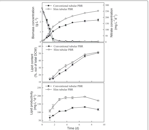
As shown in Figure 2 and Table 1, the growth rate in-creased when using the slim-type tubular PBR, resulting in a higher CO2fixation rate of 754 mg L−1d−1, which was
up to 40% higher than that achieved with a conventional-type tubular PBR. This phenomenon might have been due to the higher surface area to volume (S:V) ratio and shorter light path of the slim-type tubular PBR. The slim-type tubular PBR appears to have a greater light-receiving area and greater light penetration efficiency. Similar results were also observed in other research, demonstrating that a PBR with a high S:V ratio (80 to 100 m2m−3) has a shorter light path and eliminates dark areas in the cell broth, thus lead-ing to increased biomass concentration [28]. With respect to lipid production, Figure 2 shows that the type of PBR had no significant effect on the lipid content of Chlamydo-monassp. JSC4. Lipid productivity was thus positively pro-portional to biomass productivity. The maximum lipid productivity achieved with the slim-type tubular PBR was 194 mg L−1d−1(Table 1), which was significantly higher than that obtained with a conventional PBR. The above re-sults indicate that the slim-type tubular PBR is more effect-ive for growing strain JSC4, and thus this type or reactor was used in subsequent experiments.
Light intensity-dependent growth kinetics of Chlamydomonassp. JSC4
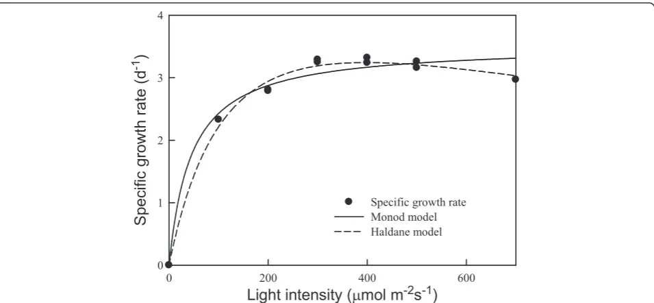
For most photoautotrophic microalgae, the growth rate depends heavily on irradiation intensity [15,16]. Consid-ering thatChlamydomonassp. JSC4 was isolated from a coastal area in tropical Taiwan, it was thought that the light intensity of 150 μmol m−2s−1used in the experi-ments described above (Figure 2) would not be optimal for growth. Therefore, strain JSC4 was cultivated in the slim-type tubular PBR illuminated at different light in-tensities to determine the optimal light intensity. As shown in Figure 3 and Table 2, the specific growth rate
of strain JSC4 increased rapidly as the light intensity in-creased from 100 to 300 μmol m−2s−1, reaching a plat-eau at light intensities greater than 300 μmol m−2 s−1. The highest specific growth rate was around 3.2 d−1 in the light-saturation region (300 to 500 μmol m−2 s−1),
which was significantly higher than that found in our previous work [7] and other related studies [15,16]. In addition, Table 2 shows that the specific growth rate of strain JSC4 at different light intensities could be fitted via Monod and Haldane models. The results fit the
Figure 2Performance (growth, lipid content, and lipid productivity) ofChlamydomonassp. JSC4 cultivated using different PBR types. Error bars indicate standard deviation of three replicated experiments (light intensity = 150μmol m−2s−1; CO2aeration = 2.0%; CO2flow rate = 0.1 vvm).
Table 1 Growth, lipid accumulation, and CO2-fixation performance of marineChlamydomonassp. JSC4
PBR type PBR
diameter (cm)
S:Varatio (m2m−3)
Light-receiving area (cm2)
Biomass productivity (mg L−1d−1)
Lipid content (%)
Lipid productivity (mg L−1d−1)
CO2fixation rate (mg L−1d−1)
Conventional 9.5 41.8 104.4 285 ± 13 46.1 ± 1.4 132 ± 2 537 ± 24
Slim 5.0 80.1 200.2 401 ± 10 48.5 ± 0.8 194 ± 2 754 ± 19
Chlamydomonassp. JSC4 cultivated for 7 days (5 days of nitrogen depletion) in different photobioreactors (PBRs). Values are the mean ± standard deviation of three replicated experiments (light intensity = 200μmol m−2
s−1
; CO2aeration = 2%; CO2flow rate = 0.1 vvm). a
Haldane model with better agreement (R2= 0.991), indi-cating that slight light inhibition may have occurred. However, due to the high KIvalue of 1,097μmol m−2s−1,
strain JSC4 appears to have a high tolerance to the inhibitory effects of high light intensity. These data indi-cate that strain JSC4 has good potential for outdoor culturing throughout the year in subtropical and trop-ical areas.
Combined effects of light intensity and nitrogen
depletion on growth, CO2fixation, and lipid accumulation inChlamydomonassp. JSC4
To enhance the economic feasibility of microalgae-based biodiesel production, lipid synthesis should be maximized to reach a satisfactory lipid content. In addition, biomass productivity should be enhanced to ensure high lipid prod-uctivity. It is known that many microalgae can rapidly ac-cumulate and store large amounts of lipids as an energy source in response to environmental stresses that are un-favorable to growth, such as high light intensity [19], nitro-gen depletion [4,18], and high salinity [7]. Both salinity and nitrogen depletion have been shown to be critical factors that affect growth and lipid accumulation in microalgae.
Our previous study indicated that cultivation of Chlamydo-monassp. JSC4 in 2% sea salt for 3 days under nitrogen-depletion conditions can enhance lipid productivity [7]. Furthermore, irradiance also strongly influences growth and lipid accumulation in some microalgae, including Sce-nedesmus [16], Nannochloropsis [19], Pavlova [29], and Desmodesmus [4]. As our previous study showed that strain JSC4 has a high potential to serve as an oil feedstock for biodiesel production [7], optimization of the irradiance conditions for this strain to achieve higher lipid productiv-ity is of great importance. Obtaining a thorough under-standing of the synergistic effects of light intensity control and nitrogen depletion is also very important. Another variable that must be evaluated is the cultivation time re-quired to achieve the highest lipid production efficiency.
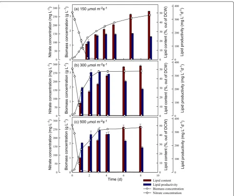
In this study, strain JSC4 was cultivated under different light intensities for 8 to 9 days, with regular monitoring of biomass and nitrate concentrations, lipid content, lipid productivity, and CO2fixation rate. As shown in Figures 4
and 5, the rates of growth and CO2fixation increased
sig-nificantly as the light intensity increased from 150 to 300 μmol m−2s−1. However, a further increase in light intensity to 500μmol m−2s−1resulted in a slight decrease in both the growth and CO2fixation rates, suggesting that
exces-sive illumination inhibits growth and CO2fixation in this
species, a phenomenon commonly known as the photo-inhibition effect. For instance, Hoet al. reported that under high light intensity (540 μmol m−2 s−1), the growth and CO2fixation rates inScenedesmus obliquusdecline
mark-edly [16]. In our experiments, a maximum CO2 fixation
rate of 2,886 mg L−1d−1was obtained at a light intensity of 300μmol m−2s−1during the optimal time period indicated
Figure 3Light intensity-dependent growth kinetics and model simulations ofChlamydomonassp. JSC4 cultivated in a slim-type tubular PBR (CO2aeration = 2.0%; CO2flow rate = 0.1 vvm).
Table 2 Estimated kinetic parameters of Monod- and Haldane-type models
Model Estimated parameters
μmax Ks KI R2
Monod 3.52 45.2 N.A. 0.972
Haldane 5.58 143.2 1,097 0.991
in Figure 5. This performance was superior to that ob-tained both in our previous study [7] and that of most re-lated studies [6]. This excellent CO2fixation performance
makes strain JSC4 a potential candidate for use in microalgae-based CO2mitigation processes for real
indus-trial fuel gases.
With respect to lipid production in strain JSC4, Figure 4 shows that lipid productivity increased slightly as the light intensity increased from 150 to 300μmol m−2s−1, due pri-marily to enhanced growth associated with the increase in light intensity. In addition, previous reports showed that high light intensity may lead to an increase in the amount of neutral storage lipids, primarily triacylglycerol (TAG) derived from fatty acids (referred to as lipids in this study) [17,29]. However, in our study, a further increase in light intensity to 500μmol m−2s−1resulted in a slight drop in
lipid content (Figure 4), indicating that excess light sity inhibits lipid biosynthesis. Moreover, at all light inten-sities we examined, lipid content increased significantly during stationary phase (during which nitrogen deficiency occurred), suggesting that lipid accumulation in strain JSC4 is not only triggered by the supply of irradiance but also by the stress of nitrogen depletion. Palet al. [19] also demonstrated that exposing microalgae to more than one stress simultaneously (for example, high salinity and nitro-gen depletion) may be necessary for further enhancement of lipid accumulation. Therefore, as shown in Figures 4 and 5, appropriately increasing the light intensity to pro-mote growth in combination with limiting the nitrogen supply to trigger lipid accumulation can lead to a simultan-eous increase in both the lipid content and productivity in strain JSC4. At a light intensity of 300μmol m−2s−1, strain
JSC4 displayed a maximum lipid productivity of 328 mg L−1d−1and lipid content of 34.1%.
From an engineering perspective, lipid productivity is absolutely one of the most important performance indi-cators for evaluating the commercialization potential of lipid-producing microalgae. Moreover, the lipid content is also important when considering the operation costs of downstream lipid extraction and purification pro-cesses [16]. Normally, the lipid content of microalgae tends to increase when the nitrogen starvation period is extended, until the maximum lipid content is reached. However, such a prolonged cultivation time leads to a decrease in lipid productivity. Therefore, it was vital to determine the optimal incubation time for strain JSC4 under nitrogen depletion as well as the timing of cell harvesting to ensure the best lipid production perform-ance. At the optimal light intensity determined in this study (300 μmol m−2 s−1), the lipid content increased sharply as the nitrogen depletion time increased, until reaching a maximum value of 53.7% after 7 days (Table 3).
However, it is difficult to optimize both lipid content and lipid productivity simultaneously, because during the nitro-gen depletion period required for triggering lipid accumu-lation, growth appeared to slow, leading to lower lipid productivity (Table 3). Therefore, based on these results, optimal lipid production (lipid content = 43.1%; lipid prod-uctivity = 312 mg L−1d−1) was obtained at a light intensity of 300μmol m−2s−1and a 3-day nitrogen depletion period. The resulting lipid productivity of 312 mg L−1d−1was sig-nificantly higher than most of the values reported in the lit-erature, as indicated in Table 4.
The effect of light intensity on lipid quality in Chlamydomonassp.
In addition to lipid content and productivity, the fatty acid composition is also important because it provides an indication of a strain’s suitability for synthesizing bio-diesel [16]. The fatty acid composition of phototrophic microalgae often changes under different irradiance con-ditions [17]. In this study, the lipid composition of Chlamydomonassp. JSC4 was monitored with respect to light intensity. As shown in Table 5, the major fatty acids present in the lipids produced by strain JSC4 were simi-lar when the cells were cultivated under different light intensities. The predominant fatty acids were palmitic acid (27% to 30%), stearic acid (14% to 15%), oleic acid (22% to 25%), and linoleic acid (16% to 19%). However, the total amount of C16/C18 fatty acids increased slightly, from 90.0% to 92.5%, when the light intensity was increased. Similar results were also reported by Sun et al. [18], who noted that the fatty acids with 16- and 18-carbon atoms inNeochloris oleoabundansHK-129 in-creased as the light intensity inin-creased from 50 to 200 μmol m−2 s−1. In addition, Table 5 shows that the
Figure 5Time-course profiles of CO2fixation rate during the growth ofChlamydomonassp. JSC4 illuminated with various light
intensities.Error bars indicate standard deviation of three replicated experiments (CO2aeration = 2.0%; CO2flow rate = 0.1 vvm).
Table 3 The effects of duration of nitrogen depletion on lipid content and lipid productivity
Duration of N depletion (d)
Lipid content (% of DCW)
Lipid productivity (mg L−1d−1)
0 13.8 ± 1.3 210 ± 4
1 26.0 ± 1.1 325 ± 20
2 34.1 ± 1.1 328 ± 1
3 43.1 ± 1.4 312 ± 2
5 52.6 ± 0.6 259 ± 8
7 53.7 ± 0.8 200 ± 7
Chlamydomonassp. JSC4 cultured at a light intensity of 300μmol m−2
s−1
proportions of the main saturated (for example, palmitic and stearic) and monounsaturated (for example, palmi-toleic and oleic) fatty acids increased slightly as light in-tensity increased, indicating that high light inin-tensity alters fatty acid biosynthesis in microalgae, resulting in the production of more of the saturated or monounsatu-rated fatty acids that primarily make up neutral lipids [17]. These phenomena were also observed in other studies demonstrating that high light intensity triggers the biosynthesis of palmitic, setaric, or oleic acids in microalgae. This lipid composition appears to be ad-equate for producing biodiesel [18,30].
The effect of light intensity on five important proper-ties (namely, kinematic viscosity, specific gravity, cetane number, iodine number, and average unsaturation) of biodiesel produced from the oil from strain JSC4 is sum-marized in Table 6. Because an increase in the degree of saturation of the oil used in biodiesel production can in-crease the cetane number and oxidative ability, whereas a higher degree of unsaturation can improve the fluidity of biodiesel at low temperatures [31,32], the average de-gree of unsaturation appears to be an essential indicator of biodiesel quality [32]. As shown in Table 6, although the biodiesel obtained from strain JSC4 grown under high light intensity had a slightly lower average degree of unsaturation, the lipid characteristics (that is, kinematic viscosity, specific gravity, cetane number, and iodine number) were not significantly affected by the illumin-ation conditions, as all of these properties showed satis-factory values based on the United States biodiesel standard ASTM D6751 and the European EN 14214 standard. Thus, regardless of the light intensity, the lipids produced by Chlamydomonas sp. JSC4 appeared to be of adequate quality for biodiesel synthesis.
Effect of specific light availability on lipid content and lipid production inChlamydomonassp. JSC4
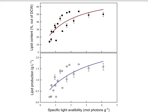
To further evaluate the feasibility of biodiesel production using Chlamydomonassp. JSC4, the influence of differ-ences in specific light availability on lipid production was investigated. Specific light availability was defined as moles of photons per gram DCW in the PBR over a des-ignated time interval [33]. According to Figure 6, both lipid content and lipid production were positively corre-lated with the specific light availability. The data clearly show that both lipid content and lipid production in-crease dramatically initially when the specific light avail-ability is increased until reaching a maximal value. It has been reported that the total amount of energy bound per gram of biomass increases with increasing lipid con-tent because the heating value of the microalgal biomass increases from 18.8 to 27.1 MJ kg−1as the lipid content increases from 10% to 50% [33]. Wilhelmet al. [34] also reported that the photon demand increases during lipid
Table 5 Fatty acid composition ofChlamydomonassp. JSC4
Chlamydomonassp. JSC4 FA composition
150μmol m−2s−1
300μmol m−2s−1
500μmol m−2s−1
Palmitic acid (C16:0) 27.3 ± 0.4 28.5 ± 0.6 29.5 ± 0.5
Palmitoleic acid (C16:1) 2.0 ± 0.2 2.5 ± 0.1 3.1 ± 0.2
Stearic acid (C18:0) 14.0 ± 0.3 14.9 ± 0.5 15.4 ± 0.3
Oleic acid (C18:1) 22.0 ± 0.1 22.9 ± 0.4 24.5 ± 0.9
Linoleic acid (C18:2) 18.7 ± 0.2 18.5 ± 0.3 15.5 ± 0.3
Linolenic acid (C18:3) 5.9 ± 0.1 5.1 ± 0.1 4.9 ± 0.2
Saturated fatty acid 41.9 ± 0.5 43.4 ± 0.9 44.9 ± 0.7
Monounsaturated fatty acid 24.0 ± 0.2 25.4 ± 0.4 27.6 ± 0.9
Polyunsaturated fatty acid 24.6 ± 0.2 23.6 ± 0.4 20.4 ± 0.4
C16 and C18 groups 90.0 ± 1.4 92.3 ± 1.8 92.5 ± 2.1
Lipid content (%) 48.5 ± 0.8 53.1 ± 1.3 48.9 ± 2.8
Chlamydomonassp. JSC4 cultured for 5 days under nitrogen depleted conditions and different light intensities. Values are the mean ± standard deviation of three replicated experiments (CO2aeration = 2.0%; CO2flow
rate = 0.1 vvm.).
Table 4 Comparison of lipid content and lipid productivity
Species Lipid
content (%)
Lipid productivity (mg L−1d−1)
Reference
Chlorella sorokinianaCY1 57.7 140.8 [2]
Chlorella vulgaris 17.3 79.1 [31]
Chlorella emersonii 18.6 54.4 [31]
Chlorella protothecoides 43.4 N.A. [46]
Chlamydomonassp. JSC4 33.1 169.1 [47]
Chlamydomonassp. JSC4 59.4 223 [7]
D. tertiolectaUTEX LB999 25.9 N.A. [48]
D. tertiolectaATCC30929 67.0 N.A. [49]
Isochrysis zhangjiangensis 46.0 136.2 [50]
Nannochloropsissp. 31.3 N.A. [27]
Nannochloropsissp. 59.9 74.2 [14]
Nannochloropsissp. F&M-M24 68.5 110.1 [51]
Nannochloropsis gaditana 73.1 51 [52]
Nannochloropsissp. F&M-M28 35.7 60.9 [53]
Nannochloropsissp. F&M-M26 29.6 61.0 [53]
T. pseudonanaCCMP1335 20.3 N.A. [48]
Tetraselmis suecicaF&M-M33 39.1 53.1 [51]
Chlamydomonassp. JSC4 43.1 312 This study
ComparedChlamydomonassp. JSC4 with that of other marine microalgae as reported in the literature.
biosynthesis in microalgae relative to the demand for primary sugar biosynthesis because of the additional reduction steps. Therefore, an increase in light energy provides more energy for both growth and lipid biosyn-thesis. However, as shown in Figure 6, increasing the
specific light availability to 3 to 4 mol photons g−1 re-sults in a slight drop in both lipid content and lipid pro-duction, suggesting that the excess illumination inhibits photosynthetic efficiency and lipid synthesis in microal-gae [35]. Thus, specific light availability is a simple but
Table 6 Biodiesel properties ofChlamydomonassp. JSC4
Biodiesel property 150μmol m−2s−1 300μmol m−2s−1 500μmol m−2s−1 US standard (ASTM D6751-08)
European standard (EN 14214)
Kinematic viscositya 4.71 4.72 4.74 1.9 to 6.0 3.5 to 5.0
Specific gravityb 0.88 0.88 0.88 0.85 to 0.90 0.86 to 0.90
Cetane numberc 57.60 57.69 57.99 min47 min51
Iodine numberd 71.54 70.53 67.24 - max120
Average unsaturation 0.79 0.78 0.73 -
-The fatty acids produced from dried biomass ofChlamydomonassp. JSC4 cultured for 5 days under conditions of nitrogen depletion and different light intensities. The relationship between average degree of unsaturation of fatty acids and biodiesel properties as reported by Songet al. [32] are shown. Values are the mean of three replicated experiments (CO2aeration = 2.0%; CO2flow rate = 0.1 vvm.).
a
Calculated from the following equation: Kinematic viscosity (mm2
s−1
, at 40°C) =−0.6316 × average unsaturation + 5.2065.
b
Calculated from the following equation: Specific gravity (kg L−1
) = 0.0055 × average unsaturation + 0.8726.
c
Calculated from the following equation: Cetane number =−6.6684 × average unsaturation + 62.876.
d
Calculated from the following equation: Iodine number (gl2g− 1
) = 74.373 × average unsaturation + 12.71.
important indicator of the relationship between light supply and lipid production in microalgae that have the potential for large-scale outdoor culture.
Metabolic profiling ofChlamydomonassp. JSC4 under light stress and nitrogen depletion
Lipid accumulation in microalgae is often affected by en-vironmental stimuli, acting either individually or in com-bination [17]. Some researchers have postulated that exposing microalgae to combined stresses (such as sim-ultaneous high salinity and nitrogen depletion) may fur-ther enhance lipid production efficiency [7,19]. However, little is known about the metabolic details of lipid bio-synthesis in response to different light intensities used for cell growth. Metabolic profiling techniques appear to be well-suited for studies aimed at elucidating the mech-anisms underlying metabolic changes in lipid biosyn-thesis in microalgae exposed to various environmental stimuli (such as irradiance and nitrogen deficiency) [23]. To the best of our knowledge, this is the first report dem-onstrating the metabolic profile of microalgae cultivated under combined light irradiation and nitrogen depletion.
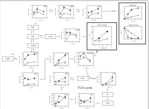
In this study, Chlamydomonas sp. JSC4 was cultivated under low and high light intensities (30 and 300μmol m−2 s−1), respectively. Intracellular metabolites were extracted and analyzed using capillary electrophoresis coupled with mass spectrometry (CE/MS), and variations in metabolite amounts over time were monitored as described previously [7]. As shown in Figure 7, cultivation at high light intensity resulted in a higher nitrogen consumption rate and a higher biomass concentration compared with cells cultured under low light intensity, suggesting that 300μmol m−2s−1 is a more suitable light intensity for lipid accumulation in Chlamydomonassp. JSC4. Thus, optimization of the light intensity provides a direct means of obtaining higher cell densities and enhanced lipid content. Notably, the levels of TAG-synthesis-related metabolites (for example, glycerol-3-phosphate [G3P] and acetyl-CoA (AceCoA)) in cells cul-tured under high light intensity were significantly higher than in cells cultured under low light intensity. This result is in good agreement with previous reports showing that ir-radiation with high-intensity light provides more energy during cultivation, which may induce microalgae to accu-mulate more TAG as the neutral storage lipid [17,36]. Moreover, it is known that the photon demand increases during lipid synthesis due to the additional reduction steps [34], and the metabolites of both G3P and AceCoA are ex-tremely important precursors for triacylglycerol formation [17,37]. The positive relationship between G3P, AceCoA, and triacylglycerol synthesis observed in this study in strain JSC4 cultured under high light intensity was therefore ex-pected. Sufficient light supply thus clearly triggers the for-mation of G3P and AceCoA and then further promotes the synthesis of TAG.
The levels of several key metabolites involved in the Cal-vin cycle (3-phosphoglyceric acid (3-PG), fructose-6-phosphate (F6P), and glucose-6-fructose-6-phosphate (G6P)) increased significantly along with cell growth, reaching maximal levels on day 2. This suggests that nutrients such as nitrogen ei-ther in the medium or inside the cells are sufficient for cell viability. However, after 2 days of nitrogen limitation (day 4), the levels of several metabolites involved in the Calvin cycle (F6P and G6P) decreased, primarily due to transformation of energy compounds into TAG (Figure 7). Moreover, re-gardless of nitrate status, an increase in the level of the starch precursor glucose-1-phosphate (G1P) was observed in cells irradiated with high-intensity light. This result indi-cates that strain JSC4 may also store considerable amounts of starch as an energy compound when exposed to certain environmental conditions. Previous reports showed that high light intensity may lead to an increase in the amount of total carbohydrate at microalgaS. obliquus, which is in good agreement with our study. In addition, several studies have shown that competition between lipid and carbohydrate synthesis occurs in microalgae under stress conditions [38,39]. Therefore, blocking the carbohydrate synthesis path-way may provide an approach for boosting TAG production. Furthermore, in the present study, cultivation under high light intensity led to a dramatic accumulation of intermedi-ates involved in glycolysis, such as 3-PG, phosphoenolpyr-uvate (PEP), and pyrphosphoenolpyr-uvate (Pyr). This may have been caused by rate-limiting conversions (that is, conversion of Pyr to AceCoA) that restricted the accumulation of lipids.
CO2is assimilated through the Calvin cycle in a reaction
catalyzed by ribulose-1,5-biphosphate carboxylase/oxygen-ase (Rubisco) to form 3-PG. Under low light intensity, the pool size (intracellular concentration) of 3-PG remained low (Figure 7), perhaps because insufficient light energy was available to drive photosynthesis, thus further restrict-ing the growth and ultimately the biosynthesis of lipids.
Analysis of metabolic turnover inChlamydomonassp. JSC4 under light stress and nitrogen depletion
As shown in Figure 4, cultivation under the dual effects of light irradiation and nitrogen depletion led to a marked increase in lipid content in Chlamydomonassp. JSC4. Metabolic profiling using CE/MS enables the iden-tification of key metabolites of interest and provides a
‘snapshot’of the metabolic status of cells at a given point in time. To further understand the dynamics of meta-bolic flux in JSC4 cells, CE/MS analysis combined with in vivo isotope labeling was employed [40]. Recent stud-ies showed that the metabolic kinetics of microalgae can be estimated by measuring changes in the abundance of isotopomers over time [20,22,26].
the presence and absence of nitrate under different light in-tensities using anin vivo13C-labeling assay. The ratio of13C to total carbon in each metabolite (that is, the13C fraction (%)) was calculated from mass isotopomer distributions de-termined by MS. By determining the ratio of the intensity of the monoisotopic ion to that of its isotopic ions, the ra-tio of stable-isotope-labeled to unlabeled metabolites can be determined. In in vivo 13C-labeling, 13C is assimilated through the Calvin cycle enzyme Rubisco in the form of
13
CO2, which is then used in the production of 3-PG. In
the presence of nitrate, the fraction of13C associated with 3-PG reached a maximum of 80% after 30 min of labeling in cells cultured under high light intensity (300μmol m−2 s−1), but it reached only 65% in cells cultured under low light intensity (30μmol m−2s−1). Irradiation with high light intensity also resulted in an increase in the13C-labeling ra-tio for other sugar phosphates, such as F6P, G6P, G1P, G3P,
PEP, and Pyr. These results indicate that sufficient irradi-ation leads to increases in both biomass concentrirradi-ation and lipid content, as shown in Figures 4 and 6. The13C fraction did not reach 100% for any metabolites in this study. This result was in agreement with previous reports showing that the13C fraction rarely reaches 100% for any metabolite in Arhrospira platensis, Nicotiana tabacum, and Quercus rubra [20,41]. This phenomenon may be the result of competition between isotope-labeled carbon and carbon derived from internal storage for assimilation into metabo-lites ultimately reaching an equilibrium phase.
Under conditions of nitrate depletion, the highest value of the13C fraction decreased for most metabolites. Notably, by limiting the nitrate supply at a fixed light intensity of 300 μmol m−2s−1, the13C fractions of malate, citrate, fumarate, and succinate declined from 85.6%, 97.1%, 75.0%, and 54.0% to 45.6%, 42.2%, 20.6%, and 6.2%, respectively, suggesting
Figure 7Time-course analysis of metabolite content ofChlamydomonassp. JSC4 cells illuminated with high light intensity (300μmol m−2s−1; closed circles) or low light intensity (30μmol m−2s−1; open circles).Error bars indicate standard deviation of three
replicated experiments (CO2aeration = 2.0%; CO2flow rate = 0.1 vvm). Abbreviations: AceCoA, acetyl-CoA; 1,3-BPG, 1,3-bisphosphoglycerate; Cit, citrate; DHAP, dihydroxyacetone phosphate; FBP, fructose-1,6-bisphosphate; F6P, fructose-6-phosphate; Fum, fumarate; GAP,
that availability of 13C for use in the TCA cycle declines when cells are confronted with nitrogen starvation, despite an increase in lipid content (Figure 8). Similar phenomena were also observed in cells cultured under low light intensity (30μmol m−2s−1). It is not clear whether these responses are common for Chlamydomonassp. and other photosyn-thetic species. However, several recent reports demonstrated that cultivation under nitrogen-deficient conditions leads to
a decrease in turnover of TCA cycle intermediates [20], which is consistent with our findings.
The initial slope of a plot of13C fraction versus time pro-vides an estimate of the turnover rate of each metabolite, as depicted in Figure 9. The turnover rates of sugar phos-phates involved in the Calvin cycle (for example, PEP and 3-PG) were significantly higher than those of organic acids, confirming that in photosynthetic microorganisms, 3-PG is
Figure 8Time-course analysis of the metabolite13C fraction of
Chlamydomonassp. JSC4 cells cultivated under high light intensity of 300μmol m−2s−1, nitrogen-rich (closed triangles) conditions; high light intensity of 300μmol m−2s−1, nitrogen-free (open triangles)
conditions; low light intensity of 30μmol m−2s−1, nitrogen-rich (closed circles) conditions; or low light intensity of 30μmol m−2s−1,
nitrogen-free (open circles) conditions.Error bars indicate standard deviation of three replicated experiments. Abbreviations: AceCoA, acetyl-CoA; Cit, citrate; F6P, fructose-6-phosphate; Fum, fumarate; G1P, glucose-1-phosphate; G3P: glycerol-3-phosphate; G6P,
rapidly generated through the action of the Calvin cycle and then further converted to PEP and other intracellular metabolites (Figure 7). Similar trends were also reported by Hasunumaet al. [20], who found that the turnover rates of sugar phosphates are significantly higher than those of or-ganic acids and amino acids inA. platensis. In addition, we observed that an increase in light intensity from 30 to 300 μmol m−2 s−1led to a sharp rise in the turnover rate of most metabolites (for example, Pyr and AceCoA), which is consistent with the efficient growth of cells and accumula-tion of lipids demonstrated in Figures 4 and 6. Further-more, under nitrogen limitation, the turnover rate of most metabolites in strain JSC4 decreased, suggesting that under nitrogen depletion, more energy is required for lipid syn-thesis, thus restricting the rate of carbon flow. Notably, the pool size of AceCoA (0.01 to 0.03 nmol mg−1), one of the most important TAG-synthesis-related metabolites, was approximately 20 to 50 times smaller than that of Pyr (0.32 to 0.78 nmol mg−1) (as shown in Additional file 1: Table S1), suggesting that lipid accumulation in strain JSC4 could be enhanced further through overexpression of related en-zymes. Some efforts have been made to increase the ex-pression of enzymes that are involved in fatty acid biosynthesis pathways, and the results are quite promising [42,43]. As a next step, knockout and overexpression of the genes involved in lipid synthesis could clarify which genes have a significant effect on lipid accumulation and lead to the development of strategies for increasing the lipid con-tent in oleaginous microalgae.
Nitrogen limitation is frequently used to increase the lipid content inChlamydomonas sp. [7]. However, little is known about the mechanism of lipid biosynthesis in these microorganisms. To the best of our knowledge, this is the first report of in vivoisotope labeling of Chlamydomonas
sp. cultivated under different light-intensity conditions coupled with nitrogen limitation. In this study, the com-bination of metabolic profiling and turnover analysis (col-lectively referred to as dynamic metabolic profiling) was applied to explore the effects of stress associated with com-bined light intensity and nitrogen depletion on carbon me-tabolism inChlamydomonassp. JSC4. The outcome of this research enhanced current understanding of the relation-ship between carbon flow and lipid biosynthesis in microal-gae cultured under combined light effect and nitrogen depletion. Using thein vivo13C-labeling assay, the dynamic metabolic profile of strain JSC4 was determined, generating a large amount of valuable information that can be applied to enhance the lipid production capacity of this organism through genetic engineering.
Conclusions
This work demonstrated that lipid production in the mar-ine microalgaChlamydomonas sp. JSC4 can be enhanced significantly by culturing the organism under conditions of appropriate light intensity (that is, 300 μmol m−2 s−1) coupled with nitrogen depletion. The highest lipid product-ivity achieved in this study was 328 mg L−1d−1, which to the best of our knowledge is the highest value reported in the literature for marine microalgae. The lipids produced under the optimal conditions contained 43.4% saturated, 25.4% monounsaturated, and 23.6% polyunsaturated fatty acids, a composition very suitable for biodiesel production. The data obtained from dynamic metabolic profiling (that is, metabolic profiling and turnover analysis) provided valuable information that increases our understanding of metabolic flow inChlamydomonassp. JSC4 when cultured in the absence of nitrate and with light of suitable intensity. By integrating cultivation strategies and dynamic metabolic
Figure 9Metabolic turnover rates of key metabolites inChlamydomonassp. JSC4 cells cultivated under high light intensity of 300μmol m−2s−1, nitrogen-rich (yellow bars) conditions; high light intensity of 300μmol m−2s−1, nitrogen-free (green bars)
conditions; low light intensity of 30μmol m−2s−1, nitrogen-rich (red bars) conditions; or low light intensity of 30μmol m−2s−1,
nitrogen-free (orange bars) conditions.Error bars indicate standard deviation of three replicated experiments. Abbreviations: AceCoA, acetyl-CoA; Cit, citrate; F6P, fructose-6-phosphate; Fum, fumarate; G1P, glucose-1-phosphate; G3P: glycerol-3-phosphate; G6P,
profiling techniques using strain JSC4, this study dem-onstrated an innovative technology for practically and markedly enhancing microalgal lipid productivity and elucidated the details of carbon metabolism related to lipid biosynthesis.
Methods
Microalgal strain and preculturing medium
The microalga used in this study wasChlamydomonassp. JSC4, which was isolated from a coastal area of southern Taiwan. The 18S rDNA sequence of this strain was depos-ited in the National Center for Biotechnology Information GenBank database under accession number KF383270. The medium used for preculturing strain JSC4 was
Modi-fied Bold 3 N medium (4.4 mM NaNO3, 0.22 mM
K2HPO4, 0.3 mM MgSO4· 7H2O, 0.17 mM CaCl2· 2H2O,
0.43 mM KH2PO4, and 0.43 mM NaCl; the levels of metals
in the medium are described in Berges et al. [44]). Cells were routinely cultured at 30°C to 33°C for 3 to 4 days with a continuous supply of 2.0% CO2 at an aeration rate of
0.05 vvm. The culture was illuminated continuously at a light intensity of approximately 250 μmol m−2 s−1. The light intensity was measured using a Li-250 Light Meter with a Li-190SA pyranometer sensor (Li-COR Inc., Lincoln, NE, USA).
PBR operation
Both conventional-type (9.5 cm in diameter; height to diameter (H:D) ratio of 0.8) and slim-type (5.0 cm in diam-eter; H:D ratio of 10) PBRs equipped with external light sources were used in this study. The S:V ratio of the con-ventional and slim-type PBRs was 41.8 and 80.1,
respect-ively. Chlamydomonas sp. JSC4 was precultured in
Modified Bold 3 N medium and inoculated into the PBRs at a concentration of 90 to 100 mg L−1. For batch cultures, microalgae were cultivated at 32°C to 35°C and pH 6.5 to 7.5 with an agitation rate of 400 rpm under various light intensities in the range of approximately 30 to 500 μmol m−2s−1. As the sole carbon source, 2.0% CO2was
continu-ously fed into the culture at a rate of 0.05 vvm. Liquid sam-ples were collected from the sealed glass vessel at designated time intervals to determine the culture density, pH, and residual nitrate concentration. The amount of CO2reduced was determined by measuring the difference
between the CO2 concentrations in the influent and
effluent streams of the PBR using a GM70 CO2detector
(Vaisala, Tokyo, Japan).
Determination of culture density
The density of microalgae in the PBR was determined regularly by measuring the optical density (OD) at a wave-length of 682 nm (denoted as OD682) using a
UVmini-1240 UV/Vis spectrophotometer (Shimadzu, Kyoto, Japan) after proper dilution with deionized water to give a range
of measurement between 0.1 and 0.9. The DCW was deter-mined by filtering 50-mL aliquots of culture through a cel-lulose acetate membrane filter (0.45-μm pore size, 47 mm diameter). Each loaded filter was then freeze-dried until the weight was invariant. The dry weight of a blank filter was subtracted from that of the loaded filter to obtain the DCW of microalgae. The OD682 values were also
con-verted to biomass concentration via appropriate calibration between OD682and DCW, and the conversion factor was
determined as 1.0 OD682= 0.7 ~ 0.9 g DCW L−1.
Measurement of residual nitrate content
Nitrate concentration was determined according to our pre-viously reported method [45]. In general, each sample col-lected from the PBR was filtered with a 0.22-μm pore-size filter and then diluted 20-fold with deionized water. The re-sidual nitrate content of the diluted samples was determined according to the OD220 with appropriate calibrations. The
conversion factor was determined as 1.0 OD220 = ~389.3
mg nitrate L−1.
Determination of growth parameters and CO2fixation performance
Change in biomass concentration (mg L−1) over time was used to calculate the specific growth rate (d−1) based on a plot of DCW (on a logarithmic scale) versus time. Biomass productivity (mg L−1 d−1) was calculated ac-cording to the following equation:
P¼ΔX
Δt ;
whereΔXrepresents the variation in biomass concentra-tion (mg L−1) over cultivation time (d).
The CO2 fixation rate (PCO2; mg L−1 d−1) was
calcu-lated according to the following equation:
PCO2ðmg=L=dÞ ¼1:88Pbiomass;
where Pbiomass represents biomass productivity (mg L−1
d−1), as described above. The typical molecular formula for microalgal biomass (CO0.48H1.83N0.11P0.01) [10] was
used in this study.
To evaluate the effect of irradiance along with bio-mass concentration on the lipid accumulation process, the specific light availability (IPAR, spec) as reported by
Munkel et al. [33] was calculated according to the fol-lowing equation:
IPAR;spec¼
Z
IPARdt
∅DWV ;
whereIPAR represents irradiance on the PBR surface
interval,V represents the total working volume, and t represents the cultivation time.
Determination of the lipid content and fatty acid profile After an appropriate amount of time for consuming ni-trogen, strain JSC4 cells were harvested from the culture medium by centrifugation (7,000 rpm for 2 min). The cells were washed twice with deionized water, lyophi-lized, and weighed. The lipid composition was deter-mined as fatty acid methyl esters (FAMEs) following direct transesterification of lipids according to the method described in Hoet al. [7]. FAMEs were analyzed by gas chromatography/mass spectrometry (GC/MS) on a GCMS-QP2010 Plus instrument (Shimadzu). Samples were injected onto a DB-23 capillary column (60 m, 0.25 mm internal diameter, 0.15-μm film thickness; Agilent Technologies, Palo Alto, CA, USA). Helium was used as the carrier gas at a flow rate of 2.3 mL min−1. The in-jector, ion source, and interface source temperatures were set at 230°C, 230°C, and 250°C, respectively. The oven temperature was initially set at 50°C for 1 min, creased from 50°C to 175°C at a rate of 25°C/min, in-creased from 175°C to 230°C at a rate of 4°C/min, and held at 230°C for 5 min. Purified FAMEs were identified based on retention time and the pattern of fragmenta-tion by electron impact analysis. Supelco 37 Component FAME Mix (Sigma-Aldrich Co., St. Louis, MO, USA) was utilized as a quantitative standard, and pentadeca-noic acid (Sigma-Aldrich Co.) was used as an internal standard.
Sampling procedures for metabolic profiling
Cell sampling was performed according to our previ-ously reported method [20], with minor modifications. Strain JSC4 cells (equivalent to 5 to 10 mg dry weight) were removed from cultivation vessels and added to a fourfold volume of prechilled (−30°C) quenching solu-tion consisting of Modified Bold 3 N medium containing 32.5% methanol, then filtered using 1-μm pore-size Omnipore filter disks (Millipore, Billerica, MA, USA). After washing with 20 mM ammonium bicarbonate pre-chilled to 4°C, cells retained on the filters were immedi-ately placed in 1 mL of prechilled (−30°C) methanol containing 12.4 μM piperazine-1,4-bis(2-ethanesulfonic acid) as an internal standard for mass analysis. Intracel-lular metabolites were extracted using cold 10:3:1 (v/v/v) methanol:chloroform:water combined with 30 min of bead-beater cell-breaking, as described previously [23]. Cells were suspended by vortexing and then 1 mL of the cell suspension was mixed with 100 μL of prechilled (4°C) water and 300μL of chloroform. The cell suspen-sion was shaken at 1,200 rpm in a model MBR-022UP incubator (TAITEC, Saitama, Japan) for 30 min at 4°C in the dark before centrifugation at 14,000 g for 5 min at
4°C. Next, 980 μL of the cell extract obtained as the supernatant was transferred to a clean tube. After adding 440 μL of water, the aqueous and organic layers were phase separated by centrifugation at 14,000g for 5 min at 4°C. After filtration through a Millipore 5-kDa cut-off filter for the removal of solubilized proteins, the aqueous-layer extracts were evaporated under vacuum using a FreeZone 2.5 Plus freeze dry system (Labconco, Kansas City, MO, USA). Dried extracts were stored at−80°C until analysis by CE/MS.
CE/MS metabolite analysis
Metabolites were determined according to the CE/MS method described by Hasunamaet al. [20]. Dried metab-olites were dissolved in 20 μL of Milli-Q water before analysis. The CE/MS experiments were performed using an Agilent G7100 CE system, an Agilent G6224AA LC/ MSD time-of-flight (TOF) mass spectrometer, and an Agilent 1200 series isocratic HPLC pump equipped with a 1:100 splitter for delivery of the sheath liquid.
Agilent ChemStation software for CE and MassHunter software for the Agilent TOF-MS system were used for system control and data acquisition, respectively. The ana-lytical conditions for anionic metabolite analyses were as described previously [20]. CE separations were performed using a fused silica capillary (1 m × 50μm i.d.) filled with 50 mM ammonium acetate (pH 9) for anionic metabolite analyses. The CE polarity was such that the electrolyte vial (inlet) was at the anode, and the electrospray ionization (ESI) probe (outlet) was at the cathode. Samples were injected into the CE system at a pressure of 50 mbar for 30 s. The voltage applied to the CE capillary was set at 30 kV, with a ramp time of 0.3 min. For anionic metabolite ana-lyses, the electrolyte solution was passed through the capil-lary using an air pump and was delivered at a pressure of 10 mbar between 0.4 and 30 min and 100 mbar between 30.1 and 49.5 min. The flow rate of the sheath liquid was set at 8μL min−1. The ESI-MS analyses were conducted in either the positive or negative ion mode using a capillary voltage of−3.5 or 3.5 kV, respectively. The TOF-MS frag-menter, skimmer, and Oct RFV were set to 100, 65, and 750 V, respectively. The flow of heated drying nitrogen gas (300°C) was maintained at 10 L min−1. Mass data were ac-quired at a rate of 1 spectrum s−1over the mass to charge ratio (m/z) range of 70 to 1,000.
13
C-labeling
To analyze metabolic turnover in Chlamydomonas sp. JSC4, in vivo 13C-labeling was performed using sodium
13
C-bicarbonate (NaH13CO3) as a carbon source. Strain
3 N or Modified Bold 0 N medium containing 25 mM NaH13CO3. After labeling for 1 to 30 min, 5 to 10 mg of
strain JSC4 cells were collected by filtration and processed as described in the previous section. Extracted intracellular metabolites were then analyzed using CE/MS. Mass spectral peaks of biological origin were identified manually by searching for mass shifts between 12C- and
13
C-mass spectra. The13C fraction of each metabolite and the metabolic turnover rate were determined as described previously [40].
Additional file
Additional file 1: Table S1.Comparison of key metabolite pools. Comparison of key metabolite pools inChlamydomonassp. JSC4 under cells cultivated under low light intensity of 30μmol m−2s−1, nitrogen-rich conditions; high light intensity of 300μmol m−2s−1, nitrogen-rich conditions; or high light intensity of 300μmol m−2s−1, nitrogen-free conditions. Values are the mean ± standard deviation of three replicated experiments.
Abbreviations
3-PG:3-phosphoglycerate; AceCoA: Acetyl-CoA; CE/MS: Capillary electrophoresis coupled with mass spectrometry; DCW: Dry cell weight; ESI: Electrospray ionization; F6P: Fructose-6-phosphate; FAMEs: Fatty acid methyl esters; G1P: Glucose-1-phosphate; G3P: Glycerol-3-phosphate; G6P: Glucose-6-phosphate; GC/MS: Gas chromatography/mass spectrometry; OD: Optical density; PBR: Photobioreactor; PEP: Phosphoenolpyruvate; Pyr: Pyruvate; TAG: Triacylglycerol; TOF: Time-of-flight.
Competing interests
The authors declare that they have no competing interests.
Authors’contributions
SHH performed and analyzed all experiments and drafted the manuscript. AN assisted in analysis of the metabolic experimental data. XY participated in metabolite analysis and contributed to the draft manuscript. JSC assisted in drafting the manuscript and revising it critically before submission. CYC assisted in analysis of growth kinetics and model simulations. TH and AK co-coordinated the study and helped in drafting the manuscript. All authors read and approved the final manuscript.
Acknowledgements
The authors would like to thank Mrs. Mika Kukuda for technical support. This work was supported through project P07015 from the New Energy and Industrial Technology Development Organization (NEDO).
Author details
1Organization of Advanced Science and Technology, Kobe University, 1-1 Rokkodai, Nada-ku, Kobe 657-8501, Japan.2Department of Chemical Science and Engineering, Graduate School of Engineering, Kobe University, 1-1 Rokkodai, Nada-ku, Kobe 657-8501, Japan.3Department of Chemical Engineering, National Cheng Kung University, Tainan 701, Taiwan.4Research Center for Energy Technology and Strategy, National Cheng Kung University, Tainan 701, Taiwan.5Center for Bioscience and Biotechnology, National Cheng Kung University, Tainan 701, Taiwan.6Biomass Engineering Program, RIKEN, 1-7-22 Suehiro-cho, Tsurumi-ku, Yokohama, Kanagawa 230-0045, Japan.
Received: 17 November 2014 Accepted: 19 February 2015
References
1. Ho S-H, Li P-J, Liu C-C, Chang J-S. Bioprocess development on microalgae-based CO2fixation and bioethanol production usingScenedesmus obliquusCNW-N. Bioresour Technol. 2013;145:142–9.
2. Chen C-Y, Chang J-S, Chang H-Y, Chen T-Y, Wu J-H, Lee W-L. Enhancing microalgal oil/lipid production fromChlorella sorokinianaCY1 using deep-sea water supplemented cultivation medium. Biochem Eng J. 2013;77:74–81. 3. Gouveia L, Oliveira AC. Microalgae as a raw material for biofuels production.
J Ind Microbiol Biotechnol. 2009;36:269–74.
4. Ho S-H, Chang J-S, Lai Y-Y, Chen C-NN. Achieving high lipid productivity of a thermotolerant microalgaDesmodesmussp. F2 by optimizing environmental factors and nutrient conditions. Bioresour Technol. 2014;156:108–16. 5. Wang B, Li YQ, Wu N, Lan CQ. CO2bio-mitigation using microalgae.
Appl Microbiol Biotechnol. 2008;79:707–18.
6. Ho S-H, Chen C-Y, Lee D-J, Chang J-S. Perspectives on microalgal CO2-emission mitigation systems—a review. Biotechnol Adv. 2011;29:189–98.
7. Ho S-H, Nakanishi A, Ye X, Chang J-S, Hara K, Hasunuma T, et al. Optimizing biodiesel production in marineChlamydomonassp. JSC4 through metabolic profiling and an innovative salinity-gradient strategy. Biotechnol Biofuels. 2014;7:97.
8. Chen C-Y, Yeh K-L, Aisyah R, Lee D-J, Chang J-S. Cultivation, photobioreactor design and harvesting of microalgae for biodiesel production: a critical review. Bioresour Technol. 2011;102:71–81.
9. Gouveia L, Marques AE, da Silva TL, Reis A.Neochloris oleabundansUTEX# 1185: a suitable renewable lipid source for biofuel production. J Ind Microbiol Biotechnol. 2009;36:821–6.
10. Chisti Y. Biodiesel from microalgae. Biotechnol Adv. 2007;25:294–306. 11. Cho S, Luong TT, Lee D, Oh Y-K, Lee T. Reuse of effluent water from a municipal
wastewater treatment plant in microalgae cultivation for biofuel production. Bioresour Technol. 2011;102:8639–45.
12. Siaut M, Cuiné S, Cagnon C, Fessler B, Nguyen M, Carrier P, et al. Oil accumulation in the model green algaChlamydomonas reinhardtii: characterization, variability between common laboratory strains and relationship with starch reserves. BMC Biotechnol. 2011;11:7.
13. Ho S-H, Chen W-M, Chang J-S.Scenedesmus obliquusCNW-N as a potential candidate for CO2mitigation and biodiesel production. Bioresour Technol. 2010;101:8725–30.
14. Jiang L, Luo S, Fan X, Yang Z, Guo R. Biomass and lipid production of marine microalgae using municipal wastewater and high concentration of CO2. Appl Energ. 2011;88:3336–41.
15. Liu J, Yuan C, Hu G, Li F. Effects of light intensity on the growth and lipid accumulation of microalgaScenedesmussp. 11–1 under nitrogen limitation. Appl Biochem Biotechnol. 2012;166:2127–37.
16. Ho S-H, Chen C-Y, Chang J-S. Effect of light intensity and nitrogen starvation on CO2fixation and lipid/carbohydrate production of an indigenous microalga
Scenedesmus obliquusCNW-N. Bioresour Technol. 2012;113:244–52. 17. Hu Q, Sommerfeld M, Jarvis E, Ghirardi M, Posewitz M, Seibert M, et al.
Microalgal triacylglycerols as feedstocks for biofuel production: perspectives and advances. Plant J. 2008;54:621–39.
18. Sun X, Cao Y, Xu H, Liu Y, Sun J, Qiao D, et al. Effect of nitrogen-starvation, light intensity and iron on triacylglyceride/carbohydrate production and fatty acid profile ofNeochloris oleoabundansHK-129 by a two-stage process. Bioresour Technol. 2014;155:204–12.
19. Pal D, Khozin-Goldberg I, Cohen Z, Boussiba S. The effect of light, salinity, and nitrogen availability on lipid production byNannochloropsissp. Appl Microbiol Biotechnol. 2011;90:1429–41.
20. Hasunuma T, Kikuyama F, Matsuda M, Aikawa S, Izumi Y, Kondo A. Dynamic metabolic profiling of cyanobacterial glycogen biosynthesis under conditions of nitrate depletion. J Exp Bot. 2013;64:2943–54.
21. Baran R, Reindl W, Northen TR. Mass spectrometry based metabolomics and enzymatic assays for functional genomics. Curr Opin Microbiol. 2009;12:547–52. 22. Ito T, Sugimoto M, Toya Y, Ano Y, Kurano N, Soga T, et al. Time-resolved
metabolomics of a novel trebouxiophycean alga using13CO2feeding. J Biosci Bioeng. 2013;116:408–15.
23. Bölling C, Fiehn O. Metabolite profiling ofChlamydomonas reinhardtiiunder nutrient deprivation. Plant Physiol. 2005;139:1995–2005.
24. Renberg L, Johansson AI, Shutova T, Stenlund H, Aksmann A, Raven JA, et al. A metabolomic approach to study major metabolite changes during acclimation to limiting CO2inChlamydomonas reinhardtii. Plant Physiol. 2010;154:187–96. 25. Ito T, Tanaka M, Shinkawa H, Nakada T, Ano Y, Kurano N, et al. Metabolic and morphological changes of an oil accumulating trebouxiophycean alga in nitrogen-deficient conditions. Metabolomics. 2013;9:178–87.
27. Wahidin S, Idris A, Shaleh SRM. The influence of light intensity and photoperiod on the growth and lipid content of microalgae
Nannochloropsissp. Bioresour Technol. 2013;129:7–11.
28. Posten C. Design principles of photo-bioreactors for cultivation of microalgae. Eng Life Sci. 2009;9:165–77.
29. Guedes AC, Meireles LA, Amaro HM, Malcata FX. Changes in lipid class and fatty acid composition of cultures ofPavlova lutheri, in response to light intensity. J Am Oil Chem Soc. 2010;87:791–801.
30. Guihéneuf F, Mimouni V, Ulmann L, Tremblin G. Combined effects of irradiance level and carbon source on fatty acid and lipid class composition in the microalgaPavlova luthericommonly used in mariculture. J Exp Mar Biol Ecol. 2009;369:136–43.
31. Talebi AF, Mohtashami SK, Tabatabaei M, Tohidfar M, Bagheri A,
Zeinalabedini M, et al. Fatty acids profiling: a selective criterion for screening microalgae strains for biodiesel production. Algal Res. 2013;2:258–67. 32. Song M, Pei H, Hu W, Ma G. Evaluation of the potential of 10 microalgal
strains for biodiesel production. Bioresour Technol. 2013;141:245–51. 33. Münkel R, Schmid-Staiger U, Werner A, Hirth T. Optimization of outdoor
cultivation in flat panel airlift reactors for lipid production by Chlorella vulgaris. Biotechnol Bioeng. 2013;110:2882–93.
34. Wilhelm C, Jakob T. From photons to biomass and biofuels: evaluation of different strategies for the improvement of algal biotechnology based on comparative energy balances. Appl Microbiol Biotechnol. 2011;92:909–19. 35. Richmond A, Cheng-Wu Z, Zarmi Y. Efficient use of strong light for high
photosynthetic productivity: interrelationships between the optical path, the optimal population density and cell-growth inhibition. Biomol Eng. 2003;20:229–36.
36. Khotimchenko SV, Yakovleva IM. Lipid composition of the red alga
Tichocarpus crinitusexposed to different levels of photon irradiance. Phytochemistry. 2005;66:73–9.
37. Husic HD, Tolbert N. Effect of osmotic stress on carbon metabolism in
Chlamydomonas reinhardtiiaccumulation of glycerol as an osmoregulatory solute. Plant Physiol. 1986;82:594–6.
38. Li Y, Han D, Hu G, Sommerfeld M, Hu Q. Inhibition of starch synthesis results in overproduction of lipids inChlamydomonas reinhardtii. Biotechnol Bioeng. 2010;107:258–68.
39. Ramazanov A, Ramazanov Z. Isolation and characterization of a starchless mutant ofChlorella pyrenoidosaSTL‐PI with a high growth rate, and high protein and polyunsaturated fatty acid content. Phycol Res. 2006;54:255–9. 40. Hasunuma T, Harada K, Miyazawa S-I, Kondo A, Fukusaki E, Miyake C.
Metabolic turnover analysis by a combination of in vivo13C-labelling from 13CO
2and metabolic profiling with CE-MS/MS reveals rate-limiting steps of the C3 photosynthetic pathway inNicotiana tabacumleaves. J Exp Bot. 2010;61:1041–51.
41. Delwiche C, Sharkey T. Rapid appearance of13C in biogenic isoprene when 13CO
2is fed to intact leaves. Plant Cell Environ. 1993;16:587–91. 42. Radakovits R, Jinkerson RE, Darzins A, Posewitz MC. Genetic engineering of
algae for enhanced biofuel production. Eukaryot Cell. 2010;9:486–501. 43. Dunahay TG, Jarvis EE, Roessler PG. Genetic transformation of the diatoms
Cyclotella crypticaandNavicula saprophila. J Phycol. 1995;31:1004–12. 44. Berges JA, Franklin DJ, Harrison PJ. Evoluation of an artificial sea water
medium: improvements in enriched seawater, articial water over the last two decades. J Phycol. 2001;37:1138–45.
45. Ho S-H, Kondo A, Hasunuma T, Chang J-S. Engineering strategies for improving the CO2fixation and carbohydrate productivity ofScenedesmus
obliquusCNW-N used for bioethanol fermentation. Bioresour Technol. 2013;143:163–71.
46. Campenni L, Nobre BP, Santos CA, Oliveira A, Aires-Barros M, Palavra A, et al. Carotenoid and lipid production by the autotrophic microalgaChlorella protothecoidesunder nutritional, salinity, and luminosity stress conditions. Appl Microbiol Biotechnol. 2013;97:1383–93.
47. Nakanishi A, Aikawa S, Ho S-H, Chen C-Y, Chang J-S, Hasunuma T, et al. Development of lipid productivities under different CO2conditions of marine microalgaeChlamydomonassp. JSC4. Bioresour Technol. 2014;152:247–52.
48. Jiang Y, Yoshida T, Quigg A. Photosynthetic performance, lipid production and biomass composition in response to nitrogen limitation in marine microalgae. Plant Physiol Biochem. 2012;54:70–7.
49. Takagi M, Takagi M, Karseno Yoshida T. Effect of salt concentration on intracellular accumulation of lipids and triacylglyceride in marine microalgae
Dunaliellacells. J Biosci Bioeng. 2006;101:223–6.
50. Feng D, Chen Z, Xue S, Zhang W. Increased lipid production of the marine oleaginous microalgaeIsochrysis zhangjiangensis(Chrysophyta) by nitrogen supplement. Bioresour Technol. 2011;102:6710–6.
51. Bondioli P, Della Bella L, Rivolta G, Chini Zittelli G, Bassi N, Rodolfi L, et al. Oil production by the marine microalgaeNannochloropsissp. F&M-M24 and
Tetraselmis suecicaF&M-M33. Bioresour Technol. 2012;114:567–72. 52. San Pedro A, González-López CV, Acién FG, Molina-Grima E. Marine
microalgae selection and culture conditions optimization for biodiesel production. Bioresour Technol. 2013;134:353–61.
53. Rodolfi L, Chini Zittelli G, Bassi N, Padovani G, Biondi N, Bonini G, et al. Microalgae for oil: strain selection, induction of lipid synthesis and outdoor mass cultivation in a low-cost photobioreactor. Biotechnol Bioeng. 2009;102:100–12.
Submit your next manuscript to BioMed Central and take full advantage of:
• Convenient online submission
• Thorough peer review
• No space constraints or color figure charges
• Immediate publication on acceptance
• Inclusion in PubMed, CAS, Scopus and Google Scholar
• Research which is freely available for redistribution