UNIVERSITY OF TRENTO
International PhD Program in Biomolecular Sciences
XXVIII Cycle
Molecular communication between artificial cells
Tutor
Prof. Sheref S. Mansy
Candidate
Dario Cecchi
Armenise-Harvard Laboratory of synthetic and reconstructive biology
CIBIO (Centre for Integrative Biology)
Declaration
1 Abstract
Genetic engineering has been widely used to reprogram cells for a variety of
purposes, suggesting a wide range of possible applications in industrial and academic
research. Although the techniques available are very well established, the mechanisms of
cellular life are not completely understood. Therefore, despite offering a versatile tool,
engineered cells are prone to possible unexpected behaviors.
Investigations of more controllable systems are partially focused on the creation of
cellular mimics assembled from discrete components with defined properties. The
controlled assembly of molecules allows the creation of entities able to form
compartments in water solutions and to carry out enzymatic reactions or gene
expression.1–6 These artificial cells are able to establish communication pathways with
natural cells1,4 and may be further developed to fight pathogens or cancer cells, for
example. Despite these promising results, technological applications based on cellular
mimics necessitate further technical improvements.
A considerable defect of artificial cells is the lack of some mechanisms for
self-sustainment that are instead present in engineered living cells. Besides few strategies
aimed at energy restoration,7,8 artificial cells are not yet able to efficiently use the available
resources in their environment. Considering these technical limitations, this thesis
proposes to improve communication pathways between artificial and natural cells by
exploiting multiple kinds of cellular mimics. Artificial cells can vary in composition and if
engineered to coordinate activity, could be capable of overcoming individual weaknesses.
To investigate the possibility of creating communities of artificial cells that
collaborate with each other, the work described here was focused on establishing
molecular communication pathways between two kinds of artificial cells. The designed
communication was based on the exchange of chemical messages between two cellular
mimics resulting either in genetic regulation or enzymatic reactions. On one side, lipid
vesicles carrying gene expression through in vitro transcription and translation reactions and on the other side a novel structure composed of modified proteins, named
proteinosome, to carry out enzymatic reactions. Each part of the communication pathway
was separately investigated. Some efforts were put into the characterization of genetic
switches so as to be able to better tune gene expression. All the other components were
then singularly tested before combining together.
One way that artificial cells, either alone or in a community, can function as a
useful technology is if the artificial cells are able to sense and respond to environmental
changes. The sensing functionality can be conferred by natural or synthetic transcriptional
2
messages released by natural cells. The second part of the thesis summarizes two
distinct works aimed at developing two kinds of biosensors with potential applications
within artificial cells.
Several technical problems arose while testing the communication pathway, and it
was necessary to change the initial strategy to include engineered cells. Nonetheless, the
work presented here offers a method for the establishment of molecular communication
pathways within communities of artificial cells that could serve as the basis for future
3 Table of contents
Abstract 1
Table of contents 3
Abbreviations list 6
Chapter 1 Building life-like entities 7
1.1 In vitro transcription and translation systems 1.2 Compartments
1.3 Molecular communication
1.4 The need for artificial cells communities
9
10
15
16
Aims of the thesis 19
Part A Communication pathways for artificial cells 20
Chapter 2 Genetic engineering for use in artificial cell 21
2.1 Materials and methods 25
2.1.1 Reagents and general supplies
2.1.2 Instruments
2.1.3 Water treatment with DEPC for nuclease inhibition:
2.1.4 Plasmids and cloning
2.1.5 DNA purification with phenol:chloroform mix
2.1.6 In vitro gene expression with PURE system
2.1.7 In vitro gene expression with home-made S30 E. coli extract 2.1.8 Fluorescence standard curve and fluorescence data normalization
2.1.9 Luciferase assay
2.1.10 Chemically competent E. coli cells 2.1.11 Transformation of E. coli cells 2.1.12 Purification of the repressor EsaR
25 25 25 26 31 31 32 32 33 33 33 33
2.2 Results 34
2.2.1 Design of genetic circuits and tests of the transcriptional repressors
in PURE system
2.2.2 Deep characterization of LacI circuit in PURE system and some
attempts at improvement
2.2.3 Genetic circuits based on T7 promoter showed a high background
activation
2.2.4 A genetic circuit based on E. coli promoters and the regulator LuxR was a valid alternative to T7-based genetic circuits
34
36
38
41
Chapter 3 Towards artificial cells consortia 43
4 3.1.1 Reagents and general supplies
3.1.2 Instruments
3.1.3 Plasmids and cloning
3.1.4 LuxI purification
3.1.5 Preparation of vesicles with the FDEL method
3.1.6 Permeability test for glucose
3.1.7 In vitro gene expression with home-made S30 E. coli extract 3.1.8 Pore formation tests with calcein
3.1.9 Pore formation tests with Amplex red
3.1.10 Gene expression inside lipid vesicles prepared with FDEL method
3.1.11 Gene expression inside lipid vesicles prepared with Pautot’s
method 47 47 47 50 50 50 51 51 51 52 52
3.2 Results 53
3.2.1 LuxI purification and test for activity
3.2.2 Glucose permeability
3.2.3 Pore formation tests on liposomes
3.2.4 Glucose release from liposomes
3.2.5 A consortium involving bacteria
54
55
57
58
61
Part B Development of biosensors for possible integration with artificial cell 65
Chapter 4 Selection for a malachite green DNA aptamer for use in a sensor molecule 66
4.1 Materials and methods 69
4.1.1 DNA sequences
4.1.2 RNA sequences
4.1.3 Reagents and general supplies
4.1.4 Buffers and solutions
4.1.5 Affinity resin for malachite green
4.1.6 Selection cycles
4.1.7 Cloning in TOPO-TA and sequencing
4.1.8 In vitro transcription with T7 RNA polymerase 4.1.9 RNA purification through polyacrylamide gel
4.1.10 Fluorescence tests on DNA and RNA aptamers
69 70 70 71 71 71 72 73 73 73
4.2 Results 75
4.2.1 Preliminary steps to verify the validity of the protocol
4.2.2 Selection from a randomized DNA library
4.2.3 Degenerated libraries
77
79
84
5
5.1 Materials and methods 90
5.1.1 Reagents and general supplies
5.1.2 Plasmids and cloning
5.1.3 In silico analysis of TrpR
5.1.4 In vitro gene expression and fluorescence measurements 5.1.5 In vivo gene expression and fluorescence measurements
90
90
93
94
94
5.2 Results 95
Chapter 6: Conclusions 104
6.1 Future perspectives 107
Bibliography 110
6 Abbreviations list
3OC6 HSL HSL = N-3-(oxohexanoyl) homoserine lactone
αHL = alpha-hemolysin
ACP = acyl carrier protein
AHLs = acyl homoserine lactones
APS = ammonium persulfate
aTc = anhydrotetracycline
DEPC = Diethyl pyrocarbonate
DFHBI = (Z)-4-(3,5-difluoro-4-hydroxybenzylidene)-1,2-dimethyl-1H-imidazol-5(4H)-one
DMF = N,N-dimethylformamide
DTT = dithiothreitol
FDEL = Freeze-Dried Empty Liposomes
GOx = glucose oxidase
HRP = horseradish peroxidase
IPA = indole-3-propionic acid
IPTG = isopropyl ß-D-1-thiogalactopyranoside
LLO = listeryolisin O
MBP = maltose binding protein
MG = malachite green
MGI = malachite green isothiocyanate
mRNA = messenger RNA
Ni-NTA = nickel-nitrilotriacetic acid
PCR = polymerase chain reaction
PEG = polyethylene glycol
PFO = perfringolysin O
PNIPAAm = poly(N-isopropylacrylamide)
POPC = 1-palmitoyl-2-oleoyl-sn-glycero-3-phosphocholine
QS = quorum sensing
RBS = ribosome binding site
SAM = S-adenosylmethionine
TEMED = N,N,N′,N′-Tetramethylethylenediamine
tRNA = transfer RNA
TX/TL = transcription/translation
UTR = untranslated region
w/o = water-in-oil
7
Chapter 1
8
Our current understanding of the chemistry and physics of life is far from complete,
thus complicating efforts in formulating an explicit set of rules that describe life.
Nonetheless, what is known is sufficient to engineer new phenotypes of existing, living
cells. That is, biology is understood well enough to allow for some type of intervention with
limited predictability, because collateral effects may arise from any direct modification, but
much room for improvement remains.
Cells engineered to sense and respond to the environment have some useful
application. Any organism can actively sense external stimuli at a cellular level, e.g.
chemical messages released from other cells or changes in pH or temperature. Cells can
consequently respond to these variations by releasing other signals that can be
intercepted by other cells. The regulation of these signaling pathways can be
enzymatically driven and regulated at a genetic level. By knowing the molecular details of
these processes, it is possible to rationally design and exploit these communication
pathways. Some attempts in this direction produced engineered bacteria that are able to
fight cancer cells or pathogens.9–14
Despite these promising applications, our incomplete understanding of biology
results in an inability to predict the activity of engineered pathways. The newly engineered
device could lead to unexpected and possibly detrimental effects. One reason is that the
engineered pathways typically exploit biological parts that are normally used for other
purposes inside of the cell. As a consequence, there may be crosstalk or a competition of
resources, leading to reduced efficacy.
One approach to avoiding any possible cross-interference with the host genetic
pathways is to modify natural cells with orthogonal elements. Novel nucleic acids
polymers might be exploited for genetic inheritance15 and DNA plasmids containing new
base pairs can be successfully replicated in bacteria.16 Unnatural amino acids can be
included in polypeptides by means of orthogonal ribosomes17,18 and modifications of
tRNAs based either on a suppressed stop codon to carry a novel amino acid19 or on the
recognition of a quadruplet codon.20
Problems can also arise from interactions with the environment. A cell engineered
to fight a disease may be stopped by the action of the immune system, for example. One
recent attempt to circumvent such problems exploited the implantation in mice of
engineered cells in a semipermeable compartment that protects the engineered cells from
the action of immune cells.21
The promising examples described above are not yet sufficient to offer an efficient
system free of any possible unexpected effects. The orthogonal elements available for
9
implants only allow the use of cells for a period of time limited to their life cycle. These
aspects strongly limit the control of designed interactions with other cells.
To reduce the number of unmanageable outcomes, alternative devices may be
constructed by the assembly of simpler components with defined properties. Cellular
mimics can be assembled from individually purified molecules to perform interactions with
natural cells similarly to what was described for engineered cells. The use of a simplified
system, whose properties are well characterized, may result in better control. In other
words, reducing the complexity of the system should also reduce the number of collateral
effects. The elements composing the artificial cells are chosen to have no interference
with natural cells. Furthermore, the artificial cells are designed to carry specific functions
and are not able for self-replication nor have any homeostatic mechanism to react to
environmental changes. For these reasons the artificial cells ensure a higher degree of
control and should not produce any unwanted interactions with natural cells.
An artificial cell is conceived to contain all the elements required for molecular
communication with natural cells confined to a compartment that allows also for some
exchange with the external environment. All of the components may be assembled by
taking inspiration from what can be found in nature but do not require to be an exact
reproduction. Communication pathways can be regulated at the genetic level through the
use of cell-free transcription and translation systems and compartments can be based
either on lipid bilayers as natural cells or on other elements able to create a selectively
permeable membrane.
1.1 In vitro transcription and translation systems
Genetic regulation can be established in artificial compartments through reaction
mixtures for in vitro transcription and translation (TX/TL). Cell-free protein synthesis was first described in the 1950s as a tool for the characterization of the mechanisms involved
in gene expression22,23 before modern technologies for the introduction of recombinant
DNA in E. coli were available.24–26 To date, several optimized protocols were developed based either on eukaryotic or on prokaryotic cells,27,28 and systems based on E. coli are among the most widely used in synthetic biology.29
There are mainly two different reaction mixtures based on E. coli machinery, one based on a combination of each single component needed for gene expression and the
other on a crude cell extract. The first was initially developed by Ueda and colleagues30 as
a mixture composed of enzymes, transcription and translation factors recombinantly
overproduced and purified. The system was named “Protein synthesis Using
10
groups and biotech companies. George Church developed stable E. coli strains to facilitate the purification process by grouping more than a single component in the same
strain.31 An E. coli extract capable of protein synthesis was shown to be functional much prior to the development of the PURE system22 and the original protocol underwent
several optimization processes.24 The protocol for the preparation of an E. coli extract is usually referred to as S30, for the centrifugation conditions (30,000 g for 30 min) initially
used to separate the clarified cytosolic content from the membrane debris after cell lysis.22
The two systems offer different advantages and disadvantages. A system
composed of individually purified elements is free from endogenous nucleases, metabolic
enzymes and energy-consuming factors.29 Nonetheless, the presence of extra factors
confers added value to the S30 system. All the enzymes involved in glycolysis can help
the restoration of energy resources and allow for longer-lasting gene expression.8
Enzymes involved in fatty acid synthesis resulted useful in a pathway described in the
following chapters (see § 3.2.1). Even nucleases and proteases turned out to increase the
final protein yield, presumably because of a higher turnover of resources.32
Being a fully defined system composed of reconstituted minimal components, it
has been argued that the PURE system can be more easily modeled.33,34 Despite the
higher information on the reaction components of the PURE system compared to the S30,
it is possible to define some basic rules for the design of genetic elements both for the
PURE system35,36 and for the S30 extract.37,38 Furthermore, even if a better control degree
can be obtained for the PURE system because of the higher probability to get a complete
model of the reaction, the S30 extract leaves room for specific modifications by the use of
different E. coli strains that can result in different factor’s mixtures.24 The systems used in this work were the PURE system from New England BioLabs and a home made S30
extract prepared according to the protocols of Noireaux and colleagues.39
1.2 Compartments
Natural cells have evolved within compartments mainly composed of lipids,
proteins and carbohydrates. A simpler compartment can be obtained in water by the mere
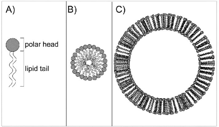
use of amphipathic lipids, consisting of hydrophilic (“head”) and hydrophobic (“tail”) parts
(A). The hydrophobic force generally results in the hydrophobic chains aggregating
together away from water molecules and the hydrophilic moieties mediating contacts with
water.40 This kind of interactions can give rise to the formation of two different
supramolecular assemblies: micelles or vesicles (Figure 1 - 1). Micelles are spherical
structures composed of a polar surface and a hydrophobic internal core where
11
lipids form a double-layered membrane. In this structure, lipid tails point towards the
center of the double layer and polar heads interact with water molecule present both on
the external and on the internal side (lumen) of vesicles. This membrane is able to
separate a water compartment from the external environment. Lipid vesicles can be
prepared by several methods and with several lipid compositions.41 Different kinds of
lipids influence the formation of the bilayer and what determines the formation of micelles
or vesicles is related to factors such as the surface of the polar head group and the length
of the hydrophobic tail.42 Among the methods described, there are two which are mainly
exploited with in vitro TX/TL systems.
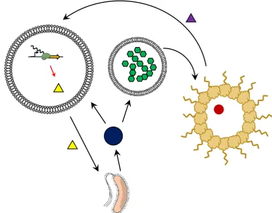
Figure 1 - 1. Schematic representation of an amphipathic lipid and the possible supramolecular assembly in water solutions. A) An amphipathic lipid is mainly composed of a polar group (named “head”) and a lipid moiety (named “tail”). When dispersed in water, lipid tails tend to interact with each other to exclude water molecules, resulting in different kinds of supramolecular assembly. B) A micelle is formed when lipid tails point towards the center of a spherical structure. C) A liposome is a spherical vesicle formed when lipids assemble in a bilayer sheet where the tails point towards the center of the sheet. A typical biological membrane mainly contains phospholipids, composed of a phosphate group (head) joined to two fatty acid chains (tails).
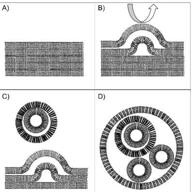
The Freeze-Dried Empty Liposomes (FDEL)43 method (Figure 1 - 2) involves the
formation of a thin lipid film after the evaporation of the organic solvent where lipids are
dispersed, typically chloroform. The film is resuspended in water to allow for the formation
of vesicles containing multiple lipid bilayers and homogenized by mechanical stirring or
extrusion. Vesicles are then lyophilized and later resuspended with the TX/TL
12
Figure 1 - 2. Freeze-Dried Empty Liposomes (FDEL). A) A thin film of lipids consisting of multiple bilayers is created by solvent evaporation. Typically, lipids are dispersed in organic solvents like chloroform and then evaporated to allow the formation of a homogeneous film. B) The resuspension of the film with water induces the swelling of lipid bilayers, from which C) multilamellar vesicles detach, to D) generate a heterogeneous dispersion of lipid vesicles.
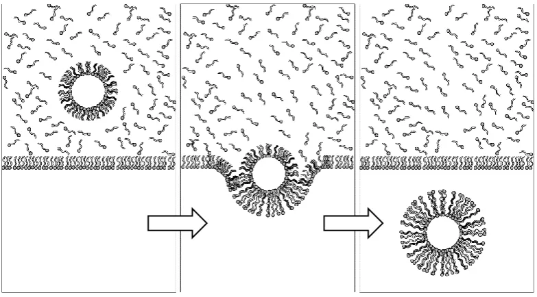
The other method was first described by Pautot et al. and involves the formation of one lipid leaflet at a time (Figure 1 - 3).46 A solution of mineral oil containing phospholipids
is deposited on top of a water solution. Phospholipids partition to the interface of the two
phases with polar head groups facing the water phase and the hydrophobic tails
submerged in the oil phase. Separately, a water-in-oil (w/o) emulsion is created by
mechanical agitation of a mixture of water and mineral oil that is stabilized by lipids. The
w/o emulsion is later placed on top of the biphasic solution. The water droplets are then
forced from the oil to the water phase by mild centrifugation. The passage through the
interface allows the formation of a second lipid leaflet surrounding the monolayer that
stabilize the w/o emulsion droplet, thus generating a lipid bilayer. The process is
13
The contents of each vesicle may vary regardless of the vesicle generation
protocol used. Therefore, gene expression through TX/TL will likely vary among the lipid
vesicles. For the method developed by Pautot et al., a heterogeneous distribution of components was confirmed and that the DNA template concentration inside each vesicle
was indicated as one of the most critical factors influencing gene expression.51 Therefore,
variability between compartments can be partially overcome by increasing the DNA
concentration to ensure that the minimal amount of genetic elements required for function
are present in each droplet.
Figure 1 - 3. A method for the creation of liposomes based on emulsion droplets.
Water-in-oil emulsion droplets are stabilized by a phospholipid monolayer and forced to cross a phospholipid interphase between oil and water. Another phospholipid leaflet is formed around the droplet during this passage. Lipid vesicles carrying enzymatic reactions and in vitro TX/TL systems can be formed with this method by the encapsulation of the desired reaction mixture in lipid emulsion droplets.
In addition to lipid membranes, it is possible to create other kinds of compartments
in aqueous solutions where enzymatic reactions or in vitro gene expression can occur.
Compartmentalization can be driven by diverse forces that can be divided into two main
groups. One type of compartments consists of semipermeable membranes obtained by
means of diverse interactions such as ion bonds or Pickering emulsions. The second type
of compartments is obtained by the separation of two liquid phases so that droplets of a
phase are dispersed in the other. These droplets typically contain a content in water much
lower than the external phase and offer a membrane-free compartment where several
kinds of small and big molecules can be absorbed. The number of possible systems
available constitutes an interesting scenario of potential artificial cells with different
14
Pickering emulsions can be generated through a process in which emulsion
droplets are stabilized by the localization of particles to the interface of two immiscible
phases.52 Particles showing an equilibrium between hydrophobic and hydrophilic
properties can behave as surfactants and isolate water droplets in an organic phase.
These includes colloidal nanoparticles made of a mixture of hydrophobic and hydrophilic
molecules to create colloidosomes3,53 and amphipathic protein-polymer conjugates can be
made to create structures called proteinosomes.5,6 The basic procedure for the creation of
these compartments involves an emulsification of aqueous solutions in an organic phase
together with colloidal particles or amphipathic protein-polymer conjugates. The surfactant
behavior of these particles creates a porous membrane surrounding the water droplet that
is later stabilized by intermolecular covalent bonding. The cross-linked membrane can be
safely transferred to water solutions, where it could potentially be in contact with natural
cells.
Porous membranes can also be formed by the aggregation of molecules through
ionic interactions occurring at the interface of two aqueous solutions. One example is
given by alginate microcapsules, resulting from the precipitation of anionic polysaccharide
alginate in presence of specific counterions. Complexes with sodium alginate are soluble,
while other cations such as calcium or the polysaccharide chitosan induce precipitation.
The addition of sodium alginate solution to a solution containing one of these cations
triggers salt precipitation at the interface, resulting in the separation of two aqueous phase
through an ionic membrane. The dimension of the compartments can be controlled by
controlling the dimensions of sodium alginate droplets added to the cation solution.
Enzymes or TX/TL machineries can be mixed together with sodium alginate to obtain
cellular mimics capable of catalysis or gene expression.4,54 Similarly to phospholipid
bilayers, despite the different composition and chemical properties, this type of membrane
allow for the passage of small molecules while retaining macromolecules,4 and some
methods are available to adjust the permeability to a certain extent.55
A good example of membrane-free compartments is given by the process of
coacervation, which involves the separation of two liquid phases as a result of the
attraction of oppositely charged macro-ions. The interaction is strong enough to reduce
the binding with water molecules but do not result in an arrangement as stable and
ordered as salt crystals. Therefore, the attractive forces give rise to phase separation
rather than salt precipitation. Coacervate droplets were initially proposed as a model of
protocells crucial for the origins of life,56 and some applications as cellular mimics were
recently reported.57,58 The absence of a membrane and the charge of these compartments
15
the molecularly crowded environment of coacervate droplets has been reported to
enhance enzymatic activity60 and gene expression.57
It is also possible to have cellular mimics composed of multiple compartments, that
allows for the specific localization of protein expression or enzymatic reactions. Water
solutions differing in density can be mixed together in a multiphasic system, and
water-in-oil emulsion droplets or lipid vesicles can be made to include these multiple
compartments.61,62 In aqueous two-phase systems, for example, it has been shown how a
protein synthesized in these systems can preferentially localize to one of the two
phases.63 Multiple compartmentalizations can also be achieved in liposomes obtained by
Pautot’s method where different emulsion droplets are merged together. Every
compartment can carry separate gene expression reactions.48 Separate compartments
can be put in contact to communicate with each other and allow the free diffusion of small
molecules, resulting in a cascade of enzymatic reactions where the major players involved
are confined to different spaces.64 The possibility to physically separate each single
component of an enzymatic reaction could allow for the optimization of each single step
with a reduced waste in resources. Therefore, multiple compartments offer a good tool for
the improvement of artificial cells.
1.3 Molecular communication
Although gene expression and enzymatic reactions have been widely shown to
occur in confined compartments, there are only a few examples of artificial cells actively
interacting with their environment. A chitosan-alginate capsule can synthesize and release
by passive diffusion quorum sensing molecules and deliver a signal to bacteria.4 Small
molecules can induce a response in liposome-based artificial cells through genetic
regulatory elements, such as riboswitches1,2 or transcriptional repressors65,66.
Despite the limited number of mechanisms described, some attempts to control
communication between artificial and natural cells have been accomplished. In a work
carried out in our group,1 it was shown how it is possible to translate an inert chemical
message for E. coli into a meaningful signal able to induce a response. The communication pathway involved the expression of a pore-forming protein inside of lipid
vesicles, under the control of the theophylline riboswitch. In the presence of theophylline,
the riboswitch bound to the molecule undergoes a conformational change unveiling a
hidden ribosome binding site (RBS) thus allowing for the expression of α-hemolysin. The
pores formed by this protein released IPTG outside of liposomes and activated the
16
obtained by a Pickering emulsion of a phospholipid monolayer surrounding water-in-oil
emulsion droplets. These phospholipid emulsion droplets were tested for their ability to set
communication with bacteria exploiting genetic regulatory elements derived from quorum
sensing machinery.67 Although the system is not so versatile because it requires bacteria
to be included in emulsion droplets, it demonstrates the feasibility of establishing
communication between artificial cells and bacteria in both directions, either by sensing or
by sending a chemical message.
1.4 The need for artificial cells communities
The potential interactions between natural and artificial cells are limited by several
constraints. Artificial cells displaying more complex behavior require more energy
demanding circuitry beyond what is easily possible with current technology. Natural cells
have evolved pathways for energy resource management and regeneration, while artificial
systems are still far from complete self-sufficiency. Nonetheless, some progress has been
made.
Attempts at restoring energy resources for TX/TL reactions in cellular mimics
resulted in increased protein yield and prolonged activity of the system. The selectivity of
the membranes is chosen to retain all the elements required for protein synthesis inside of
the artificial cell so that small molecules cannot be directly supplemented from the
external solution to replenish the reaction. To increase the availability of resources some
elements can be added to the TX/TL reaction, such as maltose that was reported for
glucose regeneration and ATP production through the Krebs cycle.8 Amino acids and
nucleotides can also be regenerated by the degradation of nascent RNA and protein. It
was shown how an increased turnover of these resources by the addition of specific
RNases and proteases increases the final yield of the protein of interest.32 It is also
possible to allow the passage of small molecules, like amino acids and nucleotides, that
are not permeable to certain type of lipid membranes such as
1-palmitoyl-2-oleoyl-sn-glycero-3-phosphocholine (POPC) through α-hemolysin pores resulting in prolonged
protein expression.7
If artificial cells were able to self-assemble, all the elements required to perform a
specific function could be easily generated thus providing a much more efficient system. A
complete duplication process in a cellular mimic has not been described yet but some
examples of cellular division processes were conducted in liposomes. The process of
growth and division was first shown to occur through physical processes consequent to
the addition of micelles68 and change in osmotic pressure62 or redox status.69 It is clear
17
system, but some attempts on the reconstitution of the natural machinery responsible for
E. coli cell divisions have been made as well. A combination of the purified proteins involved in the mechanism are able to form rings at the center of liposomes,70 eventually
leading to division.71 It is also possible to reconstitute basic elements of the eukaryotic
cytoskeleton that are involved in cellular division such as actin filaments.72,73 Although
there are no reports of division, it is possible to modulate the shape of the membrane
through the combination with myosin.74 Further, it has been shown that by combining PCR
and ionic interactions between DNA and lipid membrane, it is possible also to force the
segregation of vesicles with an equal distribution of DNA molecules. Artificial cells may
then duplicate together with genetic information.75
Despite the absence of a completely self-sustainable cellular mimic, the scenario
of artificial cell is broadening over the years offering an increasing number of artificial cells
suitable for different kind of applications. Every system has a diverse set of advantages
and disadvantages and their combination may complement reciprocal defects. We can
then envision a mixed population of artificial cells assigned with different tasks to be
fulfilled as the result of a collaboration. The success of the desired function will be
entrusted to the community rather than to the individual cellular mimic.
The creation of artificial cell communities is strictly related to the construction of
molecular communication pathways. In collaboration with Prof. Stephen Mann at the
University of Bristol, we are currently in the process of creating interactions in a mixed
population of cells consisting of liposomes carrying in vitro gene expression systems, proteinosomes with enzymatic activity and E. coli. In the meantime, his group managed to show how it is possible to have interactions between different kinds of artificial cells in a
predator-prey system where a protease-loaded coacervate seeks and destroys a
proteinosome.58 The project carried in this collaboration is instead focused on cooperative
interactions between artificial cells, possibly expanding the range of communication
pathways between artificial and natural cells by means of the exchange of chemical
messages.
The PhD project described in this dissertation is organized in two main sections
further divided into chapters. The first part reports the work aimed at establishing a
communication pathway between two different populations of artificial cells: liposomes
able for gene synthesis and proteinosomes with catalytic activity. The designed network
foresaw the delivery of a chemical message from liposomes under the control of a genetic
switch. Proteinosomes would have triggered gene expression by enzymatic synthesis of
the inducer molecule and performed enzymatic activity using the chemical message
18
the research of a genetic circuit able for the tightest control on the chemical message
delivery from liposomes. Several transcriptional regulators were tested in in vitro TX/TL reactions and the best was integrated within artificial cells. Consequently, all of the steps
of the molecular communication pathway were separately analyzed. Both the delivery of a
chemical message from liposomes and the enzymatic production of the molecule
responsible for gene induction were tested separately. The number of problems arose
while setting the artificial cells network led to reconsider the whole design so to include
some engineered bacteria.
The second part of the thesis summarizes the trials carried in engineering two
novel biosensors that could possibly be integrated within artificial cells. A DNA-based
sensor was designed to carry two aptamer sequences, one for the binding of an analyte
and another one for the binding of a fluorescent molecule. The fluorescence quantum
yield of the fluorophore would be consistently increased when bound to the aptamer. The
folding of this aptamer would depend on the presence of the analyte: the binding of the
analyte to the respective aptamer would induce a conformational change on the DNA
sensor allowing the binding of the fluorophore. In order to develop a DNA aptamer able for
binding a fluorophore, three in vitro evolution strategies were performed but none of them succeeded, therefore it was not possible to develop the designed biosensor.
The second biosensor was based on the rational design of a transcriptional
regulator able to control gene expression according to the presence of an analyte. The
protein was inspired to a natural transcriptional repressor whose activity is regulated by
the presence of a small molecule. The engineering process was based on the change of
the binding affinity to a molecule similar to the natural ligand. The protein would have
been consequently integrated into a genetic circuit and allow the detection of the analyte,
i. e. the novel ligand, through the regulation of the expression of a fluorescent reporter
protein. Attempts at changing the affinity of the regulator to a novel ligand were performed
after in silico analysis of the interactions between the protein and the natural ligand. Mutants were designed and tested in vitro for gene regulation under the control of both the natural and the novel ligand. Finally, some information on the interactions between protein
19
Aims of the thesis
The work carried in this PhD project is divided into 4 experimental sections
described in the following chapters.
Chapter 2, “Genetic engineering for use in artificial cell”, describes a part of the
project aimed at finding the best regulatory system for use in artificial cells composed of
liposomes carrying in vitro gene expression systems. Four genetic circuits were assembled to carry a transcriptional regulator and a reporter gene. The efficacy of each
regulator was tested by means of in vitro TX/TL reactions;
Chapter 3, “Towards artificial cells consortia”, is centered on a step-by-step
analysis of a molecular communication pathway designed between two kinds of artificial
cells: liposomes carrying in vitro transcription and translation reaction and proteinosomes carrying enzymatic reactions. Liposomes were tested for the ability to perform gene
expression and for the delivery of a chemical message to proteinosomes through
fluorescent reporters. One of the enzymes carried by proteinosomes was tested for the
ability to produce a chemical message capable of triggering gene expression in
liposomes;
Chapter 4, “Selection for a malachite green DNA aptamer for use in a sensor
molecule”, reports some attempts at developing a biosensor based on DNA. Engineering
this biosensor foresaw the in vitro evolution of a DNA aptamer able to bind and increase the fluorescence yield of the fluorophore malachite green. Three strategies aimed at
evolving the aptamer were designed and tested;
Chapter 5, “Engineering TrpR to sense the neurotransmitter serotonin”,
summarizes some trials focused on the development of another biosensor based on the
transcriptional repressor TrpR. Some mutants were rationally designed to switch the
affinity of the protein from the original ligand tryptophan to serotonin, that has a very
similar chemical structure. Similarly to what was performed for the transcriptional
regulators in chapter 2, these mutants were cloned in a genetic circuit and tested through
20
Part A
21
Chapter 2
22
One basic requirement for an artificial cell with potential application is tunability. A
desired function must be activated only when needed, so these systems must include a
switch mechanism capable of changing from an “on” to an “off” state. An artificial cell
based on lipid vesicles encapsulating E. coli transcription and translation machinery can be engineered by following the same methods applied to natural cells, by intervening at a
genetic level.
Gene expression can be regulated through several mechanisms, many of which
occur through interactions between small molecules and macromolecules, such as
proteins and nucleic acids. The effect of these molecules can be both positive or negative
on gene expression. Previous work carried out in our group demonstrated the possibility
to control gene expression in artificial cells by means of a riboswitch.1,2 These sequences
at the 5’-untranslated region (UTR) of mRNAs can mask or unveil the ribosome binding
site according to the presence or absence of a ligand specific for the aptameric domain of
the riboswitch. Although effective, this regulation turned out to be not strict enough and
showed much background expression in the off state, in the absence of the inducer
molecule.
One of the major disadvantages of the system was thought to be due to the fact
that the regulation occurs at a translational level. The gene of interest is actively
transcribed under the control of a highly processive polymerase (T7 RNA Polymerase)76
and even a small percentage of mRNA may result in a high background expression.
Therefore, regulation at the level of transcription seemed a better alternative to obtain a
stricter off state. One very simple example of this kind of genetic regulation is given by
transcriptional repressors. The major feature shared by these protein factors is to bind
specific DNA sequences, called operators, that are close or enclosed in promoter regions
and prevent the binding of RNA polymerases by steric hindrance. The action of these
proteins is regulated by the presence of a cofactor, a small molecule able to induce an
allosteric change that can forbid or allow DNA binding (Figure 2 - 1). In order to find a
good genetic regulation for use in artificial cells, three repressors from E. coli were taken into consideration: 1) LacI, involved in the transport and metabolism of lactose through the
regulation of the Lac operon;77 2) TetR, regulating the expression of a transmembrane
pump aimed at expelling the antibiotic tetracycline;78 3) TrpR, regulating the synthesis of
the amino acid tryptophan.79
All of the repressors can exist in a form bound to their ligand or in a free form. The
first form is known as holorepressor while the second is the aporepressor. The effects
induced by the switch from apo- to holorepressor is different for the four repressors taken
23
undergoes a positive regulation. In the absence of the cofactors, LacI and TetR repress
gene expression while the binding to the ligand forbids the binding to the operators
(Figure 2 - 1 A). TrpR has instead an opposite mechanism, being active only as a
holorepressor (Figure 2 - 1 B). The natural ligand for LacI is allolactose, a catabolite
created by the enzyme β-galactosidase in presence of lactose.80
The catabolite induces
the expression of all the genes required for its metabolism. TetR is instead involved in the
resistance to the antibiotic tetracycline. When the antibiotic is present, E. coli cells react with the production of a pump to expel the drug.78 TrpR regulates a whole operon involved
in the synthesis of tryptophan shutting down the expression of the enzymes required in
presence of high levels of the amino acid. Interestingly, this operon is subjected to a
secondary regulation that involves the coding sequence trpL whose peculiarity is to induce different folding of the mRNA sequence according to the levels of tryptophan. The mRNA
includes two tryptophan codons responsible for this change: in presence of high levels of
tryptophan the mRNA forms a transcriptional terminator downstream of the peptide and
none of the following genes is transcribed, while low levels of tryptophan causes a stall in
ribosome that induces an alternative folding in the downstream RNA preventing the
formation of a terminator and allowing the transcription of the downstream genes.81–83
Figure 2 - 1. Transcriptional regulatory pathways tested. A) Transcriptional repressor with negative regulation (TetR, EsaR, LacI). The repressor binds to the operator sequences close to the promoter thus blocking RNA polymerase binding through steric hindrance. The binding to the ligand induces a conformational change that clears the promoter sequence to start gene transcription. B) Transcriptional repressors with positive regulation are active for repression only in presence of the ligand (TrpR). C) Transcriptional activators promote the binding of RNA polymerase rather than blocking it. The activity depends on the presence of small cofactors that induce the necessary allosteric change to lead RNA polymerase binding (LuxR).
Not all of the natural cofactors can be exploited for in vitro analysis, but alternative ligands are commercially available. Allolactose can be substituted by the cheaper and
more efficient isopropyl β-D-1-thiogalactopyranoside (IPTG) which is not hydrolyzable by
24
β-galactosidase and has been regularly used for the induction of recombinant protein
expression.84 Tetracycline can be substituted by the non-toxic analog anhydrotetracycline
(aTc), which was shown to have a 30-fold increased binding affinity.85 For TrpR, several
analogs are described in the literature either with a positive or a negative action on the
repressor.86 Among these, indole-3-propionic acid (IPA) was chosen because of the ability
to induce gene expression in vivo.87
The transcriptional regulators were individually tested in TX/TL reactions for their
ability to control the expression of a reporter gene. The repressor and the reporter genes
were cloned separately under the control of a T7 promoter, and the reporter gene
contained the specific operator sequence for the repressor right downstream of the
promoter. Both translation and transcription levels were simultaneously monitored by
means of a fluorescent protein and an aptamer joint at the 3’-UTR of the mRNA able to
bind and increase the fluorescence quantum yield of a fluorophore. In order to individuate
the regulator with the best difference between on and off state, the experiments were
conducted in presence or absence of the repressor and in presence or absence of the
25 2.1 Materials and methods
2.1.1 Reagents and general supplies
PURExpress® In vitro Protein Synthesis Kit, DpnI, RNase inhibitor was purchased from New England Biolabs; Wizard® SV Gel and PCR Clean-Up System Wizard® Plus
SV Minipreps DNA Purification System were purchased from Promega; indole 3-propionic
acid (IPA), isopropyl β-D-1-thiogalactopyranoside (IPTG), anhydrotetracycline (aTc),
diethyl pyrocarbonate (DEPC) were purchased from Sigma-Aldrich;
phenol:chloroform:isoamyl alcohol (25:24:1) solution was purchased from Thermo Fisher
Scientific; (Z)-4-(3,5-difluoro-4-hydroxybenzylidene)-1,2-dimethyl-1H-imidazol-5(4H)-one
(DFHBI) was purchased from Lucerna technologies; One Shot® TOP10 Chemically
Competent E. coli cells were purchased from Invitrogen; nickel- nitrilotriacetic acid agarose resin (Ni-NTA) and 0.1 ml tubes were purchased from Qiagen; E.Z.N.A.®
MicroElute RNA Clean Up Kit was purchased from Omega Biotek.
All the material used for the preparation of the home made S30 extract was
purchased according to Noireaux’s indications39 except for the following: Yeast extract
and Tryptone to prepare 2xYT medium, polyethylene glycol (PEG) 8000 Da, all of the 20
amino acids, Tris base were purchased from Sigma-Aldrich; SnakeSkin™ Dialysis Tubing
was purchased from Thermo Fisher Scientific; 10X TBE buffer was purchased from
Euroclone; E. coli BL21 Rosetta 2 (DE3) from Novagen was used in place of E. coli BL21 Rosetta 2 and was received from Prof. Friedrich C. Simmel
2.1.2 Instruments
FastPrep®-24 from MP biomedicals was used for the bead-beating step in S30
extract; Rotor-Gene Q from Qiagen was used for the measurements of protein and RNA
levels in S30 and PURE system reactions; Infinite M200 plate reader from Tecan was
used to measure luminescence.
2.1.3 Water treatment with DEPC for nuclease inhibition:
26
2.1.4 Plasmids and cloning
All of the plasmids were assembled with the Gibson method88 and DNA primers
were purchased at Eurofins MWG. Linear fragments of double-stranded DNA were
created by PCR using Phusion polymerase. A 50 µl mix contained 1X HF buffer, 0.2 mM
each dNTPs, 0.5 µM of each primer, 0.02 U/µl Phusion, ~ 0.2 ng/µl plasmid DNA
template. The reaction ran on a thermal cycler with the following protocol: 98°C for 2 min
for initial denaturation, then 29 cycles of 98 °C for 5 s, annealing temperature for 10 s and
72 °C for 15 s/kb and a final extension step at 72 °C for 10 min. The annealing
temperature was calculated for each primer by the online tool IDT oligo analyzer.
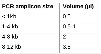
PCR fragments were treated with 0.4 U/µl of DpnI for at least 1 h at 37 °C, then
mixed together with a premixed Gibson assembly stored at -80 °C. The final volume of
reaction was 10 µl and the mixture contained: 100 mM Tris pH 7.4, 10 mM MgCl2, 0.2 mM
each dNTP, 10 mM DTT, 6.25 mM PEG 8000, 1 mM NAD. The DNA was added
according to the following table:
Table 2 - 1. Volumes for Gibson assembly.
PCR amplicon size Volume (µl)
< 1kb 0.5 1-4 kb 0.5-1
4-8 kb 2
8-12 kb 3.5
PCR products were always loaded on a 1X TBE 1% agarose gel for analysis.
When the PCR product contained some extra bands, the correct band was extracted from
the agarose gel with Promega Wizard gel and PCR clean-up kit and added to the mix to
fill the final volume. Mixes were then incubated at 50 °C and then added to E. coli TOP10 competent cells for transformation. Details of DNA sequences and assembly are
described in the following tables.
Table 2 - 2. Primers used for PCR amplification.
Primer ID Sequence (5’-3’)
DC010 GCGGATCCGAATTCAATTAGTTTGAACTTATAAGGAGAATAATCT ATGGCTTCCTCCGAAGACG
27
DC079 CGTACTAGTTAACTAGTACGCCCTATAGTGAGTCGTATTAATTTCGC DC083 GGAGATATACATATGGCTAGCATGATGGCCCAACAATCACCCTATTCA DC084 CTTGTCGACGGAGCTCGAATT TCAATCGCTTTTCAGCAACACCTCTT DC094 GGAGATATACATATGGCTAGCATGATGTCCAGATTAGATAAAAGTAAAGTG DC096 CTTGTCGACGGAGCTCGAATTTCAGGACCCACTTTCACATTTAAGTTG DC098 CTTGTCGACGGAGCTCGAATTCTGCCCGCTTTCCAGTC
DC099 CTTGTCGACGGAGCTCGAATTTCACTGCCCGCTTTCCAGTC DC100 ATGTATATCTCCTTCTTAAAGTTAAACA
DC101 GCGCAACGCAATTAATGTAAGTTAG
DC103 TCTCTATCACTGATAGGGACCCTATAGTGAGTCGTATTAATTTC DC104 GGATATAGTTCCTCCTTTCAGCAAA
DC111 ATGGCTTCCTCCGAAGAC DC133 CCCTATAGTGAGTCGTATTA
DC145 TGTTTAACTTTAAGAAGGAGATATACATATGAAACCAGTAACGTTATACG
DC146 TAATACGACTCACTATAGGGCCCCTCTAGAAATAATTTTGTTTAACTTTAAGA AGGAG
DC147 CTAACTTACATTAATTGCGTTGCGCAAGTGGCGAGCCCGATCTTCCCCAT DC157 GAGTCGTATTAACCGGCTGCAGATCTCGATCCTCTACGCCG
DC159 CAGTCGA AAGACTGGGCCTTTCGTTTTAT GTGATGTCGGCGATATAGGC
DC160 CCAGTCTTTCGACTGAGCCTTTCGTTTTAT GGATATAGTTCCTCCTTTCAGCAAA DC161 ATGGGGAAGATCGGGCTCGCCACTT
DC162 AAGTGGCGAGCCCGATCTTCCCCATTAATACGACTCACTATAGGGGAATT DC163 AGTTCAAACTAATTGAATTCGGATCCGCTCACTGCCCGCTTTCCAGTC DC199 ATTAATTGCGTTGCGCAGCAGCCCAGTAGTAGGTTG
DC200 AAGTGGCGAGCCCGATCTTCCCCATTAATACGACTCACTATAGGG DC257 CTAACTTACATTAATTGCGTTGCGC
DC258 CTAACTTACATTAATTGCGTTGCGC DC275 ATATCCGGATTGGCGAAT
DC276 TTAAGCACCGGTGGAGTG
DC277 AGCTCAGTCCTAGGTACAGTGCTAGCTACTAGAGTCACACAGGAAA DC278 TACCTAGGACTGAGCTAGCCGTCAATGAGCGCAACGCAA
DC299 AGTACTTTCCTGTGTTACTCTAGTA
DC300 TGCCTGGCTCTAGTATTATTACCTTGCTGCTGACGC
DC302 TAACACAGGAAAGTACTATGTTCTCTTTCTTCCTTGAAAACC DC306 ATTCGCCAATCCGGATAT
28
FC267 CCGGTTAATACGACTCACTATAGCCTGTACTATAGTGCAGGTGGAAGATTGT GAGCGGATAACAATTCC
JF001A fw TAACTCGAGCACCACCACCACCAC RL007 CCCCTCTAGAAATAATTTTGTTTA T9002g FW TAATAATACTAGAGCCAGGCATC
Table 2 - 3. Relevant sequences used in this section. The table reports the insert of relevant plasmids whose information is not available elsewhere.
ID Sequence (5’-3’)
pT7_lacO_mRFP1_ spinach
TAATACGACTCACTATAGGGGAATTGTGAGCGGATAACAATTCCCCT CTAGAAATAATTTTGTTTAACTTTAAGAAGGAGATATACATATGGCTT CCTCCGAAGACGTTATCAAAGAGTTCATGCGTTTCAAAGTTCGTATG GAAGGTTCCGTTAACGGTCACGAGTTCGAAATCGAAGGTGAAGGTG AAGGTCGTCCGTACGAAGGTACCCAGACCGCTAAACTGAAAGTTAC CAAAGGTGGTCCGCTGCCGTTCGCTTGGGACATCCTGTCCCCGCAG TTCCAGTACGGTTCCAAAGCTTACGTTAAACACCCGGCTGACATCCC GGACTACCTGAAACTGTCCTTCCCGGAAGGTTTCAAATGGGAACGTG TTATGAACTTCGAAGACGGTGGTGTTGTTACCGTTACCCAGGACTCC TCCCTGCAAGACGGTGAGTTCATCTACAAAGTTAAACTGCGTGGTAC CAACTTCCCGTCCGACGGTCCGGTTATGCAGAAAAAAACCATGGGTT GGGAAGCTTCCACCGAACGTATGTACCCGGAAGACGGTGCTCTGAA AGGTGAAATCAAAATGCGTCTGAAACTGAAAGACGGTGGTCACTACG ACGCTGAAGTTAAAACCACCTACATGGCTAAAAAACCGGTTCAGCTG CCGGGTGCTTACAAAACCGACATCAAACTGGACATCACCTCCCACAA CGAAGACTACACCATCGTTGAACAGTACGAACGTGCTGAAGGTCGT CACTCCACCGGTGCTTAAGCCCGGATAGCTCAGTCGGTAGAGCAGC GGCCGGACGCAACTGAATGAAATGGTGAAGGACGGGTCCAGGTGT GGCTGCTTCGGCAGTGCAGCTTGTTGAGTAGAGTGTGAGCTCCGTA ACTAGTCGCGTCCGGCCGCGGGTCCAGGGTTCAAGTCCCTGTTCGG GCGCCA
29
30
Table 2 - 4. Plasmids used in this section and relative cloning strategies.
Plasmid ID Backbone Insert Source Cloning strategy
Primers Template
pET21b Novagen
BBa_K731500 pSB1C3 pLacIq_LacI_ pTac_lacO
Registry of standard biological parts BBa_C0040 pSB1C3 TetR Registry of
standard biological parts BBa_T9002 pSB1A3 pTet_LuxR_
pLux_sfGFP
Registry of standard biological parts DC024A pET21b
(LacI removed from the backbone)
pT7_LacI::His DC057/DC101 pET21B DC147/DC133 pET21B DC145/DC098 BBa_
K731500 DC146/DC100 -
DC013A pET21b pT7_lacO_TrpR DC083/DC084 E. coli
genome DC057/DC058 pET21b DC019A pET21b pT7_lacO_TetR DC094/DC096 BBa_C0040
DC057/DC058 pET21b DC049A DC024A pT7_LacI DC099/JF001
A fw
DC024A
DC021A pET21b pT7_tetO_ mRFP1_spinach
DC103/RL007 FC013A
DC032A pET21b pT7_trpO_ mRFP1_spinach
DC079/RL007 FC013A
FC013A pET21b pT7_lacO_ mRFP1_spinach
Mansy Lab
DC076A pET21b pT7_trpO_ mRFP1_spinach _pT7_TrpR
31
FC043A pET21b pT7_lacO_His:: MBP::EsaR
Mansy Lab
DC053A DC024A pT7_esaO_ mRFP1_spinach
DC147/DC157 FC013A FC267/DC101 FC013A DC035A DC024A pT7_lacO_
mRFP1_spinach _ pT7_lacI
DC161/DC159 DC049A DC160/DC162 FC013A
DC052A DC024A pT7_lacO_lacI_ mRFP1
DC010/DC101 FC013A DC100/DC147 FC013A DC145/DC163 DC049A
2.1.5 DNA purification with phenol:chloroform mix
When DNA quality was not high enough (A260/A280 ≥ 1.8 and A260/230 ≥ 2), an
extra step of purification was required. 50 µl of DNA were diluted in 500 µl of
DEPC-treated water and mixed together with 1 volume (vol) of phenol:chloroform:isoamyl alcohol
(25:24:1). The tube was vortexed and spun at max speed for 10 min to separate the
aqueous from the organic phase. The upper aqueous phase was transferred to a new
tube containing 1 vol of chloroform and spun at max speed for 5 min. The upper part was
then mixed together with 2.5 vol of ethanol and 1/10 vol of 3 M sodium acetate pH 5.5 and
incubated at -20 °C for at least 1 h. Then DNA was precipitated by centrifugation at max
speed for 30 min at 4 °C. The supernatant was discarded and 200 µl of 70% ethanol were
added to the pellet without resuspending. The pellet was spun again at max speed for 10
min at 4 °C. The pellet was dried at 65 °C on a thermoblock and resuspended in 20 µl of
DEPC-treated water.
2.1.6 In vitro gene expression with PURE system
DNA templates were prepared by PCR amplification using Phusion polymerase
and primers DC046 and DC104. The reaction was assembled as described above and the
thermal protocol included changes in the annealing temperature (touchdown PCR): 72 °C
for 15 cycles, 68 °C for 10 cycles and 65 °C for the last 10 cycles. The extension time was
set at 15 sec.
The reactions were prepared with some modifications from the manufacturer’s
instructions. The final volume was increased by 8%, DNA was added to 12.5 nM and the
reaction mixture was supplemented with 0.75 U/µl RNase inhibitor and 60 µM DFHBI.
32
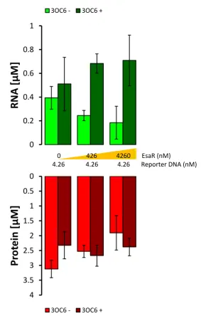
3OC6, 0.5 mM IPTG, 1 mM aTc, 0.5 mM IPA. Fluorescence kinetics of both transcription
and translation were monitored with real-time PCR cycler Rotor-Gene Q. Transcription
levels were monitored through spinach aptamer bound to DFHBI on channel Green
(excitation: 470 ± 10 nm; emission: 510 ± 5 nm), while translation levels were monitored
by the expression of the fluorescent protein mRFP1 on channel Orange (excitation: 585 ±
5 nm; emission: 610 ± 5 nm). Fluorescence levels were recorded every 5 min at 37 °C for
~13 h.
2.1.7 In vitro gene expression with home-made S30 E. coli extract
A reaction mix for in vitro transcription and translation based on a crude E. coli
extract was prepared according to the protocol of Noireaux and colleagues39 with the
modifications indicated in § 2.1.1. The final volume of the reaction was 10 µl with 20 nM of
plasmid DNA (RL082A). When needed 3OC6 HSL was supplemented to 10 µM. Gene
expression was verified by luminescence.
2.1.8 Fluorescence standard curve and data normalization
In order to convert fluorescence raw data into molar units, a standard curve was
made both for mRFP1 and for spinach aptamer together with DFHBI. mRFP1 protein was
expressed in E. coli BL21 (DE3) pLysS cells transformed with plasmid FC011A and purified with Ni-NTA column according to manufacturer’s instructions. The protein
concentration was measured by nanodrop spectrophotometer and converted according to
its extinction coefficient (ε584 nm = 44 000 M−1 cm−1).89 Spinach RNA aptamer fused at
3’-UTR of mRFP1 mRNA was transcribed in vitro using a home-made T7 RNA polymerase and plasmid DC032A in the following reaction mix prepared according to the indications
from Seelig:90 35 mM MgCl2, 2 mM spermidine, 200 mM HEPES adjusted to pH 7.5 with
KOH, 1 mg/ml BSA, 4 mM DTT, 5 mM each NTP, 1 mU/µl yeast inorganic
pyrophosphatase, 0.4 U/µl RNase inhibitor, 3 U/µl T7 RNA polymerase, 0.2 µM DNA
template. The reaction was incubated at 37 °C for 4 h, then DNase I was added to a final
concentration of 20 mU/µl together with its buffer provided by the supplier and incubated
at 37°C for 1 h. The RNA was purified with E.Z.N.A.® MicroElute RNA Clean Up Kit and
quantified at the nanodrop spectrophotometer by measuring the absorbance at 260 nm.
The concentration was converted in molarity with a dedicated online tool
(biotools.nubic.northwestern.edu/OligoCalc.html). Different concentrations of mRFP1 (0.2,
33
KCl, pH 7.6) and different concentrations of spinach mRNA (0.06, 0.15, 0.3 and 0.6 µM)
were prepared with HEPES buffer together with 60 µM DFHBI.
2.1.9 Luciferase assay
To 10 µl of S30 reaction incubated for 4 h at 30 °C, 10 µl of a 2X luciferase mix
from Promega, containing luciferin and ATP was added. Luminescence was measured on
a 384-wells plate with a Tecan Infinite M-200 with an integration time of 1 s.
2.1.10 Chemically competent E. coli cells
E. coli TOP10 stored in glycerol stocks were grown overnight in 5 ml of LB, then reinoculated in 50 ml of LB to a starting dilution of 1:100. Cells were grown up to OD600 =
0.5, then chilled on ice for 10 min and harvested by centrifugation at 5000 g for 10 min at
4 °C. The pellet was resuspended in 15 ml of transformation buffer (10 mM Tris-HCl, pH
7.0, 50 mM CaCl2), chilled on ice for 15 min and spun down again at 5000 g for 10 min at
4°C. The pellet was resuspended in 4 ml of transformation buffer and 20% glycerol. Cells
were flash frozen in liquid nitrogen and stored at -80°C.
2.1.11 Transformation of E. coli cells
One aliquot of chemically competent E. coli was thawed on ice and incubated for 30 min with the DNA to be transformed. Heat shock was for 1 min at 42 °C, placed on ice
for 2 min, then 800 µl of LB were added to the cells. The culture was incubated at 37 °C
with shaking at 220 rpm for 1 h.
2.1.12 Purification of the repressor EsaR
The coding sequence of EsaR was fused both to a maltose binding protein (MBP)
and a His tag and cloned in plasmid pET21b to create plasmid FC043A, according to the
indications of Schu et al.91E. coli BL21 (DE3) pLysS cells were transformed with FC043A and bacteria were grown at 37 °C shaking at 220 rpm until they reached OD600 = 0.5.
Protein expression was induced by the addition of 1 mM IPTG and cells were kept
growing at 37 °C shaking at 220 rpm for 4 h. Protein was then purified with Ni-NTA
34 2.2 Results
2.2.1 Design of genetic circuits and tests of the transcriptional repressors in PURE system
For all of the repressors to be tested, two plasmids were designed. One plasmid
contained the repressor coding sequence under the constitutive promoter T7, and another
plasmid containing a reporter gene under the control of a T7 promoter fused to the
specific operator close to the transcription start site: in position +3 from transcription start
site for LacI and +4 for TetR and TrpR. A similar design was previously shown to be
functional for LacI and TetR repression activity in in vitro transcription and translation reactions,92,93 while for TrpR there was no such data available. The reporter gene coded
for the fluorescent protein mRFP194 to monitor translation levels and the spinach
aptamer95 that was placed at the 3’-UTR of the mRNA to monitor transcription levels. This
aptamer was developed to bind a ligand designed to resemble the GFP chromophore:
(Z)-4-(3,5-difluoro-4-hydroxybenzylidene)-1,2-dimethyl-1H-imidazol-5(4H)-one (DFHBI). This
molecule free in solution has a very low fluorescence quantum yield that is strongly
increased by the binding of the aptamer. Such a construct was previously demonstrated to
be a good tool for the real-time monitoring of both transcription and translation levels in
the PURE system.36
All of the genetic elements were amplified by polymerase chain reaction (PCR) in
order to obtain linear fragments of double stranded DNA spanning from the promoter to
the transcriptional terminators. The DNA fragments encoding the repressors were all
tested at different concentrations in the PURE system in the presence and absence of the
respective inducer molecule (Figure 2 - 2). TrpR showed a good transcriptional
repression with no addition of the ligand tryptophan, possibly because what was contained
in the reaction mix was enough to induce the activity of the repressor (Figure 2 - 2 A).
The analog IPA though was not capable of derepression at the concentrations reported in vivo.87 Reporter genes under the control of LacI and TetR (Figure 2 - 2 C, D) both showed a lower level of gene expression when compared with the reporter gene exploited
in TrpR circuit, but while LacI could be derepressed, TetR could not in contrast to
previously reported data.93 Lac circuit showed the best level of switch between on and off
35
Figure 2 - 2. Test on transcriptional repressors LacI, TetR and TrpR. A) Repressors and reporter genes were encoded in two separate PCR fragments under the control of a T7 promoter. The constitutive expression of the repressor gene keeps the reporter gene in an off state, that is activated by the specific inducer molecule. B) - D) RNA (green bars) and protein (red bars) concentrations reached after 6 hours of incubations at 37 °C. The fluorescence values measured for spinach aptamer bound to DFHBI and mRFP1 were converted into molar concentration as described in § 2.1.8. The charts compare the expression levels of the reporter genes in the presence (“+”) or absence (“-“) of the DNA fragments encoding for the repressor and in presence or absence of inducer molecules, that were added to final concentrations indicated in § 2.1.6. All of the values reported are technical triplicates of each sample and the error bars indicates the standard deviation. Among the three repressors tested, LacI showed the best gene induction, although the difference between induced (repressor +, inducer +) and repressed (repressor +, inducer -) state was clear in transcription but not in translation. This result was thought to be caused by the ratio between repressor and reporter DNA, that slightly favors repressor gene expression (the exact ratio is 1:1.15).
A)
C)
36
2.2.2 Deep characterization of LacI circuit in PURE system and some attempts at improvement
In order to improve the difference between on and off states, the molar ratio
between the two plasmids was gradually changed to favor the repressor rather than the
reporter gene. As it can be observed in Figure 2 - 3, a 1:1 molar ratio was enough for
proper gene derepression, and increasing amounts of repressor’s DNA reduced the final
yield of protein after induction. It is possible that most of the elements required for gene
expression are directed towards the synthesis of the repressor more than the reporter
gene.
As the 1:1 molar ratio showed a good difference between on and off states, the
two genes were joined together in the same plasmid. Such a construct, rather than two
separate linear fragments, would allow for an equal encapsulation efficiency of the two
genes when used in artificial cells. Two plasmid designs were compared: one containing
the two coding sequences under the control of two separate T7 promoters, while the other
Figure 2 - 3. Effect of different molar ratios between the DNA fragment encoding the LacI repressor and the DNA fragment encoding the reporter gene under the control of lac operator. To validate whether the induction of gene expression could be improved by decreasing the amount of repressor, three different molar ratios of the DNA fragments carrying the lacI gene and the reporter gene were tested in the same genetic circuit described in Figure 2 – 2 A. The exact molar ratio of 1:1 between reporter and repressor DNA is enough to show a good difference between on and off state, both in RNA (green bars) and protein (red bars) levels. Increasing the molar ratio still provided activation but an excessive amount of repressor’s DNA resulted detrimental to the reaction, possibly because of a sequestration of all the elements required for gene expression that are