ISSN 2319-7617 (Online (An International Research Journal), IF = 4.715, www.physics-journal.org
Influence of Ag+ Fe Co-doping on the Antibacterial
Properties of ZnO Nanopowders Synthesized by
Combustion Method
P. Sathish
Post Graduate, Department of Physics, Annai Vailankanni Arts & Science College,
Thanjavur - 613 007, Tamil Nadu, INDIA. email: [email protected]
(Received on: July 7, Accepted: July 11, 2017)
ABSTRACT
(Ag+Fe) doped ZnO nanopowders have been synthesized using combustion method. Ag doping level was kept as 2 at.% and Fe doping level was varied from 3 to 6 at.% and the structural, optical, surface morphological, and antibacterial properties have been investigated. The FE-SEM images indicate that there is a gradual decrease in the grain size with the increase in the doping level of Fe. The XPS profile clearly confirms the presence of expected elemental composition. Photoluminescence studies reveal the presence of extrinsic defects in the material. Antibacterial activity of Ag and Fe doped ZnO nanopowders against Staphylococcus aureus bacteria were also investigated.
Keywords: ZnO:Ag:Fe nanopowders; FE-SEM; XPS; Antibacterial activities.
1. INTRODUCTION
ZnO is 3.37 eV wide band gap semiconductor which has been extensively studied material. It is an inexpensive and biocompatible material1. It has large free exciton binding
energy (60 meV), which makes it a good candidate for semiconductor device applications such as piezo- electric transducers and solar cell windows2. Silver is one of the well established
anti-bactericidal agents, among various metal dopants which show strong antibacterial efficiency3. In the present study Fe doping level was varied (3, and 6 at. %) and Ag doping
concentration was kept constant as 2 at.% in ZnO nanopowders and their antibacterial and structural properties were studied.
methods the combustion method is inexpensive, which offers high reaction rate. In the present work doped ZnO nanopowders are synthesized using the combustion method.
2. METHOD AND MATERIALS
2.1 Synthesis Process
ZnO:Ag:Fe nanopowders were synthesized using the combustion method. Firstly, 0.2 mol/L of zinc nitrate hexahydrate was dissolved in 200 mL of de-ionized water. Silver nitrate and ferric nitrate were used as dopant precursors for Ag (2 at. %) and Fe ( 3, and 6 at. %), respectively. Urea was used as a fuel and suitable amount of ammonia solution was added to keep the pH of the starting solution as 9. Polyethylene glycol is added as surfactant which can be used to get the gel formation and to achieve the homogeneity. The prepared mixture was magnetically stirred at room temperature and the stirring speed was maintained at 650 r/min. The solution is then shifted to heating mantle, which is maintained at 90 ºC for 2 h. After the ignition process, the setup is allowed to cool for 20 minutes. The collected ash powder is grey in colour and it is crushed using mortar and pestel until it becomes a fine powder. Now the powder is calcined at a temperature of 550 °C for 2 h using a muffle furnace to get the final product.
2.2 Characterization of ZnO:Ag:Mn:F Nanopowders
The crystalline structure of the synthesized powders was studied using x-ray powder
diffraction technique (PANalytical-PW 340/60 X’ pert PRO) using Cu-Kα radiation (λ = 1.5406 Å). Photoluminescence (PL) spectra were recorded using spectro-fluorometer
(JobinYvon_FLUROLOG-FL3-11) with xenon lamp (450 W) as the excitation source of wavelength 325 nm. The surface morphology of synthesized ZnO nanopowders was observed using scanning electron microscope (Carl Zeiss Ultra 55 FE-SEM). The analysis of the element state was done with X-ray photoelectron spectroscopy (XPS) (ESCALAB 250, Thermo).
2.3 Evaluation of Antibacterial Activity
The antibacterial activity of the synthesized ZnO nanopowders was tested against one gram positive bacteria Staphylococcus aureus using agar well diffusion method. Nutrient agar medium was used for growth of bacteria. This agar medium was sterilized in an autoclave at 121 ºC for 15 min and then loaded into petriplate and allowed to solidify in a laminar air flow chamber. After solidification, using a sterile cotton swab, fresh bacterial culture was spread over the plate using spread plate technique.
Three wells each of 5 mm in diameter were made in the agar plates with the help of sterile cork borer. The wells were inoculated with 100 µg/mL of stock solution of the product. All the plates were incubated at 37 ºC for 24 h. After incubation, the plates were observed for
3. RESULTS AND DISCUSSION
3.1 Structural Studies
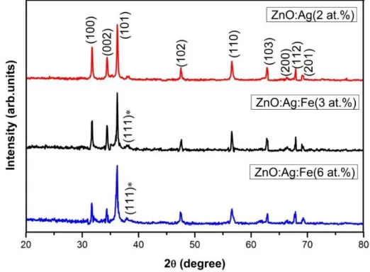
The XRD profiles show that all the samples have hexagonal wurtzite structure of ZnO along with one unmatched diffraction peak observed at 2θ = 38.065°. This peak is associated with the (111) plane of face centered cubic structured metal Ag (JCPDS card No. 04-0783) indicating the presence of secondary phase of metal Ag in the synthesized product The predominance of (101) peak is not affected by the addition of Fe and Ag, even at the highest level of Fe doping.
The crystallite size (D) of the doped ZnO nanopowders are estimated using Scherre’s formula8:
D= .
β θ (1)
where λ (1.5406 Ǻ) is the wavelength of Cu-Kߙ radiation, θ is the Bragg angle and β is the
Full width at half maximum (FWHM).
From fig. 1. It is found that the calculated values of the lattice parameter ‘a’ and ‘c’ and volume of the unit cell (V) are almost consistent with that of standard JCPDS card of ZnO material. From table it is seen that the crystallite size of Ag: ZnO decreases from ∼ 42 to ∼ 33 nm when Fe doping level increases from 3 at.% to 6 at.%. The reduction in the grain size of the doped samples may be due to the slight mismatch in the radii of the host and dopant ions9.
Fig. 1. XRD patterns of Ag (2 at.%) and Fe (3 and 6 at.%) doped ZnO nanopowders
3.2 Photoluminescence Studies
due to the band to band transition10. The increase in the Fe doping level results in a red shift
(increase in wavelength) due to sp-d exchange interactions11,12 . The peak arises at 468 nm is
owing to the presence of singly ionized oxygen vacancies13.
Fig. 2. Room temperature PL spectra of doped ZnO nanopowders.
3.3 FE-SEM Studies
In the case of ZnO:Ag(2 at.%) samples, the grain size is found to be around 40 nm. When Fe is doped with ZnO: Ag(2 at.%) nanopowders the grain size drastically reduces. When the doping level is at 3 at.% the grain size is found to be around 26 nm and the value reduces further to 22 nm, when the doping level is 6 at.%. which may be due to the formation of Fe3O4
from fig.3.14.
3.4 XPS
From fig.4. The two peaks at 1025.5 and 1046.6 eV correspond to binding energies of Zn 2p3/2and Zn 2p1/2, respectively, indicating that Zn is present in the form of Zn2+ 15. In the
XPS spectra of the O 1s region, the peak at 534.3 eV can be ascribed to O2–ions that are in
oxygen deficient regions inside the matrix of ZnO. The peaks at 716.1 and 728.7 eV are in accordance with the bind-ing energies of Fe 2p3/2and Fe 2p1/2, respectively. The XPS signals
for Fe2+and Fe3+ions are in close proximity to ∼710 eV for the BE of Fe 2p
3/2 which specify
gaussian fitting results16.
Fig. 4. XPS Spectrum of doped ZnO nanopowders
3.5 Antibacterial Activities
The antibacterial activity of Ag (2 at.%) doped and Ag (2 at.%) + Fe (3 and 6 at.%) doped ZnO nanopowders was investigated against one gram positive bacteria Staphylococcus
aureus. The observed enhancement in the inhibition zones after Fe doping indicates the better
antibacterial efficiency of the co- doped (Ag+Fe): ZnO nanopowders as shown in Fig.5. The improved antibacterial efficiency after Fe addition may be due to the increase in the carrier concentration caused by the substitution of Fe3+ ions into the Zn2+ sites. It is well known that
substitution of each Fe3+ ion results in a release of one free electron. As reported by several
researchers this increased free electrons induces the generation of ROS (superoxide anion, hydroxyl radical and hydrogen peroxide) in the system17. Moreover, the decrease in the
Fig. 5. Zone of inhibition of Ag (2 at.%) and Fe (3 and 6 at.%) doped ZnO nanopowders against (a) S. aureus.
4. CONCLUSION
The investigation of antibacterial efficiency of (Ag+Fe) doped ZnO nanopowders showed that the sample with Ag+Fe ( 2+6) at.% exhibits better antibacterial activity against S.
aureus bacterial. The reduction in the grain size and the increase in reactive oxygen species
caused by co-doping (Ag+Fe) play major role in enhancing the antibacterial efficiency of ZnO nanopowders.
REFERENCES
1. K.B. Dermenci, B. Genc, B. Ebin, T.O. Hanci and S. Gürmen, J. Alloys Compd. 586, 267 (2014).
2. R. Zamiri, B.K.Singh, D. Dutta, A. Reblo and J.M.F.Ferreira, Ceram. Int. 40, 4471 (2014). 3. C.-W. Chang, H.-T. Wu, S.-H. Huang, C.-K. Chen, I.-W. Un and T.-J, Acta Mater. 61,
6993 (2013).
4. K. Ravichandran, P. Sathish, S. Snega, K. Karthika, P. V. Rajkumar, K. Subha and B. Sakthivel, Powder Technol. 274, 250 (2015).
5. C. Huo and H. Yang, Appl. Clay. Sci. 50, 362 (2010).
6. K. Saravanakumar, B. Sakthivel and K. Ravichandran, Mater. Lett. 65, 2278 (2011). 7. F.M. Ali, M.K. Abu-Assy, S. El-Gazzar, M. Iqbal and M. Hussain, Mater. Sci.Poland.
30, 248 (2012).
8. Y.C. Lee, C.S. Yang, H.J. Huang, S.Y. Hu, J.W. Lee, C.F. Henge, C.C. Huanga, M.K. Tsai and H.C. Kuanga, J. Lumin. 130, 1756 (2010).
9. N.L. Tarwal, P.R. Jadhav, S.A. Vanalakar, S.S. Kalagi, R.C. Pawar, J.S. Shaikh, S.S. Mali, D.S. Dalavi, P.S. Shinde and P.S. Patil, Powder Technol. 208, 185 (2011).
11. K. Ravichandran, R.Mohan, N. Jabena Begum, K. Swaminathan and C. Ravidhas, J. Phys.
Chem. Solids. 74, 1794 (2013).
12. K. Ravichandran, K. Karthika, B. Sakthivel, N. Jabena Begum, S. Snega, K. Swaminathan and V. Senthamilselvi, J. Magn. Magn. Mater. 358–359, 52 (2014).
13. M. Mehedi Hassan, W. Khan, A. Azam and A.H. Naqvi, J. Lumin. 145, 160 (2014). 14. K. Ravichandran, R. Rathi, M. Baneto, K. Karthika, P.V. Rajkumar, B. Sakthivel and R.
Damodaran, Ceram. Int. 41, 3390 (2015).
15. S. Yi, J. Cui, S. Li, L. Zhang, D. Wang and Y. Lin. Appl. Surf. Sci. 319, 230 (2014). 16. J . Sawai, J. Microbiol. Methods. 54, 177 (2003).