Genes Associated with Fungal Fruiting Body Development in
Neurospora crassa
Zheng Wang,a,bFrancesc Lopez-Giraldez,bNina Lehr,bMarta Farré,cRalph Common,dFrances Trail,d,eJeffrey P. Townsenda,b,f,g
‹Department of Biostatistics,a
Department of Ecology and Evolutionary Biology,b
Program in Computational Biology and Bioinformatics,f
and Program in Microbiology,g Yale University, New Haven, Connecticut, USA; Departament de Biologia Cellular, Fisiologia i Immunologia, Universitat Autònoma de Barcelona, Barcelona, Spainc
; Department of Plant Biologyd
and Department of Plant, Soil and Microbial Sciences,e
Michigan State University, East Lansing, Michigan, USA
Fungi can serve as highly tractable models for understanding genetic basis of sexual development in multicellular organisms. Applying a reverse-genetic approach to advance such a model, we used random and multitargeted primers to assay gene expres-sion across perithecial development inNeurospora crassa. We found that functionally unclassified proteins accounted for most upregulated genes, whereas downregulated genes were enriched for diverse functions. Moreover, genes associated with develop-mental traits exhibited stage-specific peaks of expression. Expression increased significantly across sexual development for mat-ing type genemat a-1and format A-1specific pheromone precursorccg-4. In addition, expression of a gene encoding a protein similar to zinc finger,stc1, was highly upregulated early in perithecial development, and a strain with a knockout of this gene exhibited arrest at the same developmental stage. A similar expression pattern was observed for genes in RNA silencing and sig-naling pathways, and strains with knockouts of these genes were also arrested at stages of perithecial development that paralleled their peak in expression. The observed stage specificity allowed us to correlate expression upregulation and developmental pro-gression and to identify regulators of sexual development. Bayesian networks inferred from our expression data revealed previ-ously known and new putative interactions between RNA silencing genes and pathways. Overall, our analysis provides a fine-scale transcriptomic landscape and novel inferences regarding the control of the multistage development process of sexual crossing and fruiting body development inN. crassa.
S
hifts in gene expression over the course of development have been attributed a primary role in the unfolding of the devel-opmental program of animals and plants (1–4). However, the genomic basis of development in a third multicellular clade, the fungi, is arguably very different and the least well understood. Estimated as comprising 1.5 to 7.1 million species, fungi have deep evolutionary origins (5–7) and diverse body plans, ranging from highly reduced unicellular species such as microsporidia and yeasts to notoriously large hyphal mats that produce multicellular fruiting bodies such as mushrooms, which feature specialized cell types (8). To address multicellular fruiting body development from a reverse-genetic approach, model fungi can provide ideal systems, as they are easily manipulated, develop fruiting structures with a few well-characterized tissue types, and have relatively small genome sizes, so numerous fungal genomes have been se-quenced.Neurospora crassais a multicellular ascomycete fungus in the
family Sordariomycetes, which has been used as a genetic model organism due to its simple filamentous asexual stage (9,10), and which exhibits promise for revealing the molecular basis of the more complex sexual development of fungi.N. crassahas 28 mor-phologically distinct cell types, including 14 that are finely differ-entiated during the development of its sexual reproductive struc-ture (11,12). Sexual development can be induced by crossing conidia (asexual spores) from one mating type with protoperith-ecia (presexual reproductive structures) from the opposite mating type, producing a large number of perithecia (sexual reproductive structures) that develop at sufficient synchronicity. The fertilized perithecium undergoes morphogenic processes characteristic of other complex multicellular organisms, sequentially
differentiat-ing into different tissue types: a perithecium wall, ascogenous cells, paraphyses and periphyses that are sterile hyphae emergent from ascogenous cells and perithecium wall, then asci with asco-spores differentiated from ascogenous cells. These tissues and their fates within the developing perithecium represent an ideal model for studying multicellular development. From this model, shifts in gene expression related to morphological development can be revealed. However, the sexual growth ofN. crassaarises as a consequence of a communion of cells of different nuclear types; the heterokaryotic reproductive cells develop into sterile paraph-yses within the perithecium or undergo karyogamy and a short diploid phase prior to the production of haploid ascospores. This heterokaryosis has made it challenging to study sexual differenti-ation using traditional methods based on genetic screens for mu-tants (13,14), and genome-wide assays so far have yielded only limited information about the genetics underlying the production of multicellular sexual reproduction structures such as perithecia (13–23).
Previous research onN. crassahas yielded knowledge and tools that facilitate the study of the genetic basis of sexual crossing and
Received23 September 2013Accepted12 November 2013 Published ahead of print15 November 2013
Address correspondence to Jeffrey P. Townsend, [email protected].
Supplemental material for this article may be found athttp://dx.doi.org/10.1128 /EC.00248-13.
Copyright © 2014, American Society for Microbiology. All Rights Reserved.
doi:10.1128/EC.00248-13
on September 8, 2020 by guest
http://ec.asm.org/
fungal multicellular development. Successive stages of morpho-logical changes can be characterized, such as the onset of the de-velopment of the ascus and the ascospore (sexual spore). Some
Neurosporamutants that affect sexual development, such as
fe-male and fe-male fertility mutants (fmf-1), giant ascospore (prf), ab-normal ascus shape (peak), and mutants that affect meiosis, such
asmei-1,mei-2, and a postmeiotic mitosis mutant (mus-8), have
been analyzed genetically and cytologically (10). In addition, the sexual biology ofN. crassaalso presents a broadly informative model for many biological processes, such as signaling path-ways, genomic incompatibility, extensive chromosome rearrange-ments, heterochromatin silencing and DNA methylation, meiotic silencing, and repeat-induced point mutation (RIP) (24). Investi-gation of these processes inN. crassahas been galvanized by a combination of early breakthroughs and the sequencing and an-notation of theN. crassagenome (25). In addition to their roles in genome defense, all known components of meiotic silencing path-ways and some genes of the quelling pathway also affect sexual development. Phenotypes of these genes in sexual development have been observed; however, the failure of development in gene knockout strains has made it challenging to study interactions among these genes in relation to their roles in the regulatory net-works across sexual development without assaying gene expres-sion.
In animals and plants, analyses of gene expression and genetic regulatory networks in model organisms have demonstrated that genes work together in response to regulatory factors to shape metabolic processes and morphological development (2,4,23,26,
27).N. crassahas evolved various mechanisms to ensure proper
development during premating, mating, and postmating stages based on heterokaryotic and protoplasmic incompatibility, sig-naling, silencing, and secondary metabolism pathways. All of these pathways have been intensively studied for specific compo-nents of theN. crassalife cycle (24). However, studies of these components have not addressed the morphological complexity of sexual reproduction, nor have they provided an integrated under-standing that can come from a genome-wide genetic and morpho-logical characterization of sexual development.
In this study, we performed the first comprehensive genome-wide analysis of gene expression across sexual development inN.
crassa. We performed transcriptomic sequencing primed by
ran-dom and multitargeted primers for eight time points, which en-compassed the morphological changes and tissue/cellular devel-opment of maturing perithecia. Examining expression patterns for genetic markers known to play key roles in morphological development, we performed microscopic characterization of per-ithecia across the same time course. Our findings provide impor-tant knowledge about genes and pathways that are known for their regulatory roles in vegetative growth and the mating process as well as about genes in the mitogen-activated protein (MAP) ki-nase signaling pathways and genes involved in RNA silencing and DNA methylation. Examination of both the phenotypes of strains in which differentially expressed genes were knocked out and the gene expression patterns facilitated inference of Bayesian net-works (28,29) of potential interactions between genes and path-ways in controlling sexual development. Our results indicate that stage-specific gene expression correlates with developmental function during sexual development in Neurospora crassa and provide key insights into the control of the multistage
develop-ment process of sexual crossing and fruiting body developdevelop-ment in this fungus.
MATERIALS AND METHODS
Two strains of N. crassa of complementary mating types, MAT-a (FGSC4200) and MAT-A (FGSC2489), were kindly provided by the Fun-gal Genetics Stock Center (FGSC) (30). The strains were grown on carrot agar medium (CA) (31). Unlike traditionally used synthetic crossing me-dium, which often yields patches of protoperithecia, CA provided more evenly distributed protoperithecia in greater developmental synchrony. On CA, a large number of conidia are produced during the first week of growth, especially along the edge of the plate. These conidia were used for crossing to avoid different nutrient backgrounds between conidia and protoperithecia. CA was covered by a cellophane membrane to facilitate later extraction of perithecia, and cultures were maintained at a constant temperature of 26°C, exposed to constant light provided by five Ecolux bulbs (F17T8.SP41-ECO; General Electric Company). The net intensity of light exposure was 14Mol/m2S at the medium surface, measured at
wavelengths between 400 nm and 700 nm. Conidia from the MAT-a strain were collected and suspended in 2.5% Tween 60. Formation of MAT-A protoperithecia was examined using a stereomicroscope after 7 days, and areas of evenly distributed protoperithecia of similar size were carefully selected and marked for subsequent sampling. Crosses were performed by applying 1 ml of Mat-a conidial suspension (105to 106conidia/ml with
2.5% Tween 60) to the surface of the MAT-A protoperithecium plates. Subsequent sexual development occurred under the same culture condi-tions as before the crossing and was monitored using a stereomicroscope. Representative portions of cultures were carefully excised from the plates to ensure that nearly all perithecia in each sample were at the same devel-opmental stage. Tissues were collected from the plate surface using a razor blade just before the crossing and at 2 h after crossing. All tissues and perithecia collected from the same plate were counted as one biological replicate, and three to five biological replicates were prepared for each sampled time point. The color of protoperithecia changed from pale to dark after crossing, and perithecia could be identified with the naked eye by 24 h.
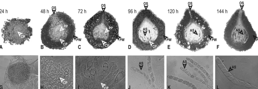
Perithecia at similar development stages (based on size and color un-der the stereomicroscope) were collected from the cellophane-covered agar surface using a razor blade at 24, 48, 72, 96, 120, and 144 h after crossing. To minimize physical disturbance, a minimal fraction of adja-cent vegetative hyphae around sampled perithecia were not removed, es-pecially at spots where multiple perithecia of the same development stage had formed gregariously. Morphological development, as indicated by size and color of perithecia as well as development of asci and ascospores, was observed for perithecia at all sampled time points (Fig. 1). Differen-tiated asci and ascospores were observed in perithecia from 144 h onward. Pieces of cellophane membrane (about 4 mm by 2 mm) carrying⬃5 to 20 perithecia of similar size were cut from cultures and fixed in 1.5 formal-dehyde and 0.025 M phosphate buffer for at least 48 h. The samples were embedded in resin and prepared for transmission electron microscopy (TEM) and light microscopy as previously described (32). Blocks were sectioned at 0.7 to 1m using a glass knife and stained with 1% toluidine blue. A Lecia DM LB microscope (Leica Microsystem Gmbh, Wetzlar, Germany) with differential interference contrast (DIC) capabilities was used to observe samples, and images were captured using a Zeiss AxioCam MRc color camera and AxioVision 4.8.2 (Göttingen, Germany). Image processing and annotation were performed using Adobe Photoshop CS3 (San Jose, CA). All tissues and perithecia were rapidly frozen in liquid nitrogen as they were sampled and stored at⫺80°C.
Tissue samples from biological replicates were pooled for RNA extrac-tion at each sampling time point. Tissues were homogenized with a Dounce glass grinder, and debris was filtered through Qiagen Qiashredder columns (Qiagen, Chatsworth, CA). Total RNA was extracted from ho-mogenized tissue using TRI Reagent (Molecular Research Center) (33), and mRNA was purified using Dynabeads oligo(dT) magnetic separation (Invitrogen). About 100 ng of purified mRNA was mixed with 10⫻
on September 8, 2020 by guest
http://ec.asm.org/
mentation buffer (Ambion) and incubated at 70°C for 5 min before addi-tion of 1l of stop buffer (Ambion). Fragmented mRNA was precipitated using 100% ethanol with glycogen (Ambion) at⫺80°C. Preparation of cDNA for sequencing followed the Illumina mRNA sequencing sample preparation guide. A multitargeted primer (MTP; VWNVNNBDKGGC) (34) primed reverse transcription of the first cDNA strand, and random hexamers (N6; Invitrogen) primed the second cDNA strand. To provide
technical replicates across stages of sexual development, cDNA was pre-pared using only N6primers for samples at all time points. After ligation of
standard adaptors for Illumina sequencing, each sample was purified us-ing a 2% low-meltus-ing-point agarose gel. A gel band correspondus-ing to processed cDNA fragments of 200 to 400 bp was cut and purified using a QIAquick gel extraction kit (Qiagen). Selected cDNA samples were en-riched by PCR using Pfx DNA polymerase (Invitrogen) and 15 cycles of amplification at 98°C for 10 s, 65°C for 30 s, and 68°C for 30 s. The quality of purified PCR products of all samples was determined using a bioana-lyzer (Agilent Technologies). Single-end 35-bp reads of MTP- and N6
-primed preparations were separately sequenced, each on eight lanes of an Illumina genome analyzer, at the Yale Center for Genomic Analysis.
We used Tophat, version 1.2.0 (35), to perform spliced alignments of the reads against theN. crassareference genome (25). As theN. crassa
genome was based on the samemat Astrain used in this study, a sequence ofmat a-1was added to the genome for estimating expression of thismat a-specific gene. Only reads that mapped to a single unique location within the genome with a maximum of two mismatches in the anchor region of the spliced alignment are reported. We used the default settings for all other Tophat options. We tallied the number of the reads that overlapped the exons of a gene using HTSeq, version 0.4.5p6 (unpublished data;http: //www-huber.embl.de/users/anders/HTSeq/doc/), and the gene structure annotation file for the reference genome. The tally for each sample was then processed using LOX, version 1.6 (36), to estimate gene expression levels at all assayed time points during sexual development. Reads per kilobase of exon model per million mapped (RPKM) reads were estimated as in the work of Mortazavi et al. (37) (see Table S3 in the supplemental material).
The Functional Catalogue (FunCat) (38) annotation scheme was used to group genes according to their cellular or molecular functions. The statistical significance of overrepresentation of gene groups in functional categories relative to the whole genome was determined using the hypergeometric distribution, facilitated by the Munich In-formation Center for Protein Sequences (MIPS) FunCat online web application. Further functional annotation of genes showing statisti-cally significant patterns of differential expression in metabolic path-ways was based on the biochemical pathway and the annotation data-base from the Kyoto Encyclopedia of Genes and Genomes (39). Enrichment analysis was performed at theN. crassagenome web page (http://www.broadinstitute.org/annotation/genome/neurospora/) as well, where significant enrichment was reported for genes responding to light stim-ulus and genes involved in gene silencing.
Bayesian networks for RNA silencing genes and pathways were in-ferred from expression data using the Bayesian Network Webserver for Biological Network Modeling (40). Input files containing fold changes between adajcent time points across perithecium development were cal-culated from LOX data based on MTP-based experiments. Global struc-ture learning settings followed the defaults and 100 high-scoring networks with a selection threshold set to 0.5 were included in model averaging without any structural constraint. Bayesian networks of different sets of genes were used to predict interactions between genes in controlling de-velopment.
Knockouts for nearly all of the 9,733N. crassagenes, including dele-tion cassettes for genes in either mating type ofN. crassa, were acquired from the Fungal Genetic Stock Center. All knockout strains used in this study were produced and verified, typically using Southern blot analysis, within the NIHNeurosporaGenome Knock-Out Project (41), and PCR analysis was used to verify genotypes by following the recently optimized approach of Lichius et al. (42). For functional studies of genes, including
het-6,het-14,het-15,mkr-5,mkr-6,qde-1,sad-1,sad-3,sms-2,sms-3, and thestc1-like gene, knockouts of bothmat aandmat Awere available for crossing and phenotyping across sexual development (see Table 2). Knockouts of opposite mating types were crossed on synthetic complete FIG 1Analysis of perithecial development inN. crassa. (A to F) Sections of perithecia demonstrate the expansion of the perithecium wall (PW), composed of thick-walled cells, and the development of thin-walled cells in the centrum parenchyma (CP), except as noted. (G to L) Light microscopic analysis of perithecial squashes show internal contents of the fruiting body, including centrum parenchyma, asci (AS), and ascospores. (A) By 24 h, the perithecia show an initial expansion of the centrum parenchyma, which is surrounded by multiple layers of thick-walled cells composing the perithecium wall. Magnification,⫻400. (B) At 48 h, early paraphyses (PP) appear as elongated cells within the expanded centrum parenchyma (⫻200). The initial ostiole (OS) forms at the apex of the fruiting body. (C) At 72 h, abundant paraphyses arise from thin-walled cells at the bottom of the centrum. The ostiole has formed and is lined with periphyses, and layers of the perithecial wall are distinct (⫻200). (D) At 96 h, early asci are beginning to appear (⫻200). (E) At 120 h, early ascospores start developing in some asci (⫻200). (F) At 144 h, melanized ascospores are visible in many asci. The beak and ostiole appear fully developed at the apex of the fruiting body (⫻200). (G) By 24 h, the perithecium has grown, but a squash does not reveal differentiation of hyphae within the perithecium (⫻200). (H) At 48 h, thin-walled cells of the centrum parenchyma have clearly differentiated (⫻400). (I) At 72 h, early paraphyses appear (⫻400). (J) At 96 h, early asci extend from thin-walled cells at the base of the centrum (⫻400). (K) At 120 h, ascospores begin to form in asci (⫻400). (L) At 144 h, eight ascospores can be distinguished in a well-developed ascus (⫻400).
on September 8, 2020 by guest
http://ec.asm.org/
medium (SCM) (33), traditionally used for inducing sexual reproduction inN. crassa, and on CA (31). All cultures were grown on SCM and CA with three replicates at 26°C under constant white light. Opposite mating types were inoculated on different sides of the same 9-cm-wide plate. Three to 4 days after inoculation, crossing occurred where mycelia from the two mating types met, predominantly across the midline of the plates. Devel-opment of conidia, protoperithecia, and perithecia was examined to re-veal differences in sexual development attributable to culture conditions. The complete raw data set generated in this study as well as the inferred expression levels are available in the supplemental material, in the fila-mentous fungal gene expression database (FFGED) (43), and on the Townsend Lab web site.
Gene expression data accession number.Raw sequencing data and estimated expression levels are available at the Gene Expression Omnibus of the National Center for Biotechnology Information (GEO;http://www .ncbi.nlm.nih.gov/geo/) under accession numberGSE41484.
RESULTS
Assaying our wild-typeNeurosporacrosses by microscopy, devel-opment of protoperithecia before crossing and of perithecia after crossing proceeded as expected (44). Except for a slight increase in size and a slight darkening in color, no obvious tissue differentia-tion was observed during the interval when protoperithecia be-came fertilized perithecia, within 24 h after crossing. Centrum parenchyma of thin-walled cells expanded with increasing perith-ecial size. Differentiated hyphal structures and croziers became detectable after 48 h to 72 h. Asci filled with vesicles emerged in samples after 96 h, along with narrow, hyphal paraphyses. Be-tween 120 h and 144 h, the top of the perithecium protruded to form a beak. Inside the perithecium, mature asci developed: asco-spores became clearly delimited. Perithecia darkened to nearly black with maturation.
Throughout the 144 h of perithecial development, the majority of perithecia in a culture exhibited no noticeable developmental divergence. Perithecia were often aggregated in patches that were especially tightly synchronized. Throughout our experiments, in-corporation of small quantities of vegetative tissue into our sam-ples was unavoidable, especially in samsam-ples collected during the early stages 48 h after crossing, when protoperithecia and
perith-ecia were very small. Past 144 h, synchronicity became difficult to ensure because developmental landmarks became less clear. Gen-erally 160 h after crossing, ascospores started being forcibly ejected and accumulated gradually onto the lower surface of petri lids. However, the intensity and duration of this long process were increasingly impacted by stochastic environmental conditions, in-cluding small fluctuations in light intensity, humidity, airflow, and position of perithecia on culture plates. Therefore, perithecial development beyond 144 h after crossing was not investigated in this study.
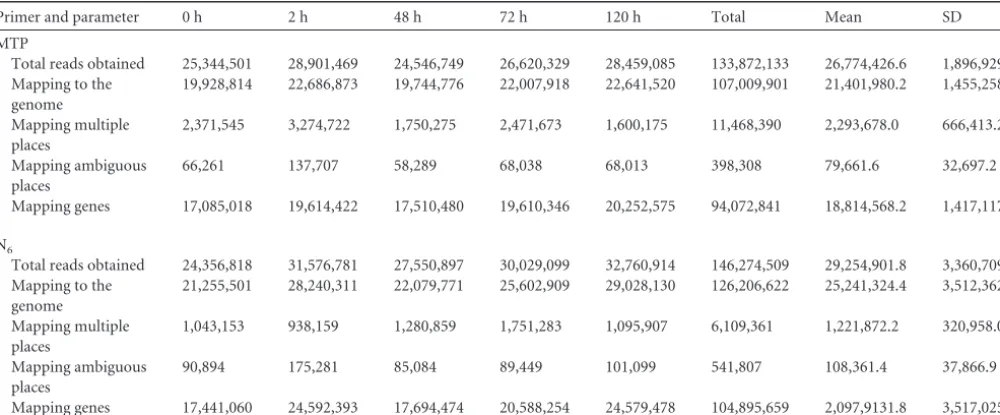
Multitargeted primer (MTP) and conventional random hex-amer (N6) priming yielded a few differences that relate to their design. A robust estimation of the genome-wide transcriptome during the eight time points of sexual development was generated by use of these two types of primers on the Illumina platform. DNA concentrations of samples prepared with N6primers for the time points 24 h, 96 h, and 144 h were much higher than other samples. These samples were diluted to prescribed levels, but they produced low-quality reads and were excluded from further anal-ysis.
As in previous microarray and transcriptomic sequencing ap-plications of MTP priming to Saccharomyces cerevisiae andN.
crassa, in which MTP priming was compared to oligo(dT)
prim-ing (34), MTP priming worked well. There were no significant differences between the total number of short reads obtained with each method (Table 1). The proportion of the total reads mapping to coding genes was significantly higher for MTP than for N6 (chi-square test,P⬍0.0001). The average number of genes signifi-cantly upregulated, or downregulated, was not different between MTP and N6priming (pairedttest,P⬎0.05 in all four cases). MTP-primed samples yielded maximum likelihood (LOX [36]) estimates of gene expression level for 9,717 genes for all eight time points. Our analyses were based on the MTP-primed data (see Table S1 in the supplemental material), which were validated as highly consistent with the N6samples (see Table S2 in the supple-mental material).
LOX estimates and RPKMs (see Table S3 in the supplemental TABLE 1Comparative performance of MTP and N6priming on samples from five development stages inN. crassasexual reproduction
Primer and parameter 0 h 2 h 48 h 72 h 120 h Total Mean SD
MTP
Total reads obtained 25,344,501 28,901,469 24,546,749 26,620,329 28,459,085 133,872,133 26,774,426.6 1,896,929.4 Mapping to the
genome
19,928,814 22,686,873 19,744,776 22,007,918 22,641,520 107,009,901 21,401,980.2 1,455,258.9
Mapping multiple places
2,371,545 3,274,722 1,750,275 2,471,673 1,600,175 11,468,390 2,293,678.0 666,413.2
Mapping ambiguous places
66,261 137,707 58,289 68,038 68,013 398,308 79,661.6 32,697.2
Mapping genes 17,085,018 19,614,422 17,510,480 19,610,346 20,252,575 94,072,841 18,814,568.2 1,417,117.0
N6
Total reads obtained 24,356,818 31,576,781 27,550,897 30,029,099 32,760,914 146,274,509 29,254,901.8 3,360,709.9 Mapping to the
genome
21,255,501 28,240,311 22,079,771 25,602,909 29,028,130 126,206,622 25,241,324.4 3,512,362.8
Mapping multiple places
1,043,153 938,159 1,280,859 1,751,283 1,095,907 6,109,361 1,221,872.2 320,958.0
Mapping ambiguous places
90,894 175,281 85,084 89,449 101,099 541,807 108,361.4 37,866.9
Mapping genes 17,441,060 24,592,393 17,694,474 20,588,254 24,579,478 104,895,659 2,097,9131.8 3,517,025.2
on September 8, 2020 by guest
http://ec.asm.org/
material) (29) from the current transcriptomic experiment were compared to a previous investigation of perithecial development using microarray analysis (22), in which more than 900 genes were measured across a completely independent experiment using the same time points of development. Observed expression patterns were generally conserved between the two experiments, especially for genes detected with high fold changes in expression as mea-sured by microarray analysis (45). Despite a typically lower power associated with microarray methods (see Table S4 in the supple-mental material), 13 genes known to regulateN. crassa sexual development were measured using both methods. Expression of these genes showed no significant discrepancies between sequenc-ing and microarray analyses at any time point across development, including for four genes (ccg-4,4hnr,mkr5, andsad-3) that exhib-ited changes of more than 20-fold based on transcriptome se-quencing that showed identical expression patterns across devel-opment as measured by our microarray analysis.
Upregulation across perithecial development was the most common pattern of gene expression. More than 30% of the genes exhibited continuous upregulation of expression during late per-ithecial development (after 48 h). Another 30% of genes were downregulated after expression peaked at an earlier stage of develop-ment. Hierarchical clustering revealed 12 main subsets correlated by expression of individual genes up- or downregulated during late per-ithecial development (Fig. 2; see also Table S5 in the supplemental material). Functional enrichment analysis for each gene cluster using the FunCat database indicated dramatic shifts of expression during sexual development, with extensive upregulation of genes not anno-tated for function (see Table S5 in the supplemental material; hereaf-ter,P⬍0.01 unless otherwise stated).
Many downregulated genes were functionally annotated, and accordingly, many functional categories were identified as being significantly enriched among downregulated genes during perithecial development. A cluster of genes was upregulated between the 2-h and 48-h stages, before differentiated paraphyses and asci were detected, and then continually upregulated after 72 h (Fig. 2A). This cluster was enriched with genes that respond to light stimulus. Another major cluster of genes peaked at 24 h and was continually upregulated after 48 h, concurrent with ascus and ascospore development (Fig. 2B). Light-responsive genes were also significantly enriched in this cluster, as was extracellular protein degradation.
A third major cluster of genes exhibited a slight increase at 2 h and then was upregulated after 48 h (Fig. 2C). Functional enrich-ment of this cluster was significant for protein metabolism, en-ergy, cell cycle, nucleic acid synthesis and processing, and signal transduction mechanisms. Smaller clusters of genes were upregu-lated at the last two sampled stages in perithecial development (Fig. 2DtoF), and most of these genes have no assigned functions. A cluster of 349 genes showed a continual down-up-down-up-down expression (Fig. 2G), and these genes were functionally enriched in cellular communication and signal transduction mechanisms, interaction with the environment, including pheromone response and sex-specific proteins, and cell type differentiation, including development of ascospores. Another cluster with a similar enrichment of functional categories ex-hibited an upregulated expression pattern during early perith-ecial development (Fig. 2H).
A cluster of genes was downregulated during ascospore matu-ration after 96 h, exhibiting functional enrichment in nucleotide and protein metabolism, rRNA processing, cellular transport, and
biogenesis of cellular components, including the cell wall (Fig. 2I). In addition, a cluster of genes was downregulated at 72 h. This cluster exhibited enrichment in extracellular metabolism, degra-dation and modification of exogenous components, cellular sens-ing and response to external stimulus, includsens-ing light, pheromone response, and mating-type determination (Fig. 2J). Even earlier downregulation manifested after 24 h and 48 h in a cluster exhib-iting enrichment of genes associated with protein synthesis and the heat shock response (Fig. 2K). A further cluster was enriched for many categories of metabolism and also in response to light stimulus (Fig. 2L). This cluster exhibited complex expression pat-terns during early perithecial development before full maturation of asci and ascospores.
More than 2,000N. crassagenes lacking homologs to other fungal genomes outside the Neurospora genus were identified based on sequence similarity. These genes were labeled
Neuro-sporaorphan genes (46). From the recently publishedSordaria
macrosporagenome (47), orthologs for 819 so-calledNeurospora
orphan genes have been identified by sequence similarity. Most of
24 0 2 24 48 72 96 120 144
Growth time (h)
0 10 20 0 10 20 0 100 200 300 0 100 200 0 10 20 30 40 0 20 40 60 80 Expression G H I J K L Expression 10 100 1k 10 100 1k 10 100 1k 10 100 1k 10 100 1k 10 100 1k
0 2 48 72 96 120 144
Growth time (h)
A B C D E F
FIG 2Gene clusters based on relative expression across sexual development. About 60% of the genes in theN. crassagenome are continuously upregulated or downregulated during late perithecium development. Based on their ex-pression profiles across earlier stages, these genes can be further classified into six upregulated clusters (A to F;yaxis logarithmically scaled) and six down-regulated clusters (G to L;yaxis linearly scaled).
on September 8, 2020 by guest
http://ec.asm.org/
these genes have no assigned function in sexual development of
Sordaria. Of theN. crassaorphan genes, 1,573 (nearly 16% of the
genome) exhibited differential expression patterns during perith-ecium development (see Fig. S1 in the supplemental material). About 20% of these orphan genes exhibit expression patterns sim-ilar to the clusters (Fig. 2CandH) that are significantly enriched in genes that function in the cell cycle, DNA processing, mRNA and RNA synthesis, mitosis, meiosis, cellular communication/signal transduction, and cell type differentiation.
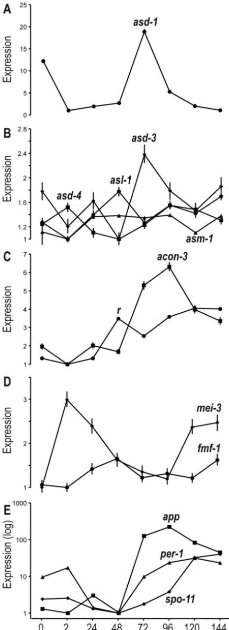
Upregulation of transcription factors and genes involved in per-ithecial development correlated with their stages of function. A num-ber of genes involved in perithecial development and meiosis and some transcription factors (TFs) exhibited diverse expression pat-terns across perithecial development (Fig. 3). These genes include three ascus development genes,asd-1,asd-3, andasd-4, and three genes ofasl-1(ascospore lethal-1),asm-1(ascospore maturation-1),
andr(round spore) with phenotypes in ascospore development (10).
Of the development-related TFs identified by Colot et al. (48), expres-sion of TFs related to sexual development (NCU00097, NCU04561, NCU09739, NCU07392, and NCU044731) showed 5- to 20-fold changes across all time points, exhibiting patterns in which upregu-lated expression is correupregu-lated with gene phenotypes observed during perithecial development (Fig. 4). For transcription factors that are not well characterized for their function in metabolism and development, we often observed increasing expression during late perithecial devel-opment (see Table S6 in the supplemental material).
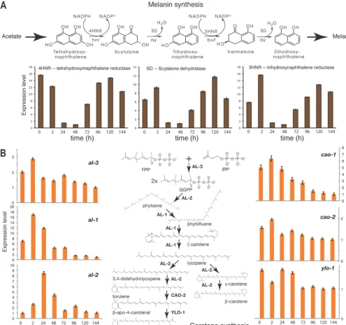
Two metabolic pathways, the melanin synthesis pathway and the carotenoid pathway, are responsible for the production of a dark pigment that increases across sexual development (melanin) and an orange pigment associated with conidiation that dissipates (carotene). Genes involved in these two pathways exhibited ex-pression patterns that were highly correspondent to the observed color changes in the cultures (Fig. 5). Generally, expression of genes with known function in sexual development agreed with expectation.
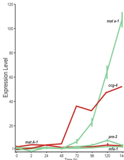
Expression of mating type loci and pheromone precursor genes was more complex than expected but in accordance with mating type specificity. Expression of mating type loci exhibited dramatic differences between theA-1,A-2, andA-3genes at the
mat Alocus and themat a-1gene at themat alocus (Fig. 6;Table
2). Expression ofmat a-1was not detectable inmat A protoper-ithecia and was almost undetectable before 48 h, but it increased continually across sexual development past 48 h, to a high of 107-fold in 144-h samples.
Differential expression was observed for four genes encoding the
mat a-specific pheromone precursor MFA-1, the pheromone
recep-tors PRE-1 (responding to pheromone encoded bymfa-1; seeTable 2) and PRE-2 (responding to pheromone encoded byccg-4), and the
mat A-specific pheromone precursor CCG-4 (Fig. 6). Whereas
ex-pression ofmfa-1,pre-1, andpre-2did not change much during
per-FIG 3Relative expression across sexual development of genes annotated as functioning in perithecial development. (A) Expression ofasd-1(ascus devel-opment) at 72 h, when asci first form. (B) Peaks of expression for the genes
asd-3,asd-4,asl-1(ascospore lethal), andasm-1(ascospore maturation) dur-ing intermediate stages of sexual development. (C) Expression ofr(round spore) andacon-3during perithecial development. (D) Expression of meiosis genemei-3and female and male fertility genefmf-1. (F) Correlated expression ofapp(abundant perithecial protein),per-1(perithecial-1), and meiosis-spe-cific geneSpo11during sexual development.
on September 8, 2020 by guest
http://ec.asm.org/
ithecial development, expression ofccg-4underwent a continuous and dramatic increase up to 45-fold. Genesmfa-1andccg-4encode prepropheromones that require a posttranscription process to be-come mature pheromones (49). The expression of genes orthologous to theS. cerevisiaeprepropheromone MF␣1 and MFa1 processing genes, including kex1 (NCU04316), kex2 (NCU03219), ste13
(NCU02515), ram2 (NCU03632), ste6 (NCU07546), ste14
(NCU00034),ste24(NCU03637), andaxl1(NCU00481), showed moderate correlation with each other (see Fig. S2 in the supple-mental material).ste13showed a continually increasing pattern after 24 h, similar toccg-4. In addition to mating loci and phero-mone genes,het(heterokaryon incompatibility) genes were also dynamically regulated during perithecium development (see Fig. S3 in the supplemental material).
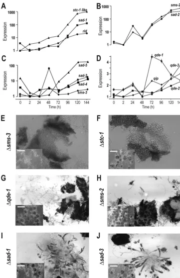
Upregulated expression was observed for genes in RNA silenc-ing pathways and genes associated with DNA methylation. We detected upregulated expression of genes that function in the veg-etative silencing (quelling), repeat-induced point mutation (RIP), and meiotic silencing pathways as well as of genes involved in facultative heterochromatin formation and DNA methylation (Fig. 7AtoD; see also Fig. S4 in the supplemental material). Up-regulation across perithecial development was observed forsad-1
(suppressor of ascus dominance-1) and the cytosine
methyltrans-ferase homologrid(RIP Defective), whose expression levels were increased 130-fold and 20-fold at 144 h, respectively (Fig. 7A). Similar to gene stc1 (siRNA to chromatin) in fission yeast, NCU01496 encodes a protein with a highly conserved Stc1 do-main (blastp E value, 1.71e⫺30) that contains eight conserved cysteines that may bind to zinc. Expression of gene NCU01496 increased almost 1,000-fold by 144 h after crossing (Fig. 7A).
Two meiotic silencing genes,sms-2andsad-2, exhibited similar regulatory dynamics, with a first peak at 24 h and a second increase after 48 h (Fig. 7B). Increases of 680-fold and 390-fold forsms-2
andsad-2occurred from 2 h to 144 h (Fig. 7B). Expression of the
sms-3(suppressor of meiotic silencing-3) andsad-3genes exhibited
dynamics similar to the expression ofsms-2, andsad-2, but with a 24-h delay (Fig. 7C). Expression levels of two recently identified MSUD (meiotic silencing by unpaired DNA) genes,sad-4and
sad-5(50), dropped at 2 h after crossing but increased significantly
starting at 48 h after crossing (11-fold forsad-4and 135-fold for
sad-5in the 144-h samples [Fig. 7C]).
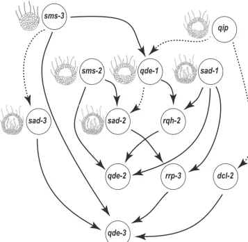
Bayesian networks constructed based on expression of genes recognized in RNA-silencing pathways (24), including qde-1,
qde-2,qde-3,qip,sad-1,sad-2,sad-3,sms-2,sms-3,rrp-3,dcl-2, and
rqh-2, always positioned genes encoding the downstream proteins
in meiotic silencing, QIP, SMS-2, and the Dicer-like RNase III enzyme SMS-3, as independent parents in the top tiers (Fig. 8). This top-tier positioning suggests that the roles of the RNA silenc-ing genes in controllsilenc-ing sexual development are played in an order that is inverted in comparison to their genetic dependence with regard to silencing. Removing any subset of the genes involved in the Quelling silencing pathway that functions only during asexual growth did not change the dependence orders among other genes in the network. The Bayesian network, includingsad-4andsad-5, suggested similar associations among RNA silencing genes, except that betweensad-2andsms-2, and was not consistent with pheno-types ofsad-4andsad-5. Additional genes that may interact with these genes during perithecial development are needed to be iden-tified and included for reconstruct the network. Addingridand
stc1to the RNA silencing genes in the Bayesian network inference demonstrated that bothridandstc1were associated with RNA silencing pathways; they were inferred to be upstream in the net-work.
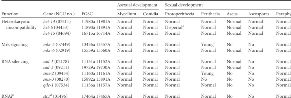
Phenotypes of knockouts were identifed for selected RNA si-lencing genes. Knockouts of many genes that were specifically up-regulated in expression during crossing and perithecial develop-ment exhibited numerous phenotypes in sexual reproduction of
N. crassa(Table 3;Fig. 7EtoJ). Knockouts of five RNA silencing
genes and the stc1-like genes all yielded visible perithecia after crossing, but in all cases perithecium development failed at vari-FIG 4Expression of transciption factors and arrested development in transcription factor knockouts. Solid lines indicate expression of known transcription factors, identified by their NCU number. Dashed lines associate peaks of expression with timing of appearance of knockout phenotype, as determined by Colot et al. (48). Illustrations depict perithecia at sequential developmental stages.
on September 8, 2020 by guest
http://ec.asm.org/
ous stages before ascospore differentiation and production (Fig. 1CandI). Knockouts ofsms-2and thestc1-like gene produced dark, normal-size perithecia without asci. Knockouts of sms-3
produced perithecia that arrested earlier, after slight increases in size and darkening of the perithecial wall prior compared to nor-mal perithecia 24 h to 48 h after crossing.
Neurospora crassasenses and responses to environmental
sig-nals, including blue light, via singaling pathways and light-respon-sive genes. Genes of the heterotrimeric G protein signaling system showed upregulation during different stages of perithecium devel-opment. Heterotrimeric G-protein signaling and cyclic AMP (cAMP) metabolism genes exhibited diverse expression patterns during perithecium development (see Fig. S5 in the supplemental
material). Blue light-responsive genes exhibited distinct expres-sion patterns across perithecial development under constant white light. Genes that putatively respond to blue light appear to be regulated in a similar pattern (see Fig. S6 in the supplemental material). A circadian pattern was not observed for the expression of the blue light sensor geneswc-1,wc-2, or related genes. Two genes encoding blue light-responsive proteins,vvdandvelvet, ex-hibited general downregulation during perithecial development, especially after 48 h.
DISCUSSION
Here, we provide an in-depth characterization of theN. crassa
transcriptome during sexual development. We observed that over FIG 5Relative expression across sexual development of genes whose products contribute to pigmentation inN. crassa. (A) Expression of three genes,4hnr,3hnr, andsd, that function in synthesis of melanin. (B) Expression patterns of genes involved in carotenoid synthesis, particularlyal-1,al-2, andcao-1.
on September 8, 2020 by guest
http://ec.asm.org/
30% of the genome was expressed during early development, fol-lowed by increasing or decreasing expression of particular genes during late perithecium development. Of the genes that were up-regulated late in the developmental process, many exhibited dra-matic upregulation. A significant portion of these genes have not been classified or annotated for a function to date, including many
Neurosporaorphan genes. The lack of homologs in other fungal
models, especially in well-annotated unicellular yeasts, makes it challenging to predict and test functions of these genes. Further-more, genes whose mutants or knockouts lead to no sexual devel-opment, including meiotic recombination, are generally hard to
study using traditional genetic approaches. Our approach has yielded important knowledge about such genes.
Our results show that the expression of genes involved in mor-phological characteristics during N. crassaperithecial develop-ment and, in particular, genes associated with the developdevelop-ment of ascogenous hyphae and young asci, such asasd-1, asd-3, asl-1,
asm-1, andround spore, peaked from 48 h to 72 h after
fertiliza-tion, when those tissues developed within perithecia. Further-more, expression of genes associated with carotenoid and melanin pigmentation contrasted across sexual development. The carote-noid biosynthetic pathway that is upregulated during asexual de-velopment inN. crassa(45) was generally downregulated during perithecium development. The expression of genes involved in the melanin synthesis pathway that produces the dark pigmentation of perithecia increased when the color of perithecia significantly darkened.
Our observations of sudden and significant shifts in the ge-nome-wide transcriptomic landscape imply the presence of key regulators or regulatory cascades that are invoked at specific de-velopment stages. For instance, many genes exhibited low expres-sion at 48 h, just before a dramatic increase. This set includes all genes encoding histones, many meiosis-specific genes, DNA methylation genes, the Cop9 signalosome (which plays major roles in hyphal growth, conidial development, and circadian func-tion [51]), and some transcription factors that are involved in the sexual development ofN. crassa(48). Two genes expected to have high expression in perithecia,app(abundantperithecialprotein, homologous toappinSordaria macrospora[52]) andper-1(
per-ithecial color [53]), increased over 100-fold after 48 h during late perithecium development. When asci and ascospores became vis-ible under a microscope at 96 h, many genes, including a large number of functionally unannotated genes as well as mating type and pheromone genes and genes involved in signal pathways, RNA silencing pathways, and heterokaryon incompatibility, were highly expressed. Full development of beak structures and first ascospore ejection usually occur from 8 to 10 days after crossing and lasts for more than a week (44), beyond the last synchronized sampling time point used in this study.
Of all developmental genes, those with perhaps the most sur-prising expression patterns were the mating and pheromone genes. The mating and pheromone genes are generally thought to function in the conidia and in hyphae involved in mating but were also highly expressed after fertilization (54,55). In our study, fer-tilization ofmat Aprotoperithecia withmat aconidia revealed differences in the regulation of the mating type locus genes over the time course of sexual development. Expression ofmat A-2and
A-3was very low, and differences over time were not statistically
20 40 60 80 100 120
mat a-1
Expression Level
0 2 24 48 72 96 120 144
Time (h)
ccg-4
1
mat A-1
mfa-1 pre-2
FIG 6Relative expression across sexual development of mating type loci and pheromone precursors and receptors in themat Afemale background. Expres-sion profile are color-coded by their nominal mating type specificity:mat Ais in red (dark) andmat ais in green (light). Expression of mating type genemat a-1increased 48 h and later after crossing betweenmat Aprotoperithecia and
mat aconidia, where expression ofmat A-1was generally unchanged across perithecial development. Among pheromone genes, expression of themat A-specific pheromone precursor geneccg-4increased during late perithecial development, and expression of the receptor PRE-2, specific for the peptide pheromone encoded byccg-4, increased from 48 h. Expression of the receptor PRE-1, specific for the peptide pheromone encoded bymfa-1, was present and steady during perithecial development (Table 2).
TABLE 2Comparative expression (in RPKM) of mating types, pheromones, and pheromone receptors acrossN. crassaperithecial development
Gene 0 h 2 h 24 h 48 h 72 h 96 h 120 h 144 h
NCU01958mat A-1 5 13 11 4 6 6 12 9
NCU01959mat A3 0 0 0 0 0 1 0 0
NCU01960mat A2 1 1 1 1 1 3 2 4
mat a-1 0 0 1 0 3 9 25 42
NCU05758pre-2 4 2 4 3 5 6 9 5
NCU00138pre-1 6 6 6 4 4 5 9 8
NCU01257amfa-1 23 18 18 16 21 19 20 25
NCU02500ccg-4 71 129 294 345 2,208 1,980 2,897 3,184
amfa-1is reannotated as NCU16992.7 in the latest annotation forN. crassa.
on September 8, 2020 by guest
http://ec.asm.org/
FIG 7Relative expression levels across sexual development and phenotypes of RNA silencing genes. Phenotypes of knockouts of RNA silencing genes were assayed on synthetic complete medium and carrot agar medium. Perithecia were crushed 6 days after a cross betweenmat Aprotoperithecia andmat aconidia. Expression for all genes is reported on a log scale. (A) Expression of thestc1-like gene,sad-1, andrid(RIP defective). (B) Expression ofsms-2(suppressor of meiotic silencing) andsad-2(suppressor of ascus dominance). (C) Expression ofsad-3,sad-4,sad-5, andsms-3. (D) Expression of Quelling-defective genes, includingqde-1,qde-2, andqde-3andqip(NCU00076,qde-2interacting protein). (E) The⌬sms-3strain produced tiny hairy perithecia without differentiation of the centrum parenchyma (magnification,⫻400). (F) The⌬stc1strain produced nomal-size perithecia with no apparent development of the centrum parenchyma (⫻400). (G and H) The⌬qde-1and⌬sms-2strains produced normal-size perithecia with undifferentiated thin-walled cells hard to detect in the centrum parenchyma area (⫻200). (I and J) The⌬sad-1and⌬sad-3strains produced normal-size perithecia with beaks and young asci without ascospores (⫻400). Scale bar⫽10 mm for perithecia overview in KO phenotyping assays. Detailed images of the wild-type crushed perithecia are presented inFig. 1GtoL.
on September 8, 2020 by guest
http://ec.asm.org/
significant. Expression ofmat A-1was meager and exhibited a net change of less than 3-fold. In contrast, expression ofmat a-1 in-creased by more than 100-fold after 48 h. Although novel haploid hyphae frommat aconidia are rarely observed on cultures densely covered withmat Amycelia,mat aconidia could not be entirely avoided in our samples. Increased expression ofmat a-1could be due to the increase ofmat anuclei during conjugate division be-fore karyogamy and meiotic and mitotic division after karyogamy. However, no corresponding increase in expression of themat a -specific pheromone precursormfa-1was observed.
A significant increase was observed for themat A-specific pher-omone precursorccg-4after crossing, especially after 48 h. This finding is consistent with microarray measurements across per-ithecial development ofN. crassaon both SCM and carrot me-dium (45). Expression ofpre-2, which encodes a receptor that recognizes the pheromone encoded byccg-4, has been reported to be high in male and female reproductive structures ofmat acells (56). Its expression increased significantly at 120 h after crossing but remained low in comparison to that ofccg-4. These patterns of mating type gene expression promise to help illuminate the bio-logical function of mating type and pheromone genes after fertil-ization and hint at roles of mating type and pheromone genes in postcrossing sexual development. Although mating loci in both
mat aandmat Astrains have been known for a number of years for
N. crassaand closely related species (57,58,82), the functions of
mating loci were unknown aside from regulating homogenic in-compatibility through highly regulated expression of pheromone genes and despite the distinct expression patterns of these genes at different stages in the life cycle ofN. crassaand the closely related
Fusarium(20).
In Neurospora and other fungal species, mating type
id-iomorphs impact diverse genes that are not directly involved in the mating process (21,23,45,59,60). The increased expression of
mat a-1andccg-4that we observed during sexual development of
N. crassacalls for further investigation using strain-specific
ex-pression assays conducted with varied crosses. Although increased transcription of the pheromone precursor geneccg-4may not re-flect a commensurate change in level of pheromone, we also ob-served increased expression of a homolog ofste-13(NCU02515), which encodes the aminopeptidase required for processing prepropheromone MF␣1 inS. cerevisiae(49). It has been shown that as long as the mating type idiomorphs appear in two different nuclei, the presence of one receptor and its compatible phero-mone is necessary and sufficient to initiate perithecium develop-ment and ascospore production (56). Consistent with these re-sults, we found low expression ofpre-2and high expression of
sms-3
qde-1
qip
sad-1
sms-2
sad-3
sad-2
rqh-2
qde-2
rrp-3
qde-3
dcl-2
FIG 8Bayesian network inference for RNA silencing genes. Arrows indicate the dependence direction between two linked nodes (genes). Edges of posterior probability higher than 50% were present, and posterior probabilities⬎90% were in solid line. Illustrations of phenotypes were based on our observations, except forqip, which was drawn based on those of Hammond et al. (50), andsad-2, which was drawn based on those of Xiao et al. (76).
on September 8, 2020 by guest
http://ec.asm.org/
ccg-4in themat Aprotoperithecia across perithecium develop-ment. Our data also demonstrate the correlated upregulation of
pre-2andccg-4expression on very different dynamic scales.
Mating type loci may control expression of pheromones and their receptors during presexual development before crossing in a mating type-specific manner (56). However, the mechanisms that regulate expression of pheromones and receptors after crossing when opposite mating types coexist in perithecia are unclear. Al-though meiosis and perithecium development can proceed in het-erokaryon-containing nuclei of the opposite mating type when GNA-1 and one compatible pheromone-receptor pair are ex-pressed, even at low levels, expression of mating types was not detected or reported in the previous study (56). Here we show the increased coexpression ofmat a-1,pre-2, andccg-4at serial time points across perithecium development after crossing between mating types. Thus, the possibility thatpre-2specifically interacts with genes inmat ato upregulateccg-4requires further investiga-tion.
A network of HMG-box transcription factors regulates mating and sexual development inPodospora anserina, a fungus that pro-duces perithecia for fruiting bodies, and the factor PaHMG5 plays a central role in regulating expression of mating types (61). InN.
crassa, NCU09387 (fmf-1) and NCU02326 are the ortholog and
inparalog of the gene encoding PaHMG5. Our data support the hypothesis advanced by Ait Benkhali et al. (61) that these two genes can regulate expression of mating type genes at different stages of sexual development. Expression of fmf-1was signifi-cantly upregulated after crossing and reached its peak at 48 h after crossing. This upregulation is consistent with the observed knock-out phenotype as well: arrested development before ascospore for-mation. In contrast, expression of the inparalog NUC02326 sig-nificantly decreased after crossing and was maintained at a low level during late perithecial development. Its regulatory role on mating type gene expression before crossing demands further in-vestigation.
As a heterothallic fungus,N. crassaemploys a self-incompati-bility mechanism termed heterokaryon incompatiself-incompati-bility (HI) to restrict hyphal fusion to genetically identical strains and to stop
growth of heterokaryotic hyphae during nonsexual development (62,63). After crossing, cells in the developingN. crassa perith-ecium can be haploid, diploid, dikaryotic, or a single nucleus in some cells of the crozier structure. Dynamic expression ofhetand
tolgenes is perhaps associated with the toggling on and off of HI during sexual development, and regulation of these genes may be responsible for proper phase changes from dikaryotic cells to hap-loid sexual spores. Further investigation of tissue-specific expres-sion of HI genes would be warranted.
Dynamic expression of genes within signaling pathways sug-gests their roles in triggering and executing sexual development. Heterotrimeric G-protein signaling, cAMP metabolism, Ras pro-teins, and G protein-coupled receptors (GPCRs) are responsible for transmitting extracellular signals to intracellular responses and are involved in sexual development inN. crassa(64–67). Expres-sion of some GPCRs and G-protein subunits, especiallygpr-1and
gna-3, increased after 48 h. A perithecial defect was observed for
both⌬bek-1and⌬gpr-1strains (66), and abnormal perithecia
with a large propotion of nonviable ascospores were found in
⌬gna-3 strains (67). Genetic tests suggest that RIC8 activates
GNA-1 and GNA-3, but not GNA-2, and positively regulates the cAMP pathway in vegetative growth of N. crassa (68). In our study, expression ofric8,cr-1,gna-2, andgpr-6was highly corre-lated. Previous studies show no apparent phenotype of⌬gna-2in asexual and sexual development ofN. crassa and suggest that
gna-2plays a minor and compensatory role forgna-1andgna-3in
the regulation of conidiation inN. crassa(65,69). Gene coexpres-sion data, especially finely sampled across time points, can provide numerous clues as to the relevant components and patterns of association that underlie complex gene networks.
Recently, NCU01496 was recognized as a homolog ofstc1in fission yeast, in which the gene is suggested to play important roles in RNA silencing and chromatin modification (70). It is possible that it fulfills a similar role inNeurospora, in which evidence sug-gests that RNA-mediated silencing and silencing by DNA methyl-ation are independent genome defense systems (71–74). InN.
crassa, diverse silencing pathways are active, including vegetative
silencing (Quelling), repeat-induced point mutation (RIP), and TABLE 3Phenotyping of knockouts for genes that exhibited upregulated expression during perithecial developmenta
Function Gene (NCU no.) FGSC
Asexual development Sexual development
Mycelium Conidia Protoperithecia Perithecia Ascus Ascospores Paraphyses
Heterokaryotic imcompatibility
het-14(07511) 11980a 11981A Normal Normal Normal Normal Normal Normal Normal
het-6(04453) 11890a 11891A Normal Normal Dispersalb Normal Normal Normal Normal
het-15(04694) 16715a 16714A Normal Normal Normal Normal Normal Normal Normal
Mrk signaling mkr-5(07449) 13456a 13457A Normal Normal Normal Youngc No No Normal
mkr-6(02919) 15559a 15560A Normal Normal Normal Normal Normal Normal Normal
RNA silencing sad-1(02178) 11151a 11152A Normal Normal Normal Normal Normal No Normal
sad-3(09211) 19729a 19730A Normal Normal Normal Normal Normal No Normal
sms-2(09434) 11160a 11161A Normal Normal Normal Young No No Normal
sms-3(08270) 15892a 15891A Normal Normal Normal No No No Normal
qde-1(07534) 11156a 11157A Normal Normal Normal Normal No No Normal
RNAib stc1d(01496) 17464a 17465A Normal Normal Normal Normal No No Normal
a
Crossings were made between KO strains for each genes from both mating types, and descriptions were based on observation on both sides of the crossing zone.
bInstead of along the crossing zone, protoperithecia were formed randomly over the plate on sides of both mating types. c
Apparent changes in size and color from protoperithecia to perithecia were observed after crossing, but no development of centrum parachyma.
dNCU01496 is homologous tostc1(siRNA to chromatin-1) in fission yeast.
on September 8, 2020 by guest
http://ec.asm.org/
meiotic silencing by unpaired DNA (MSUD). The last two path-ways function only during sexual development (24). Quelling and MSUD detect and inactivate repeated or unpaired sequences dur-ing vegetative or sexual development, whereas RIP plays a role in deactivating transposable elements in the genome. Aside from their roles in genome defense, genes functioning in RNA silencing and quelling also play critical roles in controlling development. Details about the silencing pathways have been emerging steadily with genetic studies, but how these genes and pathways regulate sexual development remains unexplored. While we may argue that these genes are likely associated in a similar pattern between the very different functions, the structures of the networks sup-porting the functions can be different in terms of their compo-nents and dependencies between genes (direction of regulation). The functional dependencies of the genes in ordinary develop-ment can accumulate in the opposite direction from the func-tional dependencies in generalized error correction or surveil-lance (which each often involves retracing steps to rectify the genomic or cellular state).
Our data support previously proposed models that RNA si-lencing pathways and genes involved in other RNA interference pathways are closely involved with meiosis and sexual develop-ment inN. crassa. According to the latest model, an MSUD com-plex includes SAD-1/SAD-3, proteins that use an aberrant RNA template to synthesize a double-stranded RNA (dsRNA) mole-cule, DCL-1 (SMS-3), that processes the dsRNA into small inter-fering RNA (siRNA); QIP (QDE-2 interacting protein)/SMS-2, which destroys complementary mRNA transcribed from the ab-errant RNA template generated from siRNA; and SAD-2, which recruits SAD-1 and possibly other proteins from the perinuclear region (75,76). Our findings of same expression pattern between SAD-3 and SMS-3 support their associated functions predicted in the initial steps of producing dsRNA and siRNA molecules. In addition, we observed similar upregulation of genes coding for SMS-2 and QIP that is consistent with their same function in destroying erroneously transcribed mRNAs. The gene pairsms-2
andsad-2and the gene pairsms-3 andsad-3exhibited similar
expression patterns, with the latter pair lagging by 24 h. However, there is not an obvious explanation for the peak expression of
sms-2andsad-2prior to the upregulation ofsms-3andsad-3. It is
possible that MSUD may be involved in RNA interference (RNAi)-induced assembly of heterochromatin inN. crassa(73), as reported in the homolog ofsad-3inSchizosaccharomyces pombe,
hrr1, which is required in fission yeast for RNAi-mediated forma-tion of heterochromatin. Our findings support this hypothesis, as we observed coexpression of thesad-1gene involved in MSUD, theridgene involved in RIP, and thestc1-like gene, whose ho-molog is also involved in RNAi and chromatin modification in fission yeast. Consistent with previous studies ofsad-1andsad-2
mutants (77,78), we observed abortive perithecia at different de-velopment stages for knockouts of some genes involved RNA si-lencing pathways. These knockout phenotypes suggest that there is an interplay between silencing pathways and ordinary develop-mental functions associated with sexual development and meio-sis. Further study on regulatory functions of different silencing pathways is needed to determine how these diverse pathways work together to regulate gene expression during sexual development of
N. crassa. Consistent with these knockout phenotypes (and
inde-pendent of them), model-averaged Bayesian network prediction based on expression data suggest thatsms-2,qip, andsms-3are
independent parents in the top tiers of the network, and expres-sion ofsad-2andsad-3is dependent on those parent genes. In-triguingly, their dependency in our sexual development time course was inferred to be causally opposite in sequence order to their dependency as a genome defense response for removal of unpaired DNAs. Includingridorstc1did not change the network structures among RNA silencing pathways.
Transcript abundance of genes involved in DNA methylation
ofN. crassaincreased in synchrony with the RNA silencing genes
in our experiment. Expression of the four DNA methylation genes,dim-2,dim-5,dim-7, andhp1(79,80), increased after 48 h, and the expression pattern of methyltransferase-coding gene
dim-2was similar to that of the RNA silencing genessms-2and
sad-2. The hp1, cdp-2, hda-1, and chap genes encode the HP1
complex (HCHC), which directs histone deacetylation and DNA methylation and is required for gene silencing in centromeric re-gions independent of DNA methylation (74). A complicated si-lencing network, the sex-induced sisi-lencing of genes by RNAi before meiosis, has recently been discovered in the yeast-like ba-sidiomyceteous fungusCryptococcus neoformans(81). Further as-sessment of methylation levels during sexual development, when asci and ascospores are differentiated, would enable stronger in-ference from upregulated expression of these genes.
In summary, we observed stage-specific expression for some known genetic markers and transcription factors that are associ-ated with morphological development of perithecia. Examining knockout strains for genes involved in RNA silencing and signal-ing pathways that were differentially expressed across develop-ment, we identified increased gene expression associated with functional stages of development, including an stc1-like gene whose homolog was recently identified as a critical link between RNAi and chromatin modification in fission yeast. Analysis of gene expression patterns during different stages of sexual devel-opment allowed us to identify conditions and develdevel-opmental stages that facilitate phenotyping genes and to discern potentially novel functions of genes during sexual development ofN. crassa. Although stage-specific expression was observed for genes in-volved in diverse functions during sexual development, the func-tions of numerous genes that were markedly upregulated during late perithecium development, when asci and ascospores develop, have not been reported. Our findings provide key insights into the functions of genes involved in key pathways such as expression of mating type genes and pheromones, pigmentation, heterokary-otic incompatibility, signaling pathways, and RNA silencing dur-ing sexual development ofN. crassa, as well as the transcription factors and other regulatory components that are responsible for the action of these pathways in sexual development.
ACKNOWLEDGMENTS
Strains and plasmids were obtained from the Fungal Genetics Stock Cen-ter (Kansas City, MO). We thank Angelika Hofmann for helpful com-ments on the manuscript.
This work was supported by funding from NSF MCB 0923797 to J.P.T. and F.T. and NIH PO1 grant GM068067 to J.P.T.
REFERENCES
1.Domazet-Lošo T, Tautz D.2010. A phylogenetically based transcriptome age index mirrors ontogenetic divergence patterns. Nature468:815– 818. http://dx.doi.org/10.1038/nature09632.
2.Freeman TC, Ivens A, Baillie JK, Beraldi D, Barnett MW, Dorward D, Downing A, Fairbairn L, Kapetanovic R, Raza S, Tomoiu A, Alberio R,