Current Multidisciplinary Studies
Available Online athttp://www.journalijcms.com
Vol. 1, Issue, 2, pp.69-73, july, 2015
RESEARCH ARTICLE
IN-VITRO ANTICANCER ACTIVITY OFEUCHEUMACOTTONIIEXTRACTS AGAINST HELA CELL LINE, HUMN LUNG CARCINOMA CELL LINE (SK-LU-1), HUMAN COLON
CARCINOMA CELL LINE (HCT-116), AND FIBROBLAST
Lee,JW*., Wang, JH., Ng, KM., Tan, CH Rabina P and Teo, SS
Department of Applied Sciences, UCSI University, No.1 JalanMenaraGading, UCSI Heights, 56000 Cheras, Kuala Lumpur, W. P. Kuala Lumpur, Malaysia
Keywords:
Red Seaweeds, E.cottonii,
Anticancer, MTT Assay, Apoptosis
Article history :
Received on June 22, 2015
Received in revised form, June 31, 2015 Accepted, July 15, 2015
Published July 28, 2015
ABSTRACT:
In recent years, much focus has been put on finding new anticancer drugs that has lower side effects.Seaweed contains high level of polysaccharides especially sulphated polysaccharides which exhibit strong biological activities such as antitumor. Eucheumacottonii, the edible marine red algae is cultivated abundantly in Sabah of East Malaysia mainly for its kappa-carrageenan production. Recent studies have shown that E. cottonii was tumour-suppressive via apoptosis induction. In this in-vitro study, the anticancer effect of E. cottonii on various cell linewere evaluated. Cancer cells were exposed to various concentrations of E. cottonii crude extract for 24 hours. The results have shown that E. cottonii crude extracts induced cytotoxicity in various cancer cells in dose-dependent manner, as measured in MTT viability assay. A complete cessation in cell proliferation was observed using highest dose of extract (20.0mg/mL) for 24 hours incubation, showing strong cytotoxic effect of E. cottonii extract. However, the extracts of E. cottonii did not showed any effect on fibroblast, a human normal cell line. Overall, this study has demonstrated that E. cottoniiextracts may exhibit potential anticancer properties against various cancer cell line yet to prevent normal human cell line to be eliminated.
INTRODUCTION: Cancer is a complex group of disease that have caused major global health problem, with significant association with death and disability. It arises from a series of mutations, as a result of genetic instability and environmental factors (Al-Hajj et al., 2003). According to World Health Organisation, the second leading cause of death in developed countries is cancer and it is also the three leading causes of death for adults in developing countries (GmbH, 2009). Cancer can be treated by surgery, radiation, chemotherapy, hormones and immunotherapy (American Cancer Society, 2011).
QUICK RESPONSE CODE Corresponding author:
Lee,JW
Department of Applied Sciences, UCSI University, No.1 JalanMenaraGading, UCSI Heights, 56000 Cheras, Kuala Lumpur, W. P. Kuala Lumpur, Malaysia
Article can be accessed online on:
www.journalijcms.com
However, there is no potent medicine in the existing cancer treatments as many of these drugs can cause side effects in many circumstances.
Various studies have been carried out to look for alternative treatment for cancer. Researchers have been focusing on the discovery of new compounds derived from the natural products which have potential anticancer properties. Antioxidant is one of the main compounds which act to protect against damages by free radicals and other reactive oxygen species. It was proven that there is a positive correlation between the amount of antioxidants in the food diet and the lowering of cancer mortality in an individual (Boopathy and Kathiresan, 2010). The possible explanation for this is that the antioxidant can cause regression of premaglinant lesions and thus, inhibit the development of these lesions into cancer.
been widely exploited by people for the development of various products including food, fragrances, insecticides, pigments and therapeutic drugs (Carte, 1996). Apart from using plant derivatives and microbial products as a source to produce medicine, researches on marine organisms have been increased drastically over the years to discover new pharmaceutical agents. It has been found that the isolated bioactive compounds from the marine organisms have much health beneficial effects as their biological activities have the potential of giving better efficacy and specificity of drugs against certain diseases. These newly isolated compounds from the marine environment have the ability to withstand extreme conditions in terms of pressure, salinity and temperature, as well as having unique structural and functional features compared to the terrestrial organisms (Boopathy and Kathiresan, 2010).
Among the marine organisms, seaweed is a major source of structurally diverse bioactive compounds. Over the years, seaweeds have been consumed in diet for its high nutrition content. In addition, their richness in sulfated polysaccharide which can be found in the cell wall also results in the diverse use of seaweed in various fields for example, cosmetics,
pharmaceutical, microbial and biotechnology
industries (Wijesekara et al., 2011). This is because sulfated polysaccharides in seaweeds have been discovered to exhibit biological activities such as anticoagulant, antiviral, antioxidative, anticancer and immunomodulating properties. These beneficial properties of seaweed have given us a promising future through the development of therapeutic drugs in the biomedical area.
Many studies have proven that E. cottonii has potential therapeutic properties due to its antioxidant properties. In a study carried out by Fard et al., (2011), E. cottonii is obtained from the coastal area of Semponia at Sabah, Malaysia. It was found that the antioxidant-rich E. cottonii had a significant effect on accelerating the wound healing process. Presence of antioxidants such as fucoxanthin, astaxanthin, carotenoid, phenolic acid, flavanoid and tannins in ethanolic E. cottonii may be the one responsible for the aceelerated wound healing properties. There was another study supported this
explanation in which flavonoids like catechol, quercitrin and myricetin were proven to cause wound contraction and increased in the rate of epithelisation during the process of wound healing (Yoshie et al., 2003). Besides, a comparison between the ethanolic and aqueous E. cottonii extracts was also carried out by Fard et al., (2011). It was found that ethanolicE.
cottonii extract had faster wound healing process as
compared to aqueous extract that has lower antioxidant and polyphenol activities.
Among all types of cancer, breast cancer is the leading cause of death among the women. Consumption of food containing antioxidant was proven to be effective in reducing cancer incidence caused by oxidative damages. In a study carried out by Namvar et al., (2012), E. cottonii samples are obtained from Kudat, the north coast of Sabah in Malaysia. E. cottoniipolyphenol-rich extract (ECME) was discovered to exhibit anti-proliferative and apoptotic effect on the oestrogen-dependent MCF-7 and oestrogen-independent MB-MDA-431 breast cancer cell lines. Besides, ECME was found to be more potent towards oestrogen-dependent MCF-7 as compared to oestrogen-independent MB-MDA-431. The mechanisms of the anti-tumour activity against MCF-7 breast cancer are via hormone modulation and apoptosis induction without cell cycle arrest. During hormonal regulation, the biosynthesis of osetrogen in the cancer cells was downregulated, thus giving it its anti-oestrogenic effect. Most importantly, ECME does not have any cytotoxic effect on normal Vero (African green monkey kidney) cells. Hence, ECME provides a better alternative to treat breast cancer and possibly to other types of cancer cells as well.
In this study, E. cottonii has been used to treat various cancer cells in vitro. In the cytotoxicity test,
E.cottonii crude extracts was evaluated using methyl
MATERIALS AND METHODS: Chemical reagents Potassium chloride, potassium dihydrogen orthophosphate, disodium hydrogen phosphate,
sodium chloride, Minimum Essential Medium
premix, sodium pyruvate, sodium bicarbonate,
penicillin/streptomycin, fetal bovine serum, trypsin, trypan blue, methanol, dimethyl sulfoxide, MTT reagents.
Samples and preparation of crude extracts from
E.cottonii Seaweed, E. cottonii samples used was
from Sabah, East Malaysia. According to Taskin et
al., (2010), seaweed samples were washed and
grinded in liquid nitrogen. A total of 10g of grinded samples were added into 150mL methanol and left for 24 hours at room temperature with stirring at 200 rpm. The solvent extracts were then filtered and the filtrate was concentrated by rotary evaporation at 45 – 50 °C. After the evaporation process, resulting extracts were dissolved in dimethyl sulfoxide (DMSO) and kept in 4 °C.
Cell Culture: HeLa (Human Cervix Adeno carcinoma), human lung carcinoma cell line (SK-LU-1), human colon carcinoma cell line (HCT-116), and Fibroblastwere used. HeLa cell line was cultured in Minimum Essential Medium (MEM) with supplemented with 5% of FBS. SK-LU-1 and HCT-116 cell line were cultured in Eagle's minimal
essential medium (EMEM) supplemented with 10%
of FBS. Fibroblast cell line was cultured in
Dulbecco's modification of Eagle's medium
(DMEM) supplemented with 10% of FBS. T-25 flask was placed in laminar flow and the spent cultured medium was discarded with a sterile Pasteur pipette. A total volume of 2mL of 1xPBS was added to the side of the T-25 flask opposite the cells to avoid dislodging the cells. Then, the cells were rinsed and the rinse was discarded afterwards. A total volume of 2mL of trypsin solution was added to the cells, ensuring the monolayer is completely covered. The T-25 flask was incubated for 5-10 minutes or the flask gently tilted until the monolayer can be seen detaching from the culture surface. Cells then observed under microscope and resulting cells should be rounded and floating. A total volume of 2mL of medium and cells were dispersed gently by repeated pipetting. Subculture was performed in a ratio of 1:4 (cell suspension:
culture medium) which 1mL of cell suspension added with 4mL of medium to a new T-25 flask for a total volume of 5mL. The T-25 flask was placed into CO2
incubator and incubated at 37 °C. HeLa cells in the T-25 flask were checked under microscope daily to monitor the cell growth.
Cell viability test with MTT [3-(4,5-Dimethylthiazol-2-yl)-2,5-diphenyltetrazolium bromide] assay Blank, positive control, negative control, solvent control, and a range of concentrations including 0.5, 1.0,5.0,10.0,15.0,and 20 mg/mL which a total 9 set of triplicate test were carried out in a 96 wells plate. Cancer cells were counted by using cell quantitation with trypan blue exclusion assay and 10,000 cells were plated per well with total volume of 200µL of medium except for blank set. The 96 wells plate was then incubated in CO2 incubator overnight. The next
day, spent cultured medium was removed by pipette it out from each well. After that, 200µL inhibitors or crude extracts of E.cottoniiwere added to each well accordingly. The 96 wells plate was then incubated in CO2incubator for 24 hours. After 24 hours, 50µL of
MTT reagent was added to each well and the aluminium foil wrapped 96 wells plate was again incubated in CO2 incubator for another 4 hours until
purple precipitate is visible. Next, all the solution in each well was discarded and 200µL of absolute DMSO was added into each well. Absorbance read at 570nm with a reference filter of 620nm by using Elisa reader and absorbance reading recorded.
RESULTS AND DISCUSSION: Cytotoxicity assay like MTT test is often used in the development of new drugs (Langdon, 2004). The effect of a potential cytotoxic agent on a population of targeted cells was determined and the final percentage of cell viability was measured. In this study, HCT-116 cells at
exponential phase were treated with
Eucheumacottonii crude extracts and the suitable
duration of exposure to this cytotoxic agent was determined.
Based on this study, the in-vitro cytotoxicity of E.
cottonii was determined using 3-(4,5-di
methylthiasol-2-yl)-2, 4,-di phenyl tetrazolium
dehydrogenase enzyme (Yedjou and Tchounwou, 2007). MTT is a water soluble tetrazolium salt which turns yellowish when dissolves in solution. When MTT was added to the cells, the dehydrogenase enzyme reduces the soluble yellow tetrazolium salt to insoluble purple formazan crystals. Crystals can besolubilised using organic solvent like dimethyl sulfoxide (DMSO) that was used in this study and then to be quantitated spectrometrically using ELISA reader. Thus, there is a linear relationship between the concentrations of purple crystals formed with the number of cells that are metabolically active. The cytotoxicity of E.
cottonii can be studied when comparison was made
between the amount of formazan crystals produced in cells treated with E. cottoniicrude extracts and the untreated control cells. This enables the effectiveness of the seaweed crude extract to be determined using a dose-response curve (Taskin et
al., 2010).
Mitochondria are the site where intracellular reduction of tetrazolium salts by metabolically active dehydrogenase enzymes are carried out. During this reaction, reducing agents such as NADH and NADPH are also generated at the same time (Selvi et al., 2011). However, dead cells do not have the ability to cleave the tetrazolium ring, hence, colour change does not occur. This is because dehydrogenase enzyme only present in the mitochondria of live cells, not dead cells. In this study, untreated cells were used as negative control whereas positive control was prepared by using 50% DMSO to kill the cancer cells.
When MTT reagent was added into the culture medium in 96-well plate, the appearance of the medium was yellowish in colour. After the 4 hours incubation, purple precipitate can be seen at the bottom of the well, together with the yellowish solution. After that, DMSO was used to solubilise the purple formazan crystals that were released from the cytosol of the cells, giving rise to the purple solution.
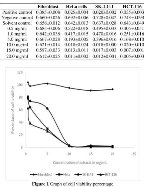
In MTT assay, different concentrations of E. cottonii crude extract were used to treat with various cancer cells and fibroblast cells. The absorbance readings at 570nm with reference filter at 630nm for 24 hours
incubation are shown in table 1 and graph was plotted as shown in figure 1.
In this study, MTT cell viability test shown that various concentration of the crude extracts from E.
cottonii inhibited the growth of the various cancer
cells for 24 hours incubated with crude extracts from
E. cottonii respectively as results shown in Table 1
and Figure 1. Cells viability percentage decreased gradually when the doses are increasing for 24 hours. On the other hands, cell viability of fibroblast did not decrease even though the highest dose of 20mg/mL was applied. In Figure 1, fibroblast cells still growing and did not shown any decreasing sign after 24 hours treated with E. Cottonii extracts. Consequently, the percentage of various cancer cells viability decreased in a dose dependent manner yet to prevent fibroblast, human normal cell lines from elimination.
When metabolic events lead to apoptosis or necrosis, it showed reduction in cell viability in MTT proliferation assay(Selvi et al., 2011). The linear relationship between cell number and signal produced is established, thus allowing an accurate quantification of changes in the rate of cell
Table 1 MTT assay of various cancer cell lines and
fibroblast cells with E. cottonii.
Fibroblast HeLa cells SK-LU-1 HCT-116 Positive control 0.085±0.008 0.025±0.004 0.020±0.002 0.035±0.003 Negative control 0.660±0.026 0.692±0.006 0.728±0.042 0.743±0.093 Solvent control 0.656±0.012 0.642±0.013 0.637±0.028 0.643±0.049 0.5 mg/ml 0.685±0.006 0.522±0.018 0.495±0.033 0.405±0.051 1.0 mg/ml 0.642±0.036 0.417±0.015 0.470±0.016 0.251±0.016 5.0 mg/ml 0.667±0.028 0.193±0.005 0.396±0.016 0.168±0.010 10.0 mg/ml 0.621±0.014 0.018±0.024 0.018±0.000 0.020±0.010 15.0 mg/ml 0.597±0.033 0.013±0.011 0.017±0.003 0.007±0.001 20.0 mg/ml 0.612±0.025 0.011±0.002 0.012±0.001 0.005±0.003
proliferation.Triplicate test was carried out in this MTT in the study. Standard deviation is important in determining the dispersion of the triplicate readings from its mean value. Therefore, outliers that were found among the absorbance readings were removed to ensure consistency of the results was established. Inaccurate cell seeding and inaccurate reagent pipetting might be the possible causes of the poor consistency of replicates. A repeating pipettor would be ideal to increase the accuracy of the cell seeding and reagent pipetting.
Cancer has a scourge on the human population for many years. Although, numerous advances have been made in prevention, diagnosis and treatment of the disease, it still continues to torment mankind (Hanahanand Weinberg, 2000). There are limited research published for the anti-cancer effect of E.
cottonii seaweed but the potential of other seaweed
species as an anti-tumor, anti-inflammatory have been well known. In previous studies, phenol-rich
extracts of E. cottonii have suppressed and
prevented breast cancer tumour. With this
background knowledge as supportive evidence, this study revealed the in vitro anticancer activity of the crude extracts from E. cottonii (Namvar et al., 2012). Thus, from the MTT analysis on the various cancer cells line revealed the crude extracts from the
E. cottonii could effectively reduce the cancer cell
proliferation. The anticancer effect of the E. cottonii may be due to the presence of the secondary metabolite compounds such as phenol or antioxidant contents which yet to be identified (Selvi et al., 2011). Those foods rich in antioxidant have been shown to play an essential role in the prevention of some human diseases especially cancer.
CONCLUSION: In conclusion, this study has demonstrated that E. cottonii red seaweed has good antiproliferative properties and strong cytotoxicity characteristic on HeLa cells, HCT-116 human colon carcinoma cell line and SK-LU-1 human lung carcinoma cell linein vitro. Although the specific bioactive compounds which responsible for the anticancer properties of E. cottonii have yet to be discovered, E. cottonii has shown its potential of contributing in the development of novel anticancer
drug based on this study. In future studies, different assay can be carried out or the same methods can be done on other cancer cell line followed by in vivo test in order to prove the anticancer effect of E. cottonii on animal model.
Acknowledgement: We wish to thankUCSI University for the supports throughout the projects.
REFERENCES:
Al-Hajj M, Wicha MS, Benito-Hernandaz A, Morrison SJ, and Clarke MF,(2003). Prospective identification of tumorigenic breast cancer cells. Proceedings of the National Academy of
Sciences, 100 (7):3983-3988.
American Cancer Society, 2011. Global Cancer Facts & Figures, 2ndEdn,Atlanta: American Cancer Society: 1-60, (2011).
Boopathy NS and Kathiresan K,(2010). Anticancer drugs from marine flora: an overview. Journal of Oncology, 2010:1-18. Carte BK,(1996). Biomedical potential of marine natural products.
BioScience,46 (4):271-286.
Fard SG, Tan RTR, Mohammed AA, Goh YM, Muhamad KS, AL-Jashamy KA, and Mohamed S,(2011). Wound healing properties of Eucheumacottonii extracts in Sprague-Dawley rats. Journal of
Medical Plants Research,5 (27):6373-6380.
Hanahan Dand Weinberg RA,(2000). The hallmark of cancer.
Cell,100:57-70.
Langdon SP, 2004. Cancer Cell Culture: Methods and Protocol. USA: Humana Press Inc:165-166, (2004)
Namvar F, Muhamed S, Fard SG,Behravan J, Mustapha NM, Alitheen NBM, and Othman F,(2012). Polyphenol-rich seaweed (Eucheumacottonii) extract suppresses breast tumour via hormone modulation and apoptosis induction. Food Chemistry, 130:376-382.
Selvi S, Umadevi P, Murugan S, and Giftson Senapathy J,(2011). Anticancer potential evoked by Pleurotusflorida and
Calocybeindica using T24urinary bladder cancer cell line. African
Journal of Biotechnology,10(37): 7279-7285.
Shetty SS, Kaushik SS, Mojamdar MM, Gogate AA, and Chaukar AP,(1996). Viability testing of homograft valves using methyl thiazol tetrazolium assay. Journal of Postgraduate Medicine,42 (3):72-75.
Taskin E, Caki Z, Ozturk M, and Taskin E,(2010). Assessment of antitumoral and antimicrobial activities of marine algae harvested from the eastern Mediterranean Sea. African Journal of
Biotechnology, 9 (27):4272-4277.
Wijesekara I, Pangestuti R, and Kim SK,(2011). Biological activities and potential health benefits of sulfate polysaccharides derived from marine algae. Carbohydrate Polymers, 84:14-21.
Yedjou CG and Tchounwou PB,(2007). In-vitro cytotoxic and genotoxic effects of arsenic trioxideon human leukemia (HL-60) cells using the MTT and alkalinesingle cell gel electrophoresis (Comet) assays. Molecular and Cellular Biochemistry, 301: 123-130.
Yoshie SY, Hsieh YP, and Suzuki T,(2003). Distribution of flavanoids and related compounds in seaweeds in Japan. Journal
of Tokyo University of Fisheries,89:1-6.