R E S E A R C H
Open Access
Effect of
Carissa opaca
leaves extract on lipid
peroxidation, antioxidant activity and
reproductive hormones in male rats
Sumaira Sahreen
1,2, Muhammad Rashid Khan
2, Rahmat Ali Khan
2,3*and Naseer Ali Shah
2Abstract
Background:Carissa opacaleaves are traditionally used in the treatment of male dysfunction and hormonal disorder as well as in oxidative stress in Pakistan and Asia. The present study was designed to assess the protective effects of methanolic extract ofCarissa opacaleaves (MLC) on carbon tetrachloride (CCl4)-induced reproductive stress in male rats and bioactive constituents responsible for the activity.
Methods:CCl4was induced in 42 male rats for eight weeks and checked the protective efficacy of methanolic extract ofCarissa opacaleaves at various hormonal imbalances, alteration of antioxidant enzymes, DNA fragmentation levels and lipid peroxidation caused testicular fibrosis in testis while High performance Liquid Chromatography (HPLC) was used for detection of bioactive components.
Results:HPLC characterization revealed the presence of isoquercitin , hyperoside , vitexin , myricetin and kaempherol. CCl4caused significant alteration in the secretion of reproductive hormones. Activity of antioxidant enzymes viz; catalase, superoxide dimutase and phase II metabolizing enzymes including glutathione peroxidase, glutathione reductase and reduced glutathione was decreased while DNA fragmentation, hydrogen per oxide contents and thiobarbituric acid reactive substances (TBARS) were increased with CCl4treatment. Co-administration of 100 mg/kg and 200 mg/kg b.w. MLC effectively ameliorated the alterations in the biochemical markers;
hormonal and molecular levels.
Conclusion:Protective effects of methanolic extract ofCarissa opacaagainst CCl4−induced antioxidant and hormonal dysfunction which might be due to bioactive compound present in extract.
Keywords:CCl4,Carissa opaca, TBARS, LH, DNA fragmentation
Background
During the last decade, considerable attention was given to the involvement of oxygen free radicals in various dis-eases. There is no doubt that reactive oxygen species (ROS) play an important role in pathological changes in the liver, particularly in the case of alcoholic and toxic liver diseases [1]. Biological membranes are particularly prone to ROS effect. The peroxidation of unsaturated fatty acids in biological membranes leads to the decrease of membrane fluidity and the disruption of membrane
integrity and function. Such peroxidation effect is impli-cated in serious pathological changes [2]. CCl4is an in-dustrial solvent causes tissue damages in various tissues of experimental animals. CCl4 requires bioactivation by phase I cytochrome P450 system to form reactive meta-bolic radicals. These free radicals can bind with polyun-saturated fatty acid (PUFA) of sperm membrane to generate lipid peroxides that are highly reactive, change enzyme activity and finally induce injury or necrosis [3,4]. Several endogenous protective mechanisms have evolved to limit ROS effect and the damage caused by them [5]. However, when this protection is not complete, or when the formation of ROS is excessive, additional pro-tective mechanisms of dietary antioxidants may be of a great importance. Therefore, many natural and synthetic * Correspondence:Rahmatgul_81@yahoo.com
2
Department of Biochemistry, Faculty of Biological Sciences, Quaid-i-Azam University, Islamabad, Pakistan
3
Department of Biotechnology, Faculty of Biological Sciences, University of Science and Technology Bannu, Khyber Pakhtunkhwa KPK 28100, Pakistan Full list of author information is available at the end of the article
agents possessing antioxidative properties have been pro-posed to prevent and treat infertility and reproductive hormonal imbalance induced by oxidative stress [6]. There is increasing evidence of the protective role of hydroxy and polyhydroxy organic compounds, particularly from vegetables, fruits and some herbs.
Plants are well-known excellent perspectives for the discovery of new therapeutical products. In recent years, an ample interest has been developed in finding natural antioxidants from commonly available wild plants, fruits and vegetables that were generally mistreated [7,8] as well as an important role in detoxification of free radi-cals induced lung injuries and fibrosis in experimental animal’s model. Carissa opaca Stapf ex Hanes a 2–3 meter tall evergreen shrub containing glabrous fruits widely found in Pakistan [9]. Traditionally this plant is used for the treatment of asthma [10], hepatitis [11], diarrhea [12] and renal dysfunction [13]. The present study was conducted to examine the toxic upshots of CCl4plus to compare the beneficial effects of plant ex-tracts on reproductive hormonal disturbance and activity of antioxidant enzymes in various experimental groups. In this respect, several parameters regarding the testicu-lar injury and fibrosis were studied.
Methods and materials Plant collection
C. opaca leaves were collected in March 2011 from the Quaid-i-Azam University Islamabad, Pakistan, recog-nized by their local names and validated by Dr. Mir Ajab Khan, Department of Plant Sciences, Quaid-i-Azam Uni-versity, Islamabad. A voucher specimen was deposited at the Herbarium of Pakistan Quaid-i-Azam University, Islamabad Pakistan for future reference.
Extract preparation
The collected plant leaves were cleaned and dried under shade for fifteen days. Willy Mill of 60-mesh size was used to prepare powder of dried samples and then 5 kg pow-dered plant sample was extracted twice with 10 L of 95% methanol at 25°C for 48 h. For filtration Whatman No. 1 filter paper was used and then filtrate was concentrated on rotary evaporator (Panchun Scientific Co., Kaohsiung, Taiwan) under reduced pressure at 40°C and dry extract was stored at 4°C for furtherin vivoinvestigation.
Phytochemical investigation
High performance liquid chromatogrhy (HPLC)
50 mg of fine powder was extracted with 6 ml of 25% hydrochloric acid and 20 ml methanol for 1 h. The ob-tained extract was filtered to a volumetric flask and di-luted to 100 ml with methanol. 10 μl was injected into HPLC column (20RBAX ECLIPSE, XDB-C18, 5 μm; 4.6 × 150 mm, Agilent USA) with UV–VIS
Spectra-Focus detector, injector-auto sampler. Solvent A (0.05% trifluoroacetic acid) and solvent B (0.038% trifluoroacetic acid in 83% acetonitrile (v/v) with the following gradient: 0–5 min, 15% B in A, 5–10 min, 70% B in A, 10–15 min, 70% B in A. The flow rate was 1 ml/min and injec-tion volume was 10 μl. Various standard compounds including rutin, myricetin, vitexin, orientin, hyperoside, isovitexin, isoquercetin, luteolin, apigenin, kaempherol, and luteolin-7-glucoside were run for comparative detec-tion and optimized. The calibradetec-tion curves were defined for each compound in the range of sample quantity 0.02-0.5μg. All samples were assayed in triplicate.
Experimental plan
Six-week-old male Sprague Dawley rats weighing 180 ± 10 g were provided with food and waterad libitumand kept at 20–22°C on a 12-h light–dark cycle. All experi-mental procedures involving animals were conducted in accordance with the guidelines of National Institutes of Health (NIH guidelines). The study protocol were ap-proved by Ethical committee of Quaid-i-Azam Univer-sity Islamabad. The rats were acclimatized to laboratory condition for 7 days before commencement of experi-ment. For chronic toxicity eight week experiment was designed. 42 male albino rats were randomly divided into seven groups (6 rats of each group). Administration of CCl4(0.5 ml/kg b.w., 20% CCl4/olive oil) was intraperi-toneally (i.p.) twice a week for eight weeks. At the same time, the rats were administered individually silymarin (50 mg/kg b.w.) and extract (100, 200 mg/kg b.w.) orally twice a week for eight weeks.
Experimental protocol
Following dosing plan was adapted for the study. Group I: the normal control received only feed
Group II: Olive oil (0.5 ml/kg b.w., i.p.) + DMSO (0.5 ml/kg b.w. orally)
Group III: CCl4twice a week (0.5 ml/kg b.w., i.p., 20% CCl4/olive oil)
Group IV: CCl4 twice a week (0.5 ml/kg b.w., i.p.) + sylimarin (50 mg/kg b.w., orally)
Group V: CCl4 twice a week (0.5 ml/kg b.w., i.p.) + MLC (100 mg/kg b.w., orally)
Group VI: CCl4 twice a week (0.5 ml/kg b.w., i.p.) + MLC (200 mg/kg b.w., orally)
treated with liquid nitrogen and stored at−80°C for fur-ther enzymatic and DNA damage analysis while the other portion was processed for histology.
Assesment of reproductive hormones and lipid profile of serum
Serum analysis of testicular hormones like FSH, LH, testos-terone, prolactin and esteradiol were radioimmunoassayed by using Marseille Cedax 9 France Kits and Czch Republic Kits from Immunotech Company. Then again, lipid profile such as Triglycerides, total cholesterol, LDL and LDH waere estimated by using standard AMP diagnostic kits (Stattogger Strasse 31b 8045 Graz, Austria).
Assessment of antioxidant enzymes
10% homogenate of tissue was prepared in 100 mM KH2PO4 buffer containing 1 mM EDTA (pH 7.4) and centrifuged at 12,000 × g for 30 min at 4°C. The super-natant was collected and used for the following parame-ters as described below.
Catalase assay (CAT)
CAT activities were determined by the method of Chance and Maehly [14] with some modifications. The reaction solution of CAT activities contained: 2.5 ml of 50 mM phosphate buffer (pH 5.0), 0.4 ml of 5.9 mM H2O2and 0.1 ml enzyme extract. Changes in absorbance of the reaction solution at 240 nm were determined after one minute. One unit of CAT activity was defined as an absorbance change of 0.01 as units/min.
Peroxidase assay (POD)
Activities of POD were determined by the method of Chance and Maehly [14] with some modifications. The POD reaction solution contained: 2.5 ml of 50 mM phosphate buffer (pH 5.0), 0.1 ml of 20 mM guaiacol, 0.3 ml of 40 mM H2O2 and 0.1 ml enzyme extract. Changes in absorbance of the reaction solution at 470 nm were determined after one minute. One unit of POD activ-ity was defined as an absorbance change of 0.01 units/min.
Superoxide dismutase assay (SOD)
SOD activity was estimated by the method of Kakkaret al. [15]. Reaction mixture of this method contained: 0.1 ml of phenazine methosulphate (186 μM), 1.2 ml of sodium pyrophosphate buffer (0.052 mM; pH 7.0), 0.3 ml of supernatant after centrifugation (1500 × g for 10 min followed by 10000 × g for 15 min) of testis homogenate was added to the reaction mixture. Enzyme reaction was initiated by adding 0.2 ml of NADH (780 μM) and stopped after 1 min by adding 1 ml of glacial acetic acid. Amount of chromogen formed was measured by record-ing color intensity at 560 nm. Results are expressed in units/mg protein.
Glutathione-S-transferase assay (GST)
Glutathione-S-transferase activity was assayed by the method of Habiget al. [16]. The reaction mixture consisted of 1.475 ml phosphate buffer (0.1 mol, pH 6.5), 0.2 ml re-duced glutathione (1 mM), 0.025 ml (CDNB) (1 mM) and 0.3 ml of homogenate in a total volume of 2.0 ml. The changes in the absorbance were recorded at 340 nm and enzymes activity was calculated as nM CDNB conjugate formed/min/mg protein using a molar extinction coeffi-cient of 9.6 × 10³M-1cm-1.
Glutathione reductase assay (GR)
Glutathione reductase activity was determined by method of Carlberg and Mannervik [17]. The reaction mixture consisted of 1.65 ml phosphate buffer: (0.1 mol; pH 7.6), 0.1 ml EDTA (0.5 mM), 0.05 ml oxidized gluta-thione (1 mM), 0.1 ml NADPH (0.1 mmol) and 0.1 ml of homogenate in a total volume of 2 ml. Enzyme activ-ity was quantitated at 25°C by measuring disappearance of NADPH at 340 nm and was calculated as nM NADPH oxidized/min/mg protein using molar extinc-tion coefficient of 6.22 × 10³M-1cm-1.
Glutathione peroxidase assay (GPx)
Glutathione peroxidase activity was assayed by the method of Mohandaset al. [18]. The reaction mixture con-sisted of 1.49 ml phosphate buffer (0.1 M; pH 7.4), 0.1 ml EDTA (1 mM), 0.1 ml sodium azide (1 mM), 0.05 ml gluta-thione reductase (1 IU/ml), 0.05 ml GSH (1 mM), 0.1 ml NADPH (0.2 mM), 0.01 ml H2O2(0.25 mM) and 0.1 ml of homogenate in a total volume of 2 ml. The disap-pearance of NADPH at 340 nm was recorded at 25°C. Enzyme activity was calculated as nM NADPH oxidized/ min/mg protein using molar extinction coefficient of 6.22 × 10³M-1cm-1.
Quinone reductase assay (QR)
The activity of quinone reductase was determined by the method of Benson et al. [19]. The 3.0 ml reaction mixture consisted of 2.13 ml Tris–HCl buffer (25 mM; pH 7.4), 0.7 ml BSA, 0.1 ml FAD, 0.02 ml NADPH (0.1 mM), and 0.l ml of homogenate. The reduction of dichlorophenolindophenol (DCPIP) was recorded at 600 nm and enzyme activity was calculated as nM of DCPIP reduced/min/mg protein using molar extinction coefficient of 2.1 × 10⁴M-1cm-1.
Reduced glutathione assay (GSH)
phosphate buffer (0.1 M; pH 7.4) and 0.2 ml DTNB (100 mM). The yellow color developed was read imme-diately at 412 nm on a SmartSpecTM plus Spectropho-tometer. It was expressed asμM GSH/g tissue.
Estimation of lipid peroxidation assay (TBARS/LPO)
The assay for lipid peroxidation was carried out following the modified method of Iqbal et al. [21]. The reaction mixture in a total volume of 1.0 ml contained 0.58 ml phosphate buffer (0.1 M; pH 7.4), 0.2 ml homogenate sample, 0.2 ml ascorbic acid (100 mM), and 0.02 ml ferric chloride (100 mM). The reaction mixture was incubated at 37°C in a shaking water bath for 1 h. The reaction was stopped by addition of 1.0 ml 10% trichloroacetic acid. Following addition of 1.0 ml 0.67% thiobarbituric acid, all the tubes were placed in boiling water bath for 20 min and then shifted to crushed ice-bath before centrifuging at 2500 × g for 10 min. The amount of TBARS formed in each of the samples was assessed by measuring optical density of the supernatant at 535 nm using spectropho-tometer against a reagent blank. The results were ex-pressed as nM TBARS/min/mg tissue at 37°C using molar extinction coefficient of 1.56 × 10⁵M-1cm-1.
Hydrogen peroxide assay (H2O2)
Hydrogen peroxide (H2O2) was assayed by H2O2-mediated horseradish peroxidase-dependent oxidation of phenol red by the method of Pick and Keisari [22]. 2.0 ml of ho-mogenate sample was suspended in 1.0 ml of solution containing phenol red (0.28 nM), horse radish peroxid-ase (8.5 units), dextrose (5.5 nM) and phosphate buffer (0.05 M; pH 7.0) and were incubated at 37°C for 60 min. The reaction was stopped by the addition of 0.01 ml of NaOH (10 N) and then centrifuged at 800 × g for 5 min. The absorbance of the supernatant was recorded at 610 nm against a reagent blank. The quantity of H2O2 produced was expressed as nM H2O2/min/mg tissue based on the standard curve of H2O2oxidized phenol red.
Molecular studies
DNA had been isolated and its fragmentation percent was quantified in molecular studies ofin vivotoxicity.
DNA fragmentation assay with diphenylamine reaction
DNA fragmentation from tissue extract was determined using the procedure of Wuet al. [23]. 100 mg tissue was homogenized in TTE solution. 0.1 ml of homogenate was labeled B, centrifuged at 200 × g at 4°C for 10 min, got supernatant labeled S. S tubes were centrifuged at 20,000 × g for 10 min at 4°C to separate intact chroma-tin, was labeled T. 1.0 ml of 25% TCA was added in all tubes T, B, S and incubated over night at 4°C. After incu-bation precipitated DNA was recovered by pelleting for 10 min at 18,000 × g at 4°C. 160 μl of 5% TCA was
added to each pellet and heated for 15 min at 90°C then 320 μl of freshly prepared DPA solution was added, vortexed and incubated for 4 hr 37°C. Optical density was read at 600 nm with a spectrophotometer (Smart spec™Plus, catalog # 170–2525).
DNA Isolations and ladder assay
DNA was isolated by using the methods of Wu et al.
[23]. 100 mg of tissue in a petri dish was washed with DNA Buffer and homogenized in 1 ml lysis buffer. 100μl of proteinase K (10 mg/ml) and 240 μl 10% SDS, shaked gently, and incubate overnight at 45°C in a water bath then 0.4 ml of phenol, was added shaked for 5–10 min, and centrifuge at 3000 rpm for 5 min at 10°C. Supernatant was mixed with 1.2 ml phenol, 1.2 ml Chloroform/isoamyl al-cohol (24:1); shaked for 5–10 min, and centrifuged at 3000 rpm for 5 min at 10°C. 25 μl of 3 M sodium acetate (pH 5.2) and 5 ml ethanol was added with supernatant, shake until DNA was precipitated. DNA was washed with 70% ethanol, dried, dissolved in TE buffer and its concen-tration checked at 260 and 280 nm. 5μg of total DNA and 0.5μg DNA standard per well were loaded on 1.5% aga-rose gel containing ethidium bromide. Electrophoresis was performed for 45 min with 100 V batteries, and DNA was observed under digital gel doc system and photographed.
Histopathological study of tissue
After weighting the portion specifies for histology small pieces of each tissue was fixed for 3–4 h in fixative sera followed by dehydration with ascending grades of alcohol (80%, 90%, and 100%) and transferred in cedar wood oil. When tissue becomes clear then all tissues were embedded in paraplast and prepared blocks for further microtomy. 3–4 μm thin slides were prepared with microtome; wax was removed, stained with hemotoxilin-eosin and photo-graphed under light microscope at 10x and 40x.
Statistical analysis
To find the different treatment effects ofin vivostudies one way analysis of variance was carried by computer software SPSS 13.0. Level of significance among the various treat-ments was determined by LSD at 0.05% level of probability.
Results
Phytochemical determination
High performance liquid chromatography (HPLC)
Table 1. The conditions used directed towards the good separation of peaks that may be identified in the chro-matogram as apigenin (Rt =4.7), myricetin (Rt =18.5), vitexin (Rt =2.5), orientin (Rt =2.75), hyperoside (Rt = 12.5), isovitexin (Rt=3.7), isoquercetin (Rt=6), rutin (Rt= 8.7), luteolin (Rt=2.01), kaempherol (Rt=3.4), luteolin-7-glucoside (Rt =1.6). A sample of 10 μl of solution was injected to the instrument and identification was done by comparing the obtained peaks of chromatogram of sam-ples with the peaks of standard flavonoids in respect to retention time and UV-spectra. The chromatogram deter-mining flavonoids components of different fractions of
C. opaca leaves in Figure 1. Table 2 summarized the flavonoids found in methanolic fraction ofC. opacaleaves. There were some peaks having different retention time could not be identified; however, based on their chromato-graphic behaviors and UV spectra, they may correspond
to unknown flavonoids compounds as presented in re-spective chromatogram.
In Vivoinvestigation
Previous studies reported that liver is not the just target organ of CCl4; it can actually distress other vital organs including kidney, lungs, heart and testis. Consequently, the intention of the current study was to assess the CCl4 as one of the male reproductive toxicant, hence alteration in the male reproductive hormones, antioxidant levels, lipid profile of serum and histopathological changes of the testis were investigated.
Effects of MLC against CCl4induced testicular toxicity in rat The current study was paying attention on the estima-tion of ameliorating potential of C. opacaleaves against testicular toxicity provoked by CCl4. The biomarkers for Table 1 Linear regression analysis of eleven standard flavonoids
Compound Retention time Regression analysis R Linear range (ppm)
Rutin 8.7 y = 12571.3x-16.62 0.9873 10-250
Myricetin 18.5 y = 9643.4x-11.07 0.9919 10-200
Vitexin 2.5 y = 23085.1x + 3.72 0.9932 10-100
Orientin 2.75 y = 36421.0x + 2.88 0.9869 25-500
Hyperoside 12.5 y = 22758.9x + 1.56 0.9865 10-250
Isovitexin 3.7 y = 31604.2x + 0.98 0.9741 5-150
Luteolin 2.01 y = 19348.6x + 2.08 0.9532 5-100
Isoquercetin 6 y = 26785.6x + 1.60 0.9616 5-500
Apigenin 4.7 y = 10623.5x-9.82 0.9765 25-250
Kaempherol 3.4 y = 26182.8x- + 2.33 0.9417 10-500
Luteolin-7-glucoside 1.6 y = 11434.6x-10.72 0.9536 5-100
Values are Mean ± SD (03 number).
Figure 11HPLC flavonoid profile of MLC, Conditions: mobile phase, ACN-dH2O; flow rate, 1 ml min−1; detection wave length, 220 nm;
testicular toxicity evaluation were based on serological studies, antioxidant enzyme levels of tissue, genotoxicity and histological variation of testis.
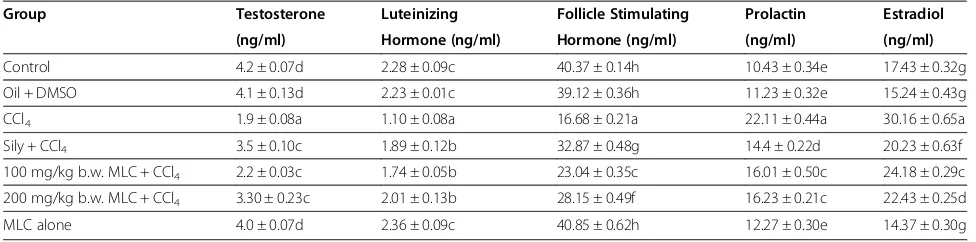
Effects of MLC on male reproductive hormones of rats
Data from the serological markers for reproductive sta-tus such as testosterone, luteinizing hormone (LH), fol-licle stimulating hormone (FSH), prolactin and estradiol is summarized in Table 3. CCl4 intoxication drastically (p < 0.05) reduced the serum level of testosterone, LH and FSH, while notably (p < 0.05) increased the intensity of prolactin and estradiol. The serum level of above said reproductive hormones was re-established (p < 0.05) by oral administration of 100 mg/kg b.w., 200 mg/kg b.w., MLC near to control group. 50 mg/kg body weight of silymarin treatment erased CCl4intoxication and re-stored the level of all tested reproductive hormones in serum of rats.
Effects of MLC on lipid panel changes
Table 4 summarizes protective effects of MLC against CCl4induced toxicity in lipids profile of serum. For lipid parameters total triglycerides, total cholesterol, HDL, and LDL were investigated.CCl4 disputation markedly in-creased the levels of triglycerides, total cholesterol, and LDL cholesterol while decreased (p < 0.01) HDL choles-terol as against the control group. Treatment of MCL can-celled the toxicity of CCl4thus, restoring the serum level of total triglycerides, total cholesterol, HDL, and LDL
towards the control group. Treatment with silymarin also produced similar results.
Effects of MLC on testis enzymatic antioxidant levels
Oxidative stress produced by CCl4 upsets the cellular antioxidant defense system. The protective effects of MLC against CCl4 toxicity on the antioxidant profile are pre-sented in Table 5. Administration of CCl4for eight weeks caused noteworthy (p < 0.05) decrease in the tissue soluble protein and CAT, POD and SOD activities as opposite to control group. Post-treatment of 100 mg/kg b.w., 200 mg/ kg b.w., MLC markedly ameliorated the affects of CCl4 in-toxication, and distinctly enhanced (p < 0.05) testicular protein and CAT, POD and SOD levels of testicular tissue. Lipid peroxidation is umpired via free radicals produced by CCl4intoxication. The significant decline in H2O2and TBARS level of testicular tissue corroborates with protect-ive power 100 mg/kg b.w., 200 mg/kg b.w., MLC against testicular CCl4induced lipid peroxidation in testis tissue. Similar protective effects were reported, while treating with silymarin.
Alterations in phase II antioxidant metabolizing en-zymes viz; GST, GPx, GR, GSH and QR as well as DNA fragmentation% testicular tissues of rat are demonstrated in Table 6. Chronic administration of CCl4, extensively (p < 0.05) abridged the glutathione status of GST, GPx, GR, GSH and QR whereas, percentage of DNA fragmentation was increased in comparison to non treated control group. Post-treatment with 100 mg/kg b.w., 200 mg/kg b.w., MLC attenuated the intoxication of CCl4 and restored the en-zymes activity near to control rats. Silymarin treatment markedly lessened the DNA fragmentation% while, in-creased the GST, GSH, GR, GPx and QR activation similar to the effects of 100 mg/kg b.w., 200 mg/kg b.w., MLC.
Effects of MLC on DNA damages (ladder assay)
Protective effects of different doses of MLC versus CCl4 induced DNA damage in the testicular tissues of rats is shown by DNA ladder assay in Figure 2. Extensive DNA breakages in testis were portrayed by the treatment of Table 2 Assessment of flavonoids in methanolic extract
ofCarissa opacaleaves extract
Compound Retention time Concentrationμg/mg of dry weight
Isoquercitin 6 0.119
Hyperoside 12.5 0.062
Vitexin 2.5 0.053
Myricetin 18.5 0.172
Kaempherol 3.4 0.08
Values are Mean ± SD (03 number).
Table 3 Effects of MLC on male reproductive hormonal level
Group Testosterone Luteinizing Follicle Stimulating Prolactin Estradiol (ng/ml) Hormone (ng/ml) Hormone (ng/ml) (ng/ml) (ng/ml)
Control 4.2 ± 0.07d 2.28 ± 0.09c 40.37 ± 0.14h 10.43 ± 0.34e 17.43 ± 0.32g
Oil + DMSO 4.1 ± 0.13d 2.23 ± 0.01c 39.12 ± 0.36h 11.23 ± 0.32e 15.24 ± 0.43g
CCl4 1.9 ± 0.08a 1.10 ± 0.08a 16.68 ± 0.21a 22.11 ± 0.44a 30.16 ± 0.65a
Sily + CCl4 3.5 ± 0.10c 1.89 ± 0.12b 32.87 ± 0.48g 14.4 ± 0.22d 20.23 ± 0.63f
100 mg/kg b.w. MLC + CCl4 2.2 ± 0.03c 1.74 ± 0.05b 23.04 ± 0.35c 16.01 ± 0.50c 24.18 ± 0.29c
200 mg/kg b.w. MLC + CCl4 3.30 ± 0.23c 2.01 ± 0.13b 28.15 ± 0.49f 16.23 ± 0.21c 22.43 ± 0.25d
MLC alone 4.0 ± 0.07d 2.36 ± 0.09c 40.85 ± 0.62h 12.27 ± 0.30e 14.37 ± 0.30g
Values are Mean ± SD (06 number), Sily = Silymarin.
CCl4to rats. Post-administration of silymarin and MLC prevented the DNA damages induced by CCl4indicating the protective effects ofC. opacaleave.
Effects of MLC on testis histoarchitecture
The histoarchitecture of testis after different treatments is presented in Figure 3. Light microscope evaluation of H & E stain showed normal testicular architecture pos-sessing normal seminiferous tubules, normal concentration of germ cells, sperms with normal morphology and concen-tration and inconspicuous sertoli cells (Figure 3A). The CCl4intoxication resulted in ruthless testicular injuries with imperative decrease in germ cells, vacuolization of germina-tive epithelium and dislocated interstitial cells away from
basement membrane and seminiferous tubules as shown in Figure 3B. The silymarin group also showed almost normal structure of testis as compare to CCl4 intoxicated group (Figure 3C). Groups treated MLC showed improved con-centration of sperms and stabilization of organized semi-niferous tubules (Figure 3D). The results obtained from histological architecture were in consistency with the hor-monal studies as well as antioxidant status.
Discussion
Oxidative stress induced by an increase in free radicals and/or decrease in antioxidant defenses is well docu-mented in animal model [24,25]. CCl4, a typical toxic agent, exerts its toxic effects by the generation of free Table 4 Effects of MLC on lipid profile
Group Triglycerides (mg/dl) Total Cholesterol (mg/dl) HDL (mg/dl) LDL (mg/dl)
Control 130.13 ± 2.97e 39.15 ± 1.43e 32.31 ± 1.08d 15.34 ± 1.34d
Oil + DMSO 128.76 ± 2.45e 40.24 ± 1.34e 30.32 ± 1.73d 13.56 ± 1.11d
CCl4 230.19 ± 2.08a 78.99 ± 1.05a 52.36 ± 1.04a 30.61 ± 2.04a
Sily + CCl4 159.32 ± 2.42d 49.23 ± 2.33d 40.33 ± 1.27c 20.76 ± 1.73c
100 mg/kg b.w. MLC + CCl4 190.06 ± 4.41b 60.73 ± 1.72b 48.23 ± 1.12b 25.26 ± 1.95b
200 mg/kg b.w. MLC + CCl4 168.99 ± 2.22c 52.84 ± 2.62c 44.84 ± 1.69c 22.33 ± 1.41c
MLC alone 127.19 ± 1.33e 39.65 ± 1.28e 29.47 ± 1.36d 12.15 ± 1.11d
Values are Mean ± SD (06 number). Sily = Silymarin.
a-e (Means with different letters) indicate significance at p < 0.05.
Table 5 Effects of MLC on tissue proteins and antioxidant enzyme levels
Group Protein CAT POD SOD TBARS H2O2
(μg/mg tissue) (U/min) (U/min) (U/mg protein) (nM/min/mg protein) (μM/ml)
Control 1.421 ± 0.023d 3.92 ± 0.22d 9.38 ± 0.13c 3.08 ± 0.23d 2.97 ± 0.16d 1.123 ± 0.010c
Oil + DMSO 1.495 ± 0.026d 4.02 ± 0.55d 9.04 ± 0.34c 3.12 ± 0.14d 3.10 ± 0.27d 1.244 ± 0.012c
CCl4 0.834 ± 0.029a 2.02 ± 0.11a 4.76 ± 0.51a 0.88 ± 0.07a 5.71 ± 0.48a 2.536 ± 0.023a
Sily + CCl4 1.312 ± 0.008c 3.54 ± 0.34c 8.58 ± 0.38b 2.75 ± 0.07c 3.80 ± 0.33c 1.560 ± 0.091b
100 mg/kg b.w. MLC + CCl4 1.27 ± 0.062c 2.9 ± 0.17b 7.79 ± 0.85c 1.95 ± 0.13c 3.74 ± 0.70b 1.30 ± 0.069c
200 mg/kg b.w. MLC + CCl4 1.344 ± 0.073c 3.25 ± 0.32c 8.37 ± 0.41b 2.29 ± 0.11b 4.01 ± 0.25c 1.644 ± 0.065b
MFC alone 1.441 ± 0.012d 3.95 ± 0.68d 10.00 ± 0.61c 3.63 ± 0.13d 2.91 ± 0.33d 1.121 ± 0.045c
Values are Mean ± SD (06 number). Sily = Silymarin.
a-h (Means with different letters) indicate significance at p < 0.05.
Table 6 Effects of MLC on phase II antioxidant enzymes and DNA fragmentation
Group GST
(nM/mg protein) GPx
(nM/mg protein) GR
(nM/mg protein) GSH (μM/g tissue)
QR
(nM/mg protein)
%DNA Injuries
Control 150.23 ± 5.34g 110.17 ± 4.46f 198.34 ± 5.78e 16.25 ± 1.11c 99.90 ± 4.97f 14.53 ± 2.57f
Oil + DMSO 157.17 ± 4.45g 105.55 ± 4.55f 204.73 ± 6.47e 15.74 ± 1.31c 107.03 ± 4.42f 13.33 ± 2.24f
CCl4 78.37 ± 4.33a 63.33 ± 3.11a 103.14 ± 4.66a 9.60 ± 1.34a 52.40 ± 4.16a 57.50 ± 3.34a
Sily + CCl4 133.67 ± 2.43f 98.46 ± 2.22e 179.56 ± 4.45d 13.75 ± 0.32b 89.14 ± 2.56e 20.18 ± 3.73e
100 mg/kg b.w. MFC + CCl4 120.54 ± 1.98c 77.39 ± 2.92b 166.07 ± 2.21c 20.05 ± 0.90c 75.25 ± 3.12d 33.30 ± 0.25d
200 mg/kg b.w. MFC + CCl4 124.12 ± 2.81e 94.65 ± 2.16e 170.68 ± 4.97d 13.29 ± 0.46b 82.56 ± 3.67d 23.56 ± 2.36e
MFC alone 159.31 ± 3.21g 112.40 ± 3.26f 201.25 ± 4.12e 17.59 ± 1.45c 109.34 ± 2.23f 12.55 ± 1.27f
Values are Mean ± SD (06 number). Sily = Silymarin.
radicals. By the activation of liver cytochromes P450, CCl4 is converted into free radicals which immediately react with cell membrane [26]. This free radical not only targets liver but it can also causes free radical generation in other tissues like kidneys, heart, lung, testis, brain and blood [27,28]. In the current study, the proposed plan aimed to assess and examine the possibility of MLC to protect and reduce the lipid peroxidation and oxidative damages caused by CCl4 in testis tissue homogenate of male Sprague Dawley rats. The reduction of testosterone levels in serum indicates either direct effects of CCl4at
Leydig cell level or indirect effects by disturbing the hor-monal environmentat hypothalamo-pituitary axis [29] due to oxidative trauma in CCl4 treated rats. It was reported that abnormal level of intra testicular testoster-ones inhibits spermatogenesis [30]. Tohda et al. (2001). The production of testosterone in Leydig cells is stimu-lated by LH, which activates FSH to bind with sertoli cells to stimulate spermatogenesis [31]. CCl4 intoxicated rats show the malfunctioning of pituitary to secrete FSH and LH indicating testicular dysfunction leading to infer-tility as was reported by previous results [32]. GSH levels are dependent upon the activities of glutathione reduc-tase (GR) and NADH [33]. Glutathione system including GPx, GR, GST, as well as SOD and CAT represent a mutually loyal team of defense against ROS [34]. En-hanced lipid peroxidations expressed in terms of TBARS determine structural and functional alterations of cellu-lar membranes [35]. In the present study, administration of various fractions of plant samples in different experi-mental groups improved the activities of SOD, CAT, POD, GPx, GST, GR and QR as well as non enzymatic (GSH, TBARS and H2O2) levels of CCl4-intoxicated testis towards normalcy in warfare of oxidative trauma
in vivo. Hence, the present results regarding chronic toxicity of CCl4are in accordance with previous reports [36,37], while studying the protective effects of Sonchus asperandLauneae procumbenson testis against oxidative Figure 2Agarose gel showing DNA damage by CCl4and
protective effects of MLC in testicular tissue.Lanes from left (M) low molecular weight marker, (1) control, (2) DMSO + Olive oil group, (3) CCl4group, (4) Silymarin + CCl4group, (5) 100 mg/kg b.w.
MLC + CCl4group, (6) 200 mg/kg b.w. MLC + CCl4group.
Figure 3Microphotograph of rat testis (H & E stain).(A) Representative section of testis from the control group showing normal histology, (B) CCl4group, (C) Silymarin + CCl4group, (D) 200 mg/kg b.w. MLC + CCl4group (✦) distorted and less concentration of germ cells, (▲) displaced
stress of CCl4. Present study revealed that the activities of antioxidant enzymes were significantly reduced the toxi-cation of chemical which might be due to the presence of bioactive elements like myricetin, kaempherol, isoquer-cetin, hyperoside and vitexin, propagating free radicals like peroxyl radicals and converting the reactive free radicals to inactive products.
It was reported that CCl4 resulted in the oxidative damage to testicular proteins in rats. Oxidative damage to proteins is very important as it can contribute sec-ondary damage resulting in hampering the DNA repair enzymes and loss of reliability of damage polymerases during DNA replication. The DNA damage in various tis-sues like brain, testis and liver was reported by Manierea
et al. [38]. From the present study, it can be assumed that chronic exposure of CCl4 may cause accumulation of many toxic species in cells thus damaging both DNA and lipids. In fact, treatment with various fractions of plant samples ameliorated the toxic effects on DNA as revealed by DNA fragmentation % and ladder assay. The present study clearly augments the defensive mechanism of vari-ous samples against oxidative stress induced by CCl4and provides confirmation about its therapeutic use in repro-ductive abnormalities.
Previous studies on histomorphology of testis showed shrinkage of the tubular diameter and testicular atrophy leading to degenerative changes in the germinal epithe-lium [39] after exposure to toxic chemical. Similar de-structive effects were also accounted in CCl4 treated groups. The CCl4 challenge revealed testicular destruc-tion and degeneradestruc-tion in histological architecture like that of profenofos that was recorded by Moustafa et al. [40] who represented damaged columnar epithelial layer, vacuolated spermatogonial cells, oedematous alterations in the seminiferous tubules and extra elongated Leydig cells. Data of the present study revealed that CCl4may hamper continuing proliferative behavior of testicular cells thus obstruct reproduction. Deformities in sperm-atogenesis and partial degeneration of germ and Leydig cells have been displayed by CCl4-treated rats. However, groups administered various fractions of plant samples in different experiments demonstrated a quality active spermatogenesis, thin basement membranes and normal seminiferous tubules in most of the part of testis. Same histopathology was noticed by Manjrekar et al. [41], while evaluating the protective effects of Phyllanthus niruri Linn treatment on testis against CCl4 intoxica-tion. This paper is in continuation of our previous stud-ies Sahreen et al. [42] in which hepatoprotective effect of methanolic extract of leaves were asasessed.
Conclusion
It can be concluded from the current study that bio-active components of MLC especially flavonoids (myricetin,
kaempherol, isoquercetin, hyperoside and vitexin) have the ability to recover the metabolic enzymatic activities and repair cellular injuries, thus providing scientific evi-dence in favor of its pharmacological use in testicular dysfunctioning.
Competing interest
The authors declare that they have no competing interests.
Authors’contributions
SS made a significant contribution to acquisition of data, analysis, drafting of the manuscript. MRK and RAK have made a substantial contribution to conception and design, interpretation of data, drafting and revising the manuscript for intellectual content. All authors read and approved the final manuscript.
Author details
1Botanical Sciences Division, Pakistan Museum of Natural History, Garden
Avenue, Shakarparian, Islamabad, Pakistan.2Department of Biochemistry, Faculty of Biological Sciences, Quaid-i-Azam University, Islamabad, Pakistan. 3
Department of Biotechnology, Faculty of Biological Sciences, University of Science and Technology Bannu, Khyber Pakhtunkhwa KPK 28100, Pakistan.
Received: 14 February 2013 Accepted: 13 June 2013 Published: 20 June 2013
References
1. Das SK, Vasudevan DM:Alcohol induced oxidative stress.Life Sci2007,
81(3):177–187.
2. Cabre M, Camps J, Paternain JL, Ferre N, Joven J:Time-course of changes in hepatic lipid peroxidation and glutathione metabolism in rats with carbon tetrachlorideinduced cirrhosis.Clin Exp Pharmacol Physiol2000,
27(9):694–699.
3. Sikka SC, Rajasekaran M, Hellstrom WJ:Role of oxidative stress and antioxidants in male infertility.J Androl1995,16(6):464–468. 4. Ogeturk M, Kus I, Colakoglu N, Zararsiz I, Ilhan N, Sarsilmaz M:Caffeic
induced nephrotoxicity and protective effect of betaine in Sprague Dawley rats.Urology2005,62:353–356.
5. Sies H:Strategies of antioxidant defense.Eur J Biochem1993,215:213–219. 6. Kandasamy CS, Shimna TP, Mohammed BE, Arul RP, Gopal V,
Venkatnarayanan R:Anti-hepatotoxic activity of polyherbal formulation in carbon tetrachloride induced toxicity in rats.RJPBCS2010,1:342–346. 7. Umamaheswari M, Chatterjee TK:In vitroantioxidant activities of the
fractions ofCoccinnia grandisL.leaf extract Afric J Trad Compl Alter Med
2008,5(1):61–73.
8. Kil HY, Seong ES, Ghimire BK, Chung I-M, Kwon SS, Goh EJ, Hoe K, Kim MJ, Lim JD, Lee D, Yu CY:Antioxidant and antimicrobial activities of crude sorghum extract.Food Chem2009,115:1234–1239.
9. Nazimuddin S, Qaiser M: Apocynaceae.Flora of Pakistan. Edited by Nasir E, Ali SI; 1983:11–13. Department of Botany, University of Karachi, Karachi. 10. Jabeen A, Khan MA, Ahmad M, Zafar M, Ahmad F:Indigenous uses of
economically important flora of Margalla Hills National Park, Islamabad Pakistan.Afric J Biotechnol2009,8(5):763–784.
11. Abbasi AM, Khan MA, Ahmad M, Zafar M, Khan H, Muhammad M, Sultana S:
Medicinal plants for the treatment of jaundice and hepatitis based on socio-economic documentation.Afric J Biotechnol2009,8(8):1643–1650. 12. Adhikari BS, Babu MM, Saklani PL, Rawat GS:Distribution, use pattern and
prospects for conservation of medicinal shrubs in Uttaranchal State. India J Mountain Sci2007,4(2):155–180.
13. Ahmad SS, Mahmood F, Dogar Z-UL-H, Khan ZI, Ahmad K, Sher M, Mustafa I, Valeem EE:Priortization of medicinal plants of Margala Hills National Park, Islamabad on the basis of available information.Pakistan J Bot2000,
41(5):2105–2114.
14. Chance B, Maehly AC:Assay of catalase and peroxidases.Methods Enzymol
1955,11:764–775.
15. Kakkar P, Das B, Viswanathan PN:A modified spectrophotomateric assay of superoxide dismutase.Indian J Biochem Biophys1984,21:130–132. 16. Habig WH, Pabst MJ, Jakoby WB:Glutathione-S-transferases: the first
enzymatic step in mercapturic acid formation.J Biol Chem1974,
17. Carlberg I, Mannervik EB:Glutathione level in rat brain.J Biol Chem1975,
250:4475–4480.
18. Mohandas J, Marshal JJ, Duggin GG, Horvath JS, Tiller DJ:Differential distribution of glutathione and glutathione-related enzymes in rabbit kidney. Possible implications in analgesic nephropathy.Biochem Pharmacol1984,33:1801–1807.
19. Benson AM, Hunkeler MJ, Talalay P:Increase of NADPH, quinone reductase activity by dietary antioxidant: Possible role in protection against carcinogenesis and toxicity.Proceed National Academy Sci United State America1990,77:5216–5220.
20. Jollow DJ, Mitchell JR, Zampaglione N, Gillete JR:Bromobenzene induced liver necrosis. Protective role of glutathione and evidence for 3, 4-bromobenzene oxide as a hepatotoxic metabolite.Pharmacol1974,
1:151–169.
21. Iqbal M, Sharma SD, Zadeh HR, Hasan N, Abdulla M, Athar M:Glutathione metabolizing enzymes and oxidative stress in ferric nitrilotriacetate (Fe-NTA) mediated hepatic injury.Redox Rep1996,2:385–391.
22. Pick A, Keisari Y:Superoxide anion and hydrogen peroxide production by chemically elicited peritoneal macrophages-induction by multiple nonphagocytic stimuli.Cell Immunol1981,59:301–318.
23. Wu JH, Tung YT, Chien SC, Wang SY, Kuo YH, Shyur LF, Chang ST:Effect of phytocompounds from the Heart-wood ofAcacia confusaon inflammatory mediator production.J Agric Food Chem2008,
56:1567–1573.
24. Botsoglou NA, Taitzoglou IA, Botsoglou E, Zervos I, Kokoli A, Christaki E, Nikolaidis E:Effect of long‐term dietary administration of oregano and rosemary on the antioxidant status of rat serum, liver, kidney and heart after carbon tetrachloride‐induced oxidative stress.J Sci Food Agric2009,
89:1397–1406.
25. Marañón G, Muñoz-Escassi B, Manley W, García C, Cayado P, de la Muela MS, Olábarri B, León R, Vara E:The effect of methyl sulphonyl methane supplementation on biomarkers of oxidative stress in sport horses following jumping exercise.Acta Vet Scand2008,50:45.
26. Dashti H, Jeppsson B, Hägerstrand I, Hultberg B, Srinivas U, Abdulla M, Bengmark S:Thioacetamide-and carbon tetrachloride-induced liver cirrhosis.European Surg Res1989,21:83–91.
27. Ozturk F, Ucar M, Ozturk IC, Vardi N, Batcioglu K:Carbon tetrachloride-induced nephrotoxicity and protective effect of betaine in Sprague– Dawley rats.Urology2003,62:353–356.
28. Preethi KC, Kuttan R:Hepato and reno protective action ofCalendula officinalisL. flower extract.Indian J Exp Biol2009,47:163–168.
29. Latif R, Lodhi GM, Aslam M:Effects of amlodipine on serum testosterone, testicular weight and gonado-somatic index in adult rats.J Ayub Med College Abbottabad2008,20(4):8–10.
30. Tohda A, Matsumiya K, Tadokoro Y, Yomogida K, Miyagawa Y, Dohmae K, Okuyama A, Nishimune Y:Testosterone suppresses spermatogenesis in juvenile spermatogonial depletion (jsd) mice.Biol Repro2001,65:532–537. 31. Conn PM:The molecular basis of gonadotropin releasing hormone
action Endocrinology.Rev1986,7:3–10.
32. Khan MR, Ahmed D:Protective effects ofDigera muricata(L.) Mart on testis against oxidative stress of carbon tetrachloride in rat.Food Chem Toxicol2009,47:1393–1399.
33. Meister A, Anderson ME:Glutathione.Annals Rev Biochem1983,52:711–760. 34. Bandhopadhy U, Das D, Banerjee KR:Reactive oxygen species: Oxidative
damage and pathogenesis.Current Sci1999,77:658–665.
35. Halliwell B, Aeschbach R, Loliger J, Aruoma OI:The characterization of antioxidants.Food Chem Toxicol1990,33:601–617.
36. Khan RA, Khan MR, Sahreen S:Protective effects of Sonchus asper against KBrO3 induced lipid peroxidation in rats.Lipids Health Dis2012,27(1):164. 11. 37. Khan RA:Protective effects ofLaunaea procumbenson rat testis damage
by CCl4.Lipids Health Dis2012,11:103.
38. Manierea I, Godardb T, Doergec DR, Churchwellc MI, Guffroyd M, Laurentiea M, Poula JM:DNA damage and DNA adduct formation in rat tissues following oral administration of acrylamide.Mutation Res2005,580:119–129. 39. Debnath D, Mandal TK:Study of quinalphos (an environmental
oestrogenic insecticide) formulation (Ekalux 25 E.C.)-induced damage of the testicular tissues and antioxidant defence systems in Sprague– Dawley albino rats.J Applied Toxicol2005,20:197–204.
40. Moustafa GG, Ibrahim ZS, Hashimoto Y, M-Alkelch A, Q–Sakamoto K, Ishizuka M, Fujita S:Testicular toxicity of profenofos in matured male rats. Arch Toxicol2007,81:875–881.
41. Manjrekar AP, Jisha V, Bag PP, Adhikary B, Pai MM, Hegde A, Nandini M:
Effect ofPhyllanthus niruriLinn treatment on liver, kidney and testes in CCl4 induced hepatotoxic rats.Indian J Exp Biol2008,46:514–520. 42. Sahreen S, Khan MR, Khan RA:Hepatoprotective effects of methanol
extract ofCarissa opacaleaves on CCl4-induced damage in rat. BMC Complement Altern Med2011,11:48.
doi:10.1186/1476-511X-12-90
Cite this article as:Sahreenet al.:Effect ofCarissa opacaleaves extract on lipid peroxidation, antioxidant activity and reproductive hormones in male rats.Lipids in Health and Disease201312:90.
Submit your next manuscript to BioMed Central and take full advantage of:
• Convenient online submission
• Thorough peer review
• No space constraints or color figure charges
• Immediate publication on acceptance
• Inclusion in PubMed, CAS, Scopus and Google Scholar
• Research which is freely available for redistribution