R E S E A R C H A R T I C L E
Open Access
Association of endothelin genetic variants
and hospitalized infection complications in
end-stage renal disease (ESRD) patients
Chih-Chin Kao
1,2,3, Shih-Ying Cheng
4,5, Yu-Jia Wang
6, Shu-Chen Chien
4,5,7, Yu-Wen Hsu
8, Mei-Yi Wu
3,9,
Hsing-Fang Lu
5, Sean Nam
5, Tao Sun
10, Mai-Szu Wu
1,3,9*†and Wei-Chiao Chang
5,11,12,13*†Abstract
Background:Infection is the second most common cause of mortality for patients with end-stage renal disease (ESRD), accompanying with immune dysfunction. Endothelin (EDN) is known to be related to inflammation; however, it is unknown whether genetic variants of theEDNgene family are associated with increased risk of hospitalized infection events.
Methods:Nineteen tagging single-nucleotide polymorphisms (tSNPs) of theEDNgene family were selected for genotyping a cohort of 190 ESRD patients. Patient demographics were recorded, the subtypes of infection events were identified, and association analysis between theEDNgenetic variants and hospitalized infection events was performed.
Results:In this study, 106 patients were hospitalized for infection events. The leading events were pneumonia, bacteremia, and cellulitis. The minor allele of rs260741, rs197173, and rs926632 SNPs ofEDN3were found to be associated with reduced risk of hospitalized bacteremia events.
Conclusions:The minor allele of rs260741, rs197173, and rs926632 inEDN3were associated with reduced risk of hospitalized bacteremia events in ESRD patients.
Keywords:Endothelin, End stage renal disease, Infection, Renal failure
Background
Infection is known as a common cause of morbidity and mortality in patients with end-stage renal disease (ESRD), accounting for 20% of total deaths in these patients [1]. More than half of patients hospitalized for infection events developed an unfavorable outcome, including prolonged hospitalization, intensive care unit (ICU) stay, or even death [2,3]. Impaired innate and adaptive immunity are associ-ated with increased risk of infection in patients with renal failure [4]. A handful of cytokines was dysregulated in ESRD patients; for example, the production of interleukin
(IL)-1, tumor necrosis factor (TNF)-α, and IL-6 was in-creased, whereas the bioavailability of IL-2 was reduced [5].
Genetic variants of immune-related genes have been shown to reflect risk of infection. Ferwerda et al. reported that the genetic variants of Toll-like receptor 4, which un-derlies the differential production of cytokines may affect the immune system, which in turn influenced the suscep-tibility of infection events and the risk of gram-negative bacterial infection [6]. Moreover, another study demon-strated that anIL-9variant may influence the susceptibil-ity of respiratory syncytial virus infection [7].
Endothelin-1 (ET-1) is the major isoform of the
endothelin (EDN) family and is a potent
vasocon-strictor. It is associated with proinflammatory cyto-kine production, fibrosis, and angiogenesis [8]. The release of lipopolysaccharide from bacteria impairs the integrity of the endothelial cell, resulting in endo-thelial cell injury and cytokine release [9]. The plasma
© The Author(s). 2019Open AccessThis article is distributed under the terms of the Creative Commons Attribution 4.0 International License (http://creativecommons.org/licenses/by/4.0/), which permits unrestricted use, distribution, and reproduction in any medium, provided you give appropriate credit to the original author(s) and the source, provide a link to the Creative Commons license, and indicate if changes were made. The Creative Commons Public Domain Dedication waiver (http://creativecommons.org/publicdomain/zero/1.0/) applies to the data made available in this article, unless otherwise stated.
* Correspondence:maiszuwu@gmail.com;wcc@tmu.edu.tw †Mai-Szu Wu and Wei-Chiao Chang contributed equally to this work.
1
Graduate Institute of Clinical Medicine, College of Medicine, Taipei Medical University, Taipei, Taiwan
5Department of Clinical Pharmacy, School of Pharmacy, College of Pharmacy,
Taipei Medical University, Taipei, Taiwan
ET-1 level is increased during sepsis and is correlated with the severity of sepsis [10]. ET-1 is known to in-crease reactive oxygen species (ROS) production, and associates with activation of nuclear factor-kappaB
and inflammatory cytokines such as TNF-α, IL-1, and
IL-6 [11]. In addition, Lin et al. reported that ET-1 is able to increase cyclooxygenase-2 expression and prostaglandin E2 release [12]. These findings suggest that ET-1 is involved in the inflammatory reactions and the severity of sepsis. Regarding ET-3, Sato et al. demonstrated that low level of ET-3 reduces inflam-matory responses [13]. However, it is still unclear
whether EDN genetic variants are associated with
hospitalized infection events in ESRD patients. In this study, genetic association study was applied to
investi-gate the correlations between EDN genetic variants
and the hospitalized infection events in ESRD
patients.
Methods Study population
Patients who received dialysis for more than 3 months at Taipei Medical University Hospital between September 2013 and June 2014 were enrolled. Demographic and
la-boratory data of these patients was shown in Table 1.
The erythropoietin resistance index (ERI) was deter-mined to characterize patients’ response to erythropoi-etin. ERI was calculated by dividing the weekly body-weight-adjusted epoetin dose by the hemoglobin concentration. Kt/V was used to measure dialysis ad-equacy. Outpatient and discharge medical records were used to determine the cause of ESRD. We prospectively followed up with the patients who were hospitalized for infection events. This study was approved by the Institu-tional Review Board of Taipei Medical University (Ap-proval No. 201309026). A written informed consent form was obtained from all patients.
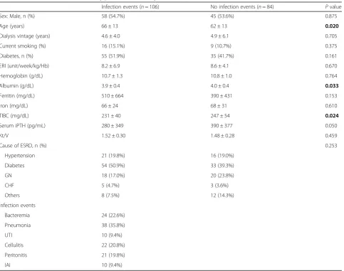
Table 1Baseline characteristics of study patients stratified by infection events
Infection events (n= 106) No infection events (n= 84) Pvalue Sex: Male, n (%) 58 (54.7%) 45 (53.6%) 0.875 Age (years) 66 ± 13 62 ± 13 0.020
Dialysis vintage (years) 4.6 ± 4.0 4.9 ± 6.1 0.705 Current smoking (%) 16 (15.1%) 9 (10.7%) 0.375 Diabetes, n (%) 55 (51.9%) 35 (41.7%) 0.161 ERI (unit/week/kg/Hb) 8.2 ± 6.9 8.6 ± 4.1 0.670 Hemoglobin (g/dL) 10.7 ± 1.3 10.8 ± 1.0 0.764 Albumin (g/dL) 3.9 ± 0.4 4.0 ± 0.4 0.033
Ferritin (mg/dL) 510 ± 664 390 ± 431 0.153 Iron (mg/dL) 66 ± 24 68 ± 31 0.610 TIBC (mg/dL) 231 ± 40 247 ± 54 0.024
Serum iPTH (pg/mL) 280 ± 349 390 ± 377 0.050 Kt/V 1.52 ± 0.30 1.48 ± 0.28 0.459
Cause of ESRD, n (%) 0.253
Hypertension 21 (19.8%) 16 (19.0%) Diabetes 54 (50.9%) 33 (39.3%) GN 18 (17.0%) 20 (23.8%) CHF 5 (4.7%) 3 (3.6%) Others 8 (7.5%) 12 (14.3%) Infection events
Bacteremia 24 (22.6%) Pneumonia 38 (35.8%) UTI 10 (9.4%) Cellulitis 22 (20.8%) Peritonitis 21 (19.8%) IAI 10 (9.4%)
ThePvalues of < 0.05 are shown in bold
Infection events
We defined hospitalized “infection events” as
bacteremia, pneumonia, cellulitis, urinary tract infec-tion (UTI), peritonitis, and intra-abdominal infecinfec-tion (IAI). Bacteremia was confirmed by a positive blood culture result, and contamination was excluded by an infection specialist. Pneumonia was defined as the presence of clinical respiratory symptoms and the findings of increased infiltration on chest radiog-raphy. Cellulitis was determined as inflammation of the skin and subcutaneous tissues. UTI was defined as the presence of clinical symptoms and the detec-tion of a pathogen in the urine. Peritonitis was diag-nosed through clinical symptom examination and peritoneal fluid analysis. IAI was confirmed by the finding of intramural inflammation of the gastro-intestinal (GI) tract without anatomic disruption. These diagnoses were ascertained by physicians and recorded in the discharge medical records.
Genotyping
Patients’ blood samples were collected, and their
genomic DNA was extracted. We selected 5 tagging
single-nucleotide polymorphisms (tSNPs) of EDN1
(i.e., rs5370, rs2070699, rs2248580, rs4714384, and
rs3087459; Fig. 1a), 4 tSNPs of EDN2 (i.e.,
rs2759257, rs11210278, rs11572340, and rs11572377; Fig. 1b), and 10 tSNPs of EDN3 (i.e., rs742650, rs260740, rs260741, rs6064764, rs197173, rs197174,
rs882345, rs926632, rs3026575, and rs11570352;
Fig. 1c). The SNPs were selected by using r2> 0.8 for linkage disequilibrium (LD) and MAF > 10% in a Beijing Han Chinese population as the setting of the Hap-loview software 4.1 (Broad Institute, Cambridge, MA, USA). The SNPs that are located in exon or untranslated regions (UTR) are defined as high priority targets to be in-cluded in selection. The LD map of target genes and SNPs
selection were shown based on r2 and D’ in
Add-itional file 1: Figures S1-S3. We performed genotyping
using a TaqMan Allelic Discrimination Assay (Applied Biosystems, Foster City, CA). A polymerase chain reaction was performed on an ABI StepOnePlus Thermal Cycler (Applied Biosystems, Foster City, CA). The fluorescence from different probes was detected and analyzed using System SDS software version 2.2.2 (Applied Biosystems, Foster City, CA).
Statistical analysis
Statistical analyses were conducted using R 3.2.0 (http:// www.r-project.org/; http://cran.r-project.org/). The chi-squared test and Student’s ttest were used to compare demographic characteristics between study (infection events) and control (no infection events) group.P< 0.05 was considered statistically significant. We used a multi-variable logistic regression model to analyze the
associ-ation of the EDN SNPs with hospitalized infection
events. Four genetic models (genotype, dominant, reces-sive, allelic model) were evaluated in this study. These models were adjusted to reduce the confounding effects, including age, sex, diabetes, hemoglobin, albumin, and
the cause of ESRD. LD was analyzed, and haplotype blocks were drawn using the default setting of the Hap-loview software 4.1 (Broad Institute, Cambridge, MA, USA).
Results
Clinical characteristics of patients
A total of 190 ESRD patients were enrolled at Taipei Medical University Hospital. Patient’s clinical character-istics were summarized in Table1. More than half of pa-tients (56%) developed hospitalized infection events during the observation period. The laboratory results
haven’t displayed a significant difference between
Table 2Analysis of association betweenEDN3single-nucleotide polymorphisms (SNPs) and hospitalized infection events
Genotype Infection (n= 106)
(%) Without infection (n= 84)
(%) Genotype model Dominant model Recessive model Allelic model
Pvalue Pvalue Pvalue Pvalue rs742650 TT 1 1.2 1 1.7 0.4215 0.8156 0.2224 0.9896
CT 18 20.9 9 15.5 CC 67 77.9 48 82.8
rs260740 GG 0 0.0 1 1.7 0.3925 0.8894 0.1864 0.9252 GT 29 30.9 14 23.3
TT 65 69.1 45 75.0
rs260741 AA 6 7.1 6 11.1 0.5823 0.4068 0.3609 0.3127 AG 30 35.3 20 37.0
GG 49 57.6 28 51.9
rs6064764 CC 2 2.1 1 1.3 0.1387 0.0568 0.9998 0.0938 CT 21 22.1 7 9.3
TT 72 75.8 67 89.3
rs197173 TT 6 6.7 9 13.8 0.5902 0.9957 0.3368 0.6674 GT 31 34.8 20 30.8
GG 52 58.4 36 55.4
rs197174 GG 2 2.1 3 4.9 0.2504 0.2100 0.1494 0.1253 GA 17 18.1 11 18.0
AA 75 79.8 47 77.0
rs882345 GG 4 4.3 0 0.0 0.0168 0.2688 0.0350 0.7671 GA 9 9.7 12 18.5
AA 80 86.0 53 81.5
rs926632 CC 3 3.2 2 2.9 0.0096 0.0095 0.6393 0.0534 CT 9 9.5 18 25.7
TT 83 87.4 50 71.4
rs3026575 AA 0 0.0 1 1.5 0.0754 0.2642 0.0269 0.1212 AG 6 6.4 5 7.7
GG 88 93.6 59 90.8
rs11570352 TT 3 2.9 5 6.0 0.3515 0.7034 0.2324 0.4364 TC 4 3.8 1 1.2
CC 97 93.3 78 92.9
infection and non-infection groups, except for the albu-min level and total iron binding capacity (TIBC). The cause of ESRD was found to be similar for both groups; diabetes mellitus was the main cause. The main hospital-ized infection events were pneumonia (35.8%), followed by bacteremia (22.6%), cellulitis (20.8%), peritonitis
(19.8%), UTI (9.4%), and IAI (9.4%). Patient’s
demo-graphics were stratified by bacteremia events, and six
bacteria species, which are Staphylococcus aureus,
Aci-netobacter baumannii,Escherichia coli,Enterococcus fae-cium, Klebsiella pneumonia, and Staphylococcus hemolyticus, were under investigation (Additional file1:
Table S1). The inflammatory markers include
procalcitonin (PCT) and c-reactive protein (CRP). The mean level of PCT was 3.6 ± 3.2 ng/mL, and CRP was 8.5 ± 9.2 mg/dL.
Associations betweenEDNgenetic variants and hospitalized infection events
The association analysis was performed for detecting the relationship ofEDNgenetic variants and hospitalized in-fection events. We haven’t observed a significant
associ-ation between the frequencies of bothEDN1 and EDN2
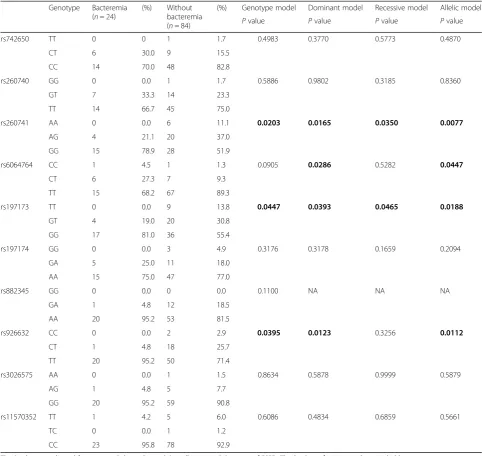
genotypes and clinical outcomes (Additional file1: Table S2 and S3), but the rs882345, rs926632 and rs3026575 Table 3Analysis of association betweenEDN3single-nucleotide polymorphisms (SNPs) and hospitalized bacteremia events
Genotype Bacteremia (n= 24)
(%) Without bacteremia (n= 84)
(%) Genotype model Dominant model Recessive model Allelic model
Pvalue Pvalue Pvalue Pvalue rs742650 TT 0 0 1 1.7 0.4983 0.3770 0.5773 0.4870
CT 6 30.0 9 15.5 CC 14 70.0 48 82.8
rs260740 GG 0 0.0 1 1.7 0.5886 0.9802 0.3185 0.8360 GT 7 33.3 14 23.3
TT 14 66.7 45 75.0
rs260741 AA 0 0.0 6 11.1 0.0203 0.0165 0.0350 0.0077
AG 4 21.1 20 37.0 GG 15 78.9 28 51.9
rs6064764 CC 1 4.5 1 1.3 0.0905 0.0286 0.5282 0.0447
CT 6 27.3 7 9.3 TT 15 68.2 67 89.3
rs197173 TT 0 0.0 9 13.8 0.0447 0.0393 0.0465 0.0188
GT 4 19.0 20 30.8 GG 17 81.0 36 55.4
rs197174 GG 0 0.0 3 4.9 0.3176 0.3178 0.1659 0.2094 GA 5 25.0 11 18.0
AA 15 75.0 47 77.0
rs882345 GG 0 0.0 0 0.0 0.1100 NA NA NA GA 1 4.8 12 18.5
AA 20 95.2 53 81.5
rs926632 CC 0 0.0 2 2.9 0.0395 0.0123 0.3256 0.0112
CT 1 4.8 18 25.7 TT 20 95.2 50 71.4
rs3026575 AA 0 0.0 1 1.5 0.8634 0.5878 0.9999 0.5879 AG 1 4.8 5 7.7
GG 20 95.2 59 90.8
rs11570352 TT 1 4.2 5 6.0 0.6086 0.4834 0.6859 0.5661 TC 0 0.0 1 1.2
CC 23 95.8 78 92.9
ofEDN3 showed a modest association with hospitalized infection events (Table2).
Associations betweenEDNgenetic variants and hospitalized bacteremia events
The rs260741, rs6064764, rs197173, and rs926632 of
EDN3 genotypes showed a significant association with
hospitalized bacteremia events (Table3). Patients
carry-ing the minor allele ofEDN3rs260741 (AA/AG vs. GG),
rs197173 (TT/TG vs. GG), and rs926632 (CC/CT vs. TT) had a lower risk of hospitalized bacteremia events
(genotype model, dominant model, and allelic model P
value < 0.05). The minor allele of rs6064764 (CC/CT vs. TT) was associated with increased risk of bacteremia
ac-cording to the dominant model and allelic model P
values (Table3). No association ofEDN1orEDN2SNPs
with hospitalized bacteremia events were detected (Additional file1: Table S4 and S5).
Haplotypes analysis ofEDN3with infection and bacteremia events
Next, we further validated the effects of haplotypes of
EDN3 gene by pairwise linkage disequilibrium (LD)
(Fig. 2). The haplotype frequency of EDN3 rs926632/
rs882345 variants among patients with hospitalized infection and bacteremia events were shown in Ta-bles 4 and 5. The EDN3 haplotype had no significant association with the risk of infection or bacteremia events. To test the possible functional roles of the
polymorphisms in EDN3, we queried expression
quantitative trait loci (eQTL) of EDN3 via
Genotype-Tissue Expression (GTEx) database in dif-ferent types of tissues [14]. Of note, the low expres-sion level of EDN3 was found in immune cells (Additional file 1: Figure S4). In addition, we analyzed the SNPs to understand the functional roles through using HaploReg V4.1. Interestingly, results indicated that the non-coding SNP rs260741 and rs6064764 of
EDN3 were potentially related to the regulation of T
cells activation by epigenetic modifications [15].
Discussion
Dialysis patients are exposed to high risk of infection, and related mortality [16, 17]. Ishigami et al. reported that a low estimated glomerular filtration rate and high albuminuria were associated with increased risk of hospitalization for infection, including bloodstream
infections, pneumonia, UTI, and cellulitis [18].
Among these infections, bacteremia was the most
critical and responsible for three-quarters of
infection-related mortality [19]. The risk factors for infection in dialysis patients include age, immunosup-pressive therapy, poor hygiene, and low performance
status [17, 20–22]. Age, albumin level, and category
of infection have been shown to associate with poor outcomes [23]. Old age and lower albumin level were the risk factors for infection events. Indeed, both in-nate and adaptive immunity are known to change with age, which may result in a persistent low-grade inflammation and tissue damage [24]. Furthermore, T-cell repertoire (TCR) complexity was revealed to predict the EPO responsiveness. Thus, TCR repertoire diversity may indicate the immune responses to infec-tion [25]. Malnutriinfec-tion, inflammainfec-tion, and atheroscler-osis syndrome, characterized by patients with renal
failure, are predictive of poor outcomes [26].
Fig. 2EDN3gene linkage disequilibrium (LD) and haplotype block structure in ESRD patients. The number on the cell is the LOD score of r2
Table 4Haplotype frequencies ofEDN3gene rs926632/ rs882345 among patients with hospitalized infection events or not
rs926632/rs882345 Case Control OR (95% CI) Pvalue T/A 91% 84% 1 Ref
C/A NA 6% – –
C/G 9% 9% 0.93(0.46–1.90) 0.8131
Haplotype frequency less than 1% was excluded Abbreviation:ORodds ratio
Table 5Haplotype frequencies ofEDN3gene rs926632/ rs882345 among patients with hospitalized bacteremia events or not
rs926632/rs882345 Case Control OR (95% CI) Pvalue T/A 97% 87% 1 Ref
C/A NA 3% – –
C/G 3% 10% 0.29 (0.04–2.02) 0.1791
Importantly, malnutrition has also been reported to associate with chronic inflammation, followed by in-creased risk of infection [27].
Correa et al. reported that EDN signaling pathway
is involved in the pathogenesis of mycobacteria
tuber-culosis infection, and that EDN receptor A or B
sig-naling is critical for the host responses to the infection [28]. Wilson et al. indicated the functional
roles of EDN genes in intestinal hypoperfusion during
bacteremia [29]. Additionally, the precursor peptide
ET-1 of EDN1 gene is correlated with the severity of
pneumonia, ICU admission, and mortality [30]. Also, ET-1 has been shown to stimulate ROS production [9] and correlate with the risk of inflammatory dis-eases such as atherosclerosis. Our previous study
showed genetic variants of EDN1 gene are associated
with an increased risk of hospitalization for a cardio-vascular event [31]. Pittet et al. demonstrated that the serum level of ET-1 was strongly correlated with re-duced cardiac output in sepsis patients [32]. In this
study, we found the correlation between EDN genetic
variants and hospitalized infection events..
A previous study on EDN3 and EDN receptor type
B (EDNRB) knockout mouse model showed that EDN3 and EDNRB were associated with severe
en-terocolitis [33]. The ET-3 peptide of EDN3 gene was
reported to evoke an attenuated inflammation by in-ducing the EDN B2 receptor and nitric oxide produc-tion [13]. Another study also proposed that ET-3 reduces platelet-activating factor (PAF)-induced in-flammation by directly binding to PAF [34]. Interest-ingly, the Human Protein Atlas showed that ET-3 is highly expressed in the GI tract and endocrine tissues [35]. During the development of enteric nervous sys-tem, ET-3 and its receptor, EDNRB, orchestrate the
signaling cascades for enteric ganglion formation [36–
38]. The enteric nervous system, composing of neu-rons and glial cells, have been shown to interact with
the outer (microbiota) and inner environments
(immune cells) [39]; the enteric microbiota is known to interact with the immune system [40]. These stud-ies indicated that ET-3 may affect the inflammation process through the modification of enteric nervous system and interaction with the microbiota. The results provided indirect evidence for supporting our findings in this study.
This study has limitations. First, our small sample size may affect the statistical power in the analysis. Second,
the gene expression and protein level of EDNwere not
measured from patients’ samples. Further validation
studies are needed in order to evaluate the associations of genetic variants with gene/protein expression. Third, a replication study should be conducted in the second population.
Conclusions
The minor allele of rs260741, rs197173, and rs926632 in
EDN3 was associated with a reduced risk of hospitalized
bacteremia events in patients with end-stage renal disease (ESRD).
Additional files
Additional file 1: Figure S1.Linkage disequilibrium (LD) map ofEDN1 gene SNPs shown (a) based on R-squared (r2) (b) based on D-prime (D’). Figure S2.Linkage disequilibrium (LD) map ofEDN2gene SNPs shown (a) based on R-squared (r2) (b) based on D-prime (D’).Figure S3.Linkage disequilibrium (LD) map ofEDN3gene SNPs shown (a) based on R-squared (r2) (b) based on D-prime (D’).Figure S4.EDN3gene expression across different tissues. Table S1.Baseline characteristics of study pa-tients stratified by bacteremia events. Table S2.Analysis of association betweenEDN1single-nucleotide polymorphisms (SNPs) and hospitalized infection events.Table S3.Analysis of association betweenEDN2 single-nucleotide polymorphisms (SNPs) and hospitalized infection events. Table S4.Analysis of association betweenEDN1single-nucleotide poly-morphisms (SNPs) and hospitalized bacteremia events.Table S5.Analysis of association betweenEDN2single-nucleotide polymorphisms (SNPs) and hospitalized bacteremia events. (DOCX 196 kb)
Abbreviations
EDN:Endothelin; ERI: Erythropoietin resistance index; ESRD: End-stage renal disease; ET-1: Endothelin-1; GTEx: Genotype-Tissue Expression; Kt/V: Adequacy of dialysis
Acknowledgements None declared.
Funding
The study was supported by the funding and grants from the Ministry of Health and Welfare (MOHW104-TDU-B-212-113001), Ministry of Science and Technology (MOST107–2314-B-038-019-MY3), and Taipei Medical University Hospital (103TMUH-NE-04). These funding was responsible for laboratory supplies, personnel expense and staff technical fees.
Availability of data and materials
All data related to this article are shown in the manuscript or are available upon request from the corresponding authors.
Authors’contributions
C-CK contributed to the study concept and design; data research; analysis and interpretation of data; drafting of the manuscript. S-YC performed mo-lecular genetic studies and researched the data. Y-JW contributed to the re-sults interpretation, concept discussion and manuscript writing. S-CC contributed to the study concept and design and data research. Y-WH ana-lyzed and interpreted the data. M-YW: study concept and design. H-FL: con-cept discussion, and researched the data. SN contributed to the manuscript writing and concept discussion. TS contributed to concept discussion. M-SW contributed to the study concept and design and approved the final version of the manuscript to be published. W-CC contributed to the study concept and experimental design, data analysis and manuscript writing.
Ethics approval and consent to participate
This study was approved by the Institutional Review Board of Taipei Medical University (Approval no. 201309026), and written informed consent was obtained from all patients.
Consent for publication Not applicable.
Competing interests
Publisher’s Note
Springer Nature remains neutral with regard to jurisdictional claims in published maps and institutional affiliations.
Author details
1Graduate Institute of Clinical Medicine, College of Medicine, Taipei Medical
University, Taipei, Taiwan.2Division of Nephrology, Department of Internal
Medicine, Taipei Medical University Hospital, Taipei, Taiwan.3Division of Nephrology, Department of Internal Medicine, School of Medicine, College of Medicine, Taipei Medical University, Taipei, Taiwan.4Department of
Pharmacy, Taipei Medical University Hospital, Taipei, Taiwan.5Department of
Clinical Pharmacy, School of Pharmacy, College of Pharmacy, Taipei Medical University, Taipei, Taiwan.6Ph.D. Program for Neural Regenerative Medicine,
College of Medical Science and Technology, Taipei Medical University, Taipei, Taiwan.7Clinical Research Center, Taipei Medical University Hospital, Taipei,
Taiwan.8Ph.D. Program for Translational Medicine, College of Medical Science and Technology, Academia Sinica, Taipei Medical University, Taipei, Taiwan.9Division of Nephrology, Department of Internal Medicine, Shuang
Ho Hospital, Taipei Medical University, New Taipei City, Taiwan.10Department
of Surgery, University of Chicago, Chicago, IL, USA.11Department of Pharmacy, Wan Fang Hospital, Taipei Medical University, Taipei, Taiwan.
12Master Program for Clinical Pharmacogenomics and Pharmacoproteomics,
School of Pharmacy, Taipei Medical University, Taipei 110, Taiwan.
13
Department of Medical Research, Shuang Ho Hospital, Taipei Medical University, New Taipei City, Taiwan.
Received: 28 March 2018 Accepted: 23 April 2019
References
1. Kato S, Chmielewski M, Honda H, Pecoits-Filho R, Matsuo S, Yuzawa Y, et al. Aspects of immune dysfunction in end-stage renal disease. Clin J Am Soc Nephrol. 2008;3(5):1526–33.
2. Allon M, Radeva M, Bailey J, Beddhu S, Butterly D, Coyne DW, et al. The spectrum of infection-related morbidity in hospitalized haemodialysis patients. Nephrol Dial Transplant. 2005;20(6):1180–6.
3. Slinin Y, Foley RN, Collins AJ. Clinical epidemiology of pneumonia in hemodialysis patients: the USRDS waves 1, 3, and 4 study. Kidney Int. 2006; 70(6):1135–41.
4. Eleftheriadis T, Liakopoulos V, Leivaditis K, Antoniadi G, Stefanidis I. Infections in hemodialysis: a concise review - part 1: bacteremia and respiratory infections. Hippokratia. 2011;15(1):12–7.
5. Descamps-Latscha B. The immune system in end-stage renal disease. Curr Opin Nephrol Hypertens. 1993;2(6):883–91.
6. Ferwerda B, McCall MB, Alonso S, Giamarellos-Bourboulis EJ, Mouktaroudi M, Izagirre N, et al. TLR4 polymorphisms, infectious diseases, and evolutionary pressure during migration of modern humans. Proc Natl Acad Sci U S A. 2007;104(42):16645–50.
7. Schuurhof A, Bont L, Siezen CL, Hodemaekers H, van Houwelingen HC, Kimman TG, et al. Interleukin-9 polymorphism in infants with respiratory syncytial virus infection: an opposite effect in boys and girls. Pediatr Pulmonol. 2010;45(6):608–13.
8. Elisa T, Antonio P, Giuseppe P, Alessandro B, Giuseppe A, Federico C, et al. Endothelin receptors expressed by immune cells are involved in modulation of inflammation and in fibrosis: relevance to the pathogenesis of systemic sclerosis. J Immunol Res. 2015;2015:147616.
9. Kowalczyk A, Kleniewska P, Kolodziejczyk M, Skibska B, Goraca A. The role of endothelin-1 and endothelin receptor antagonists in inflammatory response and sepsis. Arch Immunol Ther Exp. 2015;63(1):41–52.
10. Pan C, Wang J, Liu W, Liu L, Jing L, Yang Y, et al. Low tidal volume protects pulmonary vasomotor function from“second-hit”injury in acute lung injury rats. Respir Res. 2012;13:77.
11. Yeager ME, Belchenko DD, Nguyen CM, Colvin KL, Ivy DD, Stenmark KR. Endothelin-1, the unfolded protein response, and persistent inflammation: role of pulmonary artery smooth muscle cells. Am J Respir Cell Mol Biol. 2012;46(1):14–22.
12. Lin CC, Hsieh HL, Shih RH, Chi PL, Cheng SE, Yang CM. Up-regulation of COX-2/PGE2 by endothelin-1 via MAPK-dependent NF-kappaB pathway in mouse brain microvascular endothelial cells. Cell Commun Signal. 2013; 11(1):8.
13. Sato A, Ebina K. Endothelin-3 at low concentrations attenuates
inflammatory responses via the endothelin B2 receptor. Inf Res. 2013;62(4): 417–24.
14. Lonsdale J, Thomas J, Salvatore M, Phillips R, Lo E, Shad S, et al. The Genotype-Tissue Expression (GTEx) project. Nat Genet. 2013;45(6):580–5. 15. Kundaje A, Meuleman W, Ernst J, Bilenky M, Yen A, Heravi-Moussavi A, et al.
Integrative analysis of 111 reference human epigenomes. Nature. 2015; 518(7539):317–30.
16. Nassar GM, Ayus JC. Infectious complications of the hemodialysis access. Kidney Int. 2001;60(1):1–13.
17. Vandecasteele SJ, Boelaert JR, De Vriese AS. Staphylococcus aureus infections in hemodialysis: what a nephrologist should know. Clin J Am Soc Nephrol. 2009;4(8):1388–400.
18. Ishigami J, Grams ME, Chang AR, Carrero JJ, Coresh J, Matsushita KCKD. Risk for hospitalization with infection: the atherosclerosis risk in communities (ARIC) study. Am J Kidney Dis. 2017;69(6):752–61.
19. Bloembergen WE, Port FK. Epidemiological perspective on infections in chronic dialysis patients. Adv Ren Replace Ther. 1996;3(3):201–7. 20. Kaplowitz LG, Comstock JA, Landwehr DM, Dalton HP, Mayhall CG. A
prospective study of infections in hemodialysis patients: patient hygiene and other risk factors for infection. Infect Control Hosp Epidemiol. 1988; 9(12):534–41.
21. Teehan GS, Bahdouch D, Ruthazer R, Balakrishnan VS, Snydman DR, Jaber BL. Iron storage indices: novel predictors of bacteremia in hemodialysis patients initiating intravenous iron therapy. Clin Infect Dis. 2004;38(8):1090–4. 22. Hoen B, Paul-Dauphin A, Hestin D, Kessler M. EPIBACDIAL: a multicenter
prospective study of risk factors for bacteremia in chronic hemodialysis patients. J Am Soc Nephrol. 1998;9(5):869–76.
23. Allon M, Depner TA, Radeva M, Bailey J, Beddhu S, Butterly D, et al. Impact of dialysis dose and membrane on infection-related hospitalization and death: results of the HEMO Study. J Am Soc Nephrol. 2003;14(7):1863–70. 24. Shaw AC, Joshi S, Greenwood H, Panda A, Lord JM. Aging of the innate
immune system. Curr Opin Immunol. 2010;22(4):507–13. 25. Wong HS, Chang CM, Kao CC, Hsu YW, Liu X, Chang WC, et al. V-J
combinations of T-cell receptor predict responses to erythropoietin in end-stage renal disease patients. J Biomed Sci. 2017;24(1):43.
26. Tonbul HZ, Demir M, Altintepe L, Guney I, Yeter E, Turk S, et al. Malnutrition-inflammation-atherosclerosis (MIA) syndrome components in hemodialysis and peritoneal dialysis patients. Ren Fail. 2006;28(4):287–94.
27. Pawlaczyk K, Oko A, Lindholm B, Czekalski S. Malnutrition inflammation --atherosclerosis (MIA syndrome) in patients with renal failure. Pol Merkur Lekarski. 2003;15(88):334–41 discussion 41-3.
28. Correa AF, Bailao AM, Bastos IM, Orme IM, Soares CM, Kipnis A, et al. The endothelin system has a significant role in the pathogenesis and progression of mycobacterium tuberculosis infection. Infect Immun. 2014;82(12):5154–65. 29. Wilson MA, Steeb GD, Garrison RN. Endothelins mediate intestinal
hypoperfusion during bacteremia. J Surg Res. 1993;55(2):168–75. 30. Schuetz P, Stolz D, Mueller B, Morgenthaler NG, Struck J, Mueller C, et al.
Endothelin-1 precursor peptides correlate with severity of disease and outcome in patients with community acquired pneumonia. BMC Infect Dis. 2008;8:22. 31. Kao CC, Cheng SY, Wu MY, Chien SC, Lu HF, Hsu YW, et al. Associations of
genetic variants of endothelin with cardiovascular complications in patients with renal failure. BMC Nephrol. 2017;18(1):291.
32. Pittet JF, Morel DR, Hemsen A, Gunning K, Lacroix JS, Suter PM, et al. Elevated plasma endothelin-1 concentrations are associated with the severity of illness in patients with sepsis. Ann Surg. 1991;213(3):261–4. 33. Frykman PK, Cheng Z, Wang X, Dhall D. Enterocolitis causes profound lymphoid
depletion in endothelin receptor B- and endothelin 3-null mouse models of Hirschsprung-associated enterocolitis. Eur J Immunol. 2015;45(3):807–17. 34. Sato A, Ebina K. Common mechanism in endothelin-3 and PAF receptor
function for anti-inflammatory responses. Eur J Pharmacol. 2013;718(1–3):30–3. 35. Ponten F, Jirstrom K, Uhlen M. The Human Protein Atlas--a tool for
pathology. J Pathol. 2008;216(4):387–93.
36. Barlow A, de Graaff E, Pachnis V. Enteric nervous system progenitors are coordinately controlled by the G protein-coupled receptor EDNRB and the receptor tyrosine kinase RET. Neuron. 2003;40(5):905–16.
37. Kabouridis PS, Pachnis V. Emerging roles of gut microbiota and the immune system in the development of the enteric nervous system. J Clin Invest. 2015;125(3):956–64.
gene produce megacolon associated with spotted coat color in mice. Cell. 1994;79(7):1267–76.
39. Obata Y, Pachnis V. The effect of microbiota and the immune system on the development and organization of the Enteric Nervous System. Gastroenterology. 2016;151(5):836–44.