R E S E A R C H
Open Access
Protective effects of various ratios of DHA/
EPA supplementation on high-fat
diet-induced liver damage in mice
Tingting Shang
1†, Liang Liu
1†, Jia Zhou
1, Mingzhen Zhang
1, Qinling Hu
1, Min Fang
1, Yongning Wu
1,2,
Ping Yao
3and Zhiyong Gong
1*Abstract
Background:A sedentary lifestyle and poor diet are risk factors for the progression of non-alcoholic fatty liver disease. However, the pathogenesis of hepatic lipid accumulation is not completely understood. Therefore, the present study explored the effects of dietary supplementation of various ratios of docosahexaenoic acid (DHA) and eicosapentaenoic acid (EPA) on a high-fat diet-induced lipid metabolism disorder and the concurrent liver damage. Methods:Using high-fat diet-fed C57BL/6 J mice as the animal model, diets of various ratios of DHA/EPA (2:1, 1:1, and 1:2) with an n-6/n-3 ratio of 4:1 were prepared using fish and algae oils enriched in DHA and/or EPA and sunflower seed oils to a small extent instead of the high-fat diet.
Results:Significantly decreased hepatic lipid deposition, body weight, serum lipid profile, inflammatory reactions, lipid peroxidation, and expression of adipogenesis-related proteins and inflammatory factors were observed for mice that were on a diet supplemented with DHA/EPA compared to those in the high-fat control group. The DHA/EPA 1:2 group showed lower serum triglycerides (TG), total cholesterol (TC), and low-density lipoprotein-cholesterol levels, lower SREBP-1C, FAS, and ACC-1 relative mRNA expression, and higher Fra1 mRNA expression, with higher relative mRNA expression of enzymes such as AMPK, PPARα, and HSL observed in the DHA/EPA 1:1 group. Lower liver TC and TG levels and higher superoxide dismutase levels were found in the DHA/EPA 2:1 group. Nonetheless, no other notable effects were observed on the biomarkers mentioned above in the groups treated with DHA/EPA compared with the DHA group.
Conclusions:The results showed that supplementation with a lower DHA/EPA ratio seems to be more effective at alleviating high-fat diet-induced liver damage in mice, and a DHA/EPA ratio of 1:2 mitigated inflammatory risk factors. These effects of n-3 polyunsaturated fatty acids (PUFA) on lipid metabolism may be linked to the upregulation of Fra1 and attenuated activity of c-Jun and c-Fos, thus ultimately reducing the severity of the lipid metabolism disorder and liver damage to some extent.
Keywords:N-3 PUFA, DHA/EPA, Lipid metabolism, Liver damage, Fra1, C-Jun, C-Fos
* Correspondence:gongzycn@163.com †Equal contributors
1College of Food Science and Engineering, Wuhan Polytechnic University, 68
XueFuNan Road, Wuhan 430023, People’s Republic of China Full list of author information is available at the end of the article
Background
An unhealthy lifestyle involving poor dietary habits and low physical activity is a risk factor for the pro-gression of non-alcoholic fatty liver disease (NAFLD) [1]. Although it is generally believed that nutrient modulation is a positive means to alleviate this prob-lem [2] since the type and quantity of fat consumed regulate hepatic lipid composition and gene expres-sion [3], the pathogenesis of hepatic lipid accumula-tion is not completely understood [4].
Decreasing dietary saturated fatty acid (SFA) intake [5] and replacing it with polyunsaturated fatty acids (PUFA) [6] has been advocated as the main dietary strategy to prevent NAFLD caused by anti-inflammatory, antioxida-tive, and hypolipidemic effects of PUFAs. Meanwhile, long-chain PUFAs, mainly n-6 PUFA and n-3 PUFA, es-pecially eicosapentaenoic (C20:5 n-3, EPA) and docosa-hexaenoic (C22:6 n-3, DHA) acids, have frequently been reported to influence NAFLD risk factors [7]. Reduced hepatic lipid content with concomitant antioxidant and anti-inflammatory responses favoring insulin sensitivity in mice with high-fat diet-induced obesity are ascribed to the repletion of liver n-3 PUFA levels by n-3 PUFA dietary supplementation [7]. Nevertheless, evidence sug-gests that selectively increasing the n-3 PUFA propor-tion of the total PUFA intake [8, 9] has more significant inhibitory effects on inflammation and NAFLD than simply increasing n-3 PUFA intake [10]. Interestingly, several studies have also demonstrated the latent effects of the dietary proportion of n-6/n-3 on liver damage [10, 11], and a low n-6/n-3 ratio was shown to be more effective for the prevention of diet- or obesity-induced fatty liver than that of a high n-6/n-3 ratio. Conse-quently, a reasonable proportion of n-6/n-3 should be taken into consideration when investigating the effects of PUFAs on NAFLD [10].
Recently, there has been an increasing interest in the effects of various ratios of DHA and EPA on chronic diseases in rat models [12–15], and studies conducted to evaluate these effects suggest that both n-6/n-3 ratios and DHA/EPA ratios should be considered simultan-eously. Additionally, our research group has previously reported that DHA and EPA from fish and algal oils are more beneficial than plant oils enriched in α-linolenic acid (C18:3 n-3, ALA) at delaying atherosclerosis pro-gression in model apoE−/− mice; an intervention with higher DHA/EPA ratios was found to be beneficial for reducing high cholesterol concentrations in the blood [16]. Nevertheless, DHA and EPA differ in their physio-logical functions, and a new study indicated that DHA supplementation leads to a greater reduction in specific markers of inflammation than that by an equal dose of EPA [17]. In view of these observations, it remains un-clear whether DHA is more effective than DHA/EPA,
there is an optimal ratio of DHA and EPA supplementa-tion for reducsupplementa-tion of high-fat diet-induced liver damage, and regularizing the ratio of DHA and EPA has differen-tial effects on various symptoms.
Therefore, in the present study, we sought to investi-gate the role of dietary interventions with various ratios of DHA/EPA (2:1, 1:1, and 1:2) in high-fat diet-induced liver damage at an n-6/n-3 ratio of 4:1, which is optimal for human health as recommended by the World Health Organization (WHO). Healthy C57BL/6 J mice were employed as animal models. Mice were fed high-fat diets supplemented with sunflower seed, algae, and fish oils containing different DHA/EPA ratios (2:1, 1:1, and 1:2).
Methods
Animals and diets
water and their group-specific diet. Measurements of body weight and food intake were taken weekly. After 11 weeks, five mice from each group were anesthe-tized with isoflurane, and blood was drawn through the retro-orbital sinus puncture prior to killing the mice by decapitation after an overnight fast and used for analyses to ensure the establishment of models of steatosis and liver lesions. The rest of the mice were euthanized in the same way. Serum was collected from blood, subsequently centrifuged (4000×g, 4 °C, 10 min) after incubation at room temperature for 2 h, and then stored at −80 °C until analysis. Fresh liver tissue samples were collected for histopathology analysis or were rapidly frozen and stored at −80 °C before use.
Analysis of steatosis and liver lesions
The liver was removed and weighed immediately after the mice were euthanized. Liver fragments were sec-tioned at a thickness of 5 μm, and fixed with 4% paraformaldehyde overnight. Then the slices were stained with oil red O, in order to analyze the
steatosis and liver lesions. Images were acquired by means of an inverted fluorescence microscope (Olympus Co., Japan) equipped with a SONY DXC-970MD color video camera, and analyzed using the Image-Pro plus program.
Measurement of serum and liver lipid profiles
The serum triglyceride (TG), total cholesterol (TC), high-density lipoprotein-cholesterol (HDL-C), and low-density lipoprotein-cholesterol (LDL-C) levels were measured using similar commercial kits (Biosino Bio-technology Co., Ltd., Beijing, China) with spectrophoto-metric methods in accordance with the manufacturer’s instructions.
To determine the hepatic TC and TG levels, 50 mg of frozen liver tissue was placed in a test tube with 450 μl of isopropanol. The homogenate was incubated at 4 °C for 36 h and then centrifuged at 4000×g and 4 °C for 10 min, and the supernatant was used for analyses by means of the corresponding commercial kits (Biosino Biotechnology Co., Ltd., Beijing, China) with spectrophotometric methods in accordance with the manufacturer’s instructions.
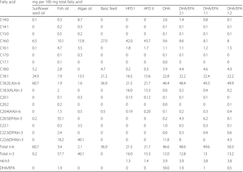
Table 1Fatty acid composition of oils and mixed diets supplemented to mice
Fatty acid mg per 100 mg total fatty acid
Sunflower seed oil
Fish oil Algae oil Basic feed HFD I HFD II DHA DHA/EPA
2:1
DHA/EPA 1:1
DHA/EPA 1:2
C14:0 0.1 0.3 8.7 0 0 0 2.6 1.4 0.8 0.1
C14:1 0 0.2 0.3 0 0 0 0.1 0.1 0.1 0.1
C15:0 0 0.5 0.2 0 0 0 0.1 0.1 0.1 0.1
C16:0 6.5 10.1 15.8 27.0 42.0 43.7 9.6 8.6 8.1 8
C16:1 0.1 4.7 3.5 0 1.8 1.7 1.1 1.1 1.2 1.5
C17:0 0 0.1 0.3 0 0 0 0.1 0.1 0.1 0
C17:1 0 0.1 0 0 0 0 0.0 0 0 0
C18:0 5.2 2.8 0 4.7 0.2 0.3 3.9 4.4 4.6 4.9
C18:1 24.9 7.9 13.5 21.2 16.5 15.6 22.8 22.2 22.4 22.2
C18:2(LA)n-6 60.7 1.9 1.6 36.9 21.5 21.7 46.4 48.4 49.3 49.9
C18:3(ALA)n-3 0 2 0 0 16.0 15.3 0.0 0.2 0.4 0.2
C20:1 0 0.1 0.3 0 0.13 0.12 0.1 0.1 0.1 0
C20:2 0 0.2 0 0 0 0 0.0 0 0 0.1
C20:4(AA)n-6 0 1.5 0.5 0.5 0.19 0.20 0.1 0.2 0.3 0.4
C20:5(EPA)n-3 0.2 35.1 0 0 0 0 0.2 4.3 6.2 8.1
C22:1 0 0.3 3.5 0 0 0 1.0 0.5 0.3 0.1
C22:5(DPA)n-3 0 2.4 0 0 0 0 0.0 0.3 0.4 0.6
C22:6(DHA)n-3 0 18.2 40.1 0 0 0 11.8 8 6 4.3
Total n-6 60.7 3.4 2.1 36.9 21.5 21.7 46.6 48.6 49.6 50.3
Total n-3 0.2 57.7 40.1 0 16.0 15.3 12.0 12.8 13 13.2
n6/n3 1.3 1.4 3.9 3.9 3.8 3.8
Measurement of serum ALT, AST, GSH, SOD, MDA, and inflammatory cytokines, and liver GSH, SOD, and MDA The serum alanine aminotransferase (ALT), aspartate aminotransferase (AST), glutathione (GSH), superoxide dismutase (SOD), and malondialdehyde (MDA) were measured by means of similar commercial kits (Nanjing Jiancheng Corporation, Nanjing, China) in accordance with the manufacturer’s recommendations. Serum tumor necrosis factor-alpha (TNF-α), interleukin-1β (IL-1β), and interleukin-6 (IL-6) were determined using the cor-responding commercial kits (Cloud-Clone Corp, Wuhan, China) by ELISA methods according to the manufac-turer’s instructions.
To analyze the hepatic levels of GSH, SOD, and MDA, 50 mg of liver tissues were homogenized with 450 μl of normal saline. After centrifugation at 4000×g and 4 °C for 10 min, the supernatant of the homogenate was used for analysis with the corresponding commercial kits (Nanjing Jiancheng Corporation) as specified by the manufacturer. In addition, protein content was mea-sured using commercial bicinchoninic acid (BCA) kits (Cloud-Clone Corp, Wuhan, China).
Real-time PCR
The total RNA was extracted from liver tissues using the RNAiso Plus reagents (TaKaRa BIO Inc., Dalian, China). Then, the cDNA was extracted using a Prime Script RT Reagent Kit (TaKaRa BIO Inc.). The mRNA was quanti-fied with a real-time PCR machine (IQ5, Bio-Rad, USA) using SYBR Premix Ex Taq (TaKaRa BIO Inc., Dalian, China) and specific primers (BGI Tech Solutions Co.,
Ltd., Shenzhen, China) in accordance with the manufac-turer’s recommendations. The forward and reverse primers for the target genes are listed in Table 2. Relative gene expression was normalized (by means of the CT
method) to that of the endogenous control β-actin, and the final results were calculated using the formula 2−ΔΔCt.
Analysis of western blots
Protein expression levels of c-Jun, c-Fos, and Fra1 were analyzed by western blotting. Briefly, the total protein was extracted in PMSF:RIPA = 1:99 (v/v), a homogenizing and lysis buffer. Equal amounts of pro-tein extracts, which were normalized to the total amount of protein, were mixed (1:3, v/v) with the loading buffer for electrophoresis using 10% SDS-PAGE gels kits (Boster Bio-engineering, Ltd., Wuhan, China). The mixture was subsequently electroblotted to a nitrocellulose transfer membrane (Millipore, USA) by a Trans-Blot SD semi-dry transfer cell (Bio-Rad, USA) as per the manufacturer’s instructions. The target protein levels were detected with specific pri-mary antibodies (Bioss Biological Technology, Ltd., Wuhan, China) against the target protein, and then incubated with the species-specific secondary anti-body, horseradish peroxidase-conjugated anti-rabbit IgG antibody (Boster Bio-engineering), as recom-mended by the manufacturer. The chemiluminescence intensity of the bands was quantified by an ECL de-veloper (Millipore, USA) using a Western Blotting Detection System (Bio-Rad, USA), and the optical densities of the bands were detected using Image Lab
Table 2Real-time quantitative PCR primer sequences
Gene Forward primer 5′–3′ Reverse primer 5′–3′
AMPK GCCATGCGCAGACTAGTT AGGATGTATGGCCGATCTTC
SREBP-1 GTGAGCCTGACAAGCAATCA GGTGCCTACAGAGCAAGAGG
PPARγ TTTCAAGGGTGCCAGTTT GAGGCCAGCATCGTGTAG
PPARα CCTCAGGGTACCACTACGGAGT GCCGAATAGTTCGCCGAA
ACC-l TACTGAACTACATCTTCTCCC AAGCAATAAGAACCTGACGA
FAS CAAATACAATGGCACCCTGA TGGCGAAGCCGTAGTTAGTT
SCD-1 GAATTCATGCCTGCGCACTTGCTACA CTCGAGTCAGCCGCTCTTGTGACTCC
CPT-1 TTAACAGCAACTACTACGCC CCAGAAGACGAATAGGTTTGAG
HSL GCCACAATGACACAGTCACTGGT CAGGCAGCGGCCGTAGAAGCA
ACOX GGTGGCTGTTAAGAAGAGTG AAGATGAGTTCCATGACCCA
c-Jun ATGACTGCAAAGATGGAAACG AAGTTGCTGAGGTTGGCG
c-Fos TACTACCATTCCCCAGCC GCTCTACTTTGCCCCTTC
Fra1 TCATCTGGAGAGGTGGGTCC CTGCGGTTCTGACTCACTCG
TNF-α AGCCCCCAGTGTGTATCCTT ACAGTCCAGGTCACTGTCCC
IL-1β CTTCAGGCAGGCAGTATCACTC TTGTTGTTCATCTCGGAGCC
IL-6 AGTTGCCTTCTTGGGACTGA TCCACGATTTCCCAGAGAAC
5.1 software (Bio-Rad). Data were corrected to elimin-ate background noise and were standardized to the optical density (OD/mm2) of β-tubulin (Boster Bio-engineering).
Statistical analysis
All data were recorded with Excel. After confirming the homogeneity of variances, differences among the groups were tested by one-way ANOVA. Means ± SEM were calculated for each group, and the figures were prepared using GraphPad Prism v6.0.1 software. The significant differences were shown by the LSD multiple comparison test using SPSS statistics 17.0 software, and any two groups without the same lowercase and uppercase let-ters, as marked in the figures, indicate a significant dif-ference ofPvalue <0.05 and <0.01, respectively.
Results
Effects of various ratios of DHA/EPA with an n-6/n-3 ratio of 4:1 on steatosis and liver cell histopathological lesions At 12 weeks post-administration, the histopathological lesions of livers in all groups were assessed. The oil red O staining patterns showed orange lipid droplets and blue nuclei, which were smaller and fewer in number in the DHA/EPA (2:1, 1:1, and 1:2) groups; while obvious numerous orange-red lipid droplets were observed in HFC-treated mice, nearly none were observed in the NC group (Fig. 1). Moreover, fewer lipid droplets were observed in mouse groups treated with DHA/EPA (ratios of 2:1, 1:1, and 1:2) compared to the group treated with DHA.
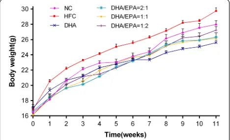
Effects of various ratios of DHA/EPA with an n-6/n-3 ratio of 4:1 on body weight, serum, and liver lipids
Compared to the HFC group, significant decreases (p< 0.05) in serum TC, TG, LDL-C levels, and liver TC and TG levels were observed in 12-week n-3 PUFA sup-plementation groups (Table 3). The growth curve of mice revealed that the body weight increased slower with n-3 PUFA supplementation than with HFC
treatment (Fig. 2), indicating the inhibitory effect of n-3 PUFA supplementation with DHA and different DHA/ EPA ratios (2:1, 1:1, and 1:2) in the mice. Moreover, mice treated with a DHA/EPA ratio of 2:1 and 1:2 showed a reduced hepatic organ coefficient with a significant dif-ference ofp< 0.01, while the statistical differences of re-duction in DHA and DHA/EPA 1:1 groups were p< 0.05 compared to those in the HFC group (Table 3). Mice treated with a DHA/EPA ratio of 1:2 showed a more appreciable decrease in serum TC, TG, and LDL-C levels, whereas mice treated with the DHA/EPA ratio of 2:1 showed lowered liver TC and TG levels to a greater degree. However, mice treated with various ratios of DHA/EPA (2:1, 1:1, and 1:2) revealed no reduction in serum TC, TG, or LDL-C levels or liver TC and TG levels in comparison with those in the DHA group. Nevertheless, as compared to the levels in the HFC group, serum HDL-C levels in DHA and DHA/EPA (2:1, 1:1, and 1:2) groups were significantly higher (p< 0.01). Furthermore, serum HDL-C levels increased more sig-nificantly in the DHA/EPA 1:2 group than in other DHA/EPA groups (p < 0.01), but the increase was not remarkable in comparison to that in the DHA group.
Effects of various ratios of DHA/EPA with an n-6/n-3 ratio of 4:1 on liver damage
Levels of serum ALT, AST, GSH, SOD, and MDA, and liver GSH, SOD, and MDA are presented in Fig. 3. The results show that treatment with n-3 PUFAs lowered the levels of serum ALT and AST and both serum and liver MDA production, and raised the levels of GSH and SOD both in serum and liver in comparison with those in the HFC group. Nonetheless, no significant differences were found in the levels of any of these biomarkers between the DHA and DHA/EPA groups. Nevertheless, supple-mentation with a DHA/EPA ratio of 1:2 resulted in greater reduction in serum ALT, AST, and MDA levels and increase of serum GSH levels among the DHA/EPA groups; however, this increase was not higher than that observed in DHA groups. Collectively, supplementation with a DHA/EPA ratio of 2:1 was the most effective among the three DHA/EPA groups at enhancing SOD levels both in serum and liver, but these levels were not higher than those in the DHA group. Particularly, mice treated at a DHA/EPA ratio of 1:1 showed increased liver GSH levels as compared to the DHA group (2.828 ± 0.279 vs. 2.612 ± 0.546 mg/g prot).
Effects of various ratios of DHA/EPA with an n-6/n-3 ratio of 4:1 on lipid metabolism genes
Gene expression variation of the enzymes and transcrip-tion factors of lipid metabolism was measured by real-time PCR as shown in Fig. 4. Compared to the HFC group, 43%, 27%, 46%, and 27% increases in AMPK
(AMP-activated protein kinase) mRNA expression, and 103%, 68%, 84%, and 92% increases in ACOX (acyl-co-enzyme A oxidase) mRNA expression, were demon-strated in DHA groups and groups with the DHA/EPA ratio of 2:1, 1:1, and 1:2, respectively. Noticeably, mice treated with a DHA/EPA ratio of 1:2 showed a greater reduction in SREBP-1C (sterol regulatory element bind-ing protein-1C), SCD-1 (stearoyl-CoA desaturase-1), FAS (fatty acid synthase), ACC-1 (acetyl CoA carboxylase-1), and PPARγ(peroxisome proliferator acti-vated receptor gamma) mRNA expression, while the DHA/EPA 1:1 group showed higher HSL (hormone sen-sitive lipase), PPARα, and CPT-1 (carnitine palmitoyl transferase-1) mRNA expression, among the DHA/EPA groups. Particularly, the group treated with the DHA/ EPA ratio of 1:2 showed lowered SCD-1 mRNA expres-sion, and the DHA/EPA 1:1 group showed enhanced CPT-1 mRNA expression, whereas no significant differ-ences were observed in the DHA group.
Effects of various ratios of DHA/EPA with an n-6/n-3 ratio of 4:1 on the inflammatory response
The protein expression levels of the three main subunits of AP-1 (c-Jun, c-Fos, and Fra1) were detected by
western blot (Fig. 5a–d). The protein expression levels of c-Jun and c-Fos were significantly lower in the DHA/ EPA groups compared to the HFC group (p < 0.01), meanwhile supplementation with a DHA/EPA ratio of 1:1 revealed greater reductions in the expression levels of these two proteins in contrast to the DHA group. Nonetheless, the protein expression levels of Fra1 were found to be significantly higher in the DHA and DHA/ EPA treated groups in comparison to the HFC group (p< 0.05). Supplementation with DHA/EPA at the ratio of 1:2 did not result in enhancement in Fra1 expression levels. The mRNA expression levels related to c-Jun, c-Fos, and Fra1 were quantified by real-time PCR (Fig. 5e–g). Supplementation with n-3 PUFA showed a significant decrease in c-Jun and c-Fos mRNA ex-pression and an increase in Fra1 mRNA exex-pression, as compared to those in the HFC group (p < 0.05). Moreover, mice treated with the DHA/EPA ratio of 1:1 and 1:2 showed lowered c-Jun and c-Fos mRNA expression, respectively, in comparison to those in the DHA group. Nevertheless, no significant increase in Fra1 mRNA expression was observed in the best per-formance group (treated with the DHA/EPA ratio of 1:2) as compared with the DHA group.
The concentrations of serum TNF-α, IL-1β, and IL-6 were all significantly lower in the DHA and the DHA/ EPA groups as compared to those in HFC group (p < 0.05) (Fig. 6). The quantitative real-time PCR assay, which roughly identified the inflammatory cyto-kine levels, revealed significantly decreased expression of TNF-α, IL-1β, and IL-6 in liver tissues of DHA/EPA-treated mice in comparison to those in HFC-DHA/EPA-treated mice (p < 0.05). Furthermore, mice treated with a DHA/EPA ratio of 2:1 revealed greater decreases in serum TNF-αcontents and its mRNA expression levels in contrast to the DHA group. Additionally, supple-mentation with a DHA/EPA ratio of 1:2 demonstrated a more obvious reduction in serum IL-1βand IL-6 con-tents, and their mRNA expression as compared to those in the DHA group.
Table 3Effects of various ratios of DHA/EPA on serum and liver lipids in high-fat diet-induced mice
NC HFC DHA DHA/EPA
2:1
DHA/EPA 1:1
DHA/EPA 1:2
Serum TC/(mmol/L) 3.693 ± 0.298aA 5.175 ± 0.368bB 3.559 ± 0.246aA 4.144 ± 0.323aAB 4.058 ± 0.181aA 3.935 ± 0.180aA
Serum TG/(mmol/L) 1.198 ± 0.045bB 1.388 ± 0.029cC 0.862 ± 0.035aA 0.981 ± 0.060aA 0.986 ± 0.028aA 0.879 ± 0.046aA
Serum HDL-C/(mmol/L) 1.144 ± 0.044cdBC 0.791 ± 0.028aA 1.270 ± 0.043dC 1.015 ± 0.017bB 1.128 ± 0.038bcBC 1.267 ± 0.029dC
Serum LDL-C/(mmol/L) 1.954 ± 0.082bcB 2.654 ± 0.055dC 1.600 ± 0.081aA 2.158 ± 0.087cB 2.070 ± 0.076bcB 1.914 ± 0.067bB
Liver TC/(mmol/L) 1.276 ± 0.099aA 2.452 ± 0.101dD 1.515 ± 0.078bAB 1.689 ± 0.055bcBC 1.811 ± 0.055cBC 1.823 ± 0.038cC
Liver TG/(mmol/L) 3.559 ± 0.184bAB 6.177 ± 0.330dD 2.693 ± 0.166aA 4.293 ± 0.100cBC 4.494 ± 0.350cC 4.538 ± 0.172cC
hepatic organ coefficient /(g/100 g) 3.835 ± 0.098aA 4.268 ± 0.037bB 4.019 ± 0.080aAB 3.934 ± 0.097aA 4.033 ± 0.037aAB 3.939 ± 0.060aA
Note: Each value is the mean ± SEM (n= 8), and any two groups without the same lowercase or uppercase letters, as marked in the table, indicate a significant difference ofp< 0.05 andp< 0.01, respectively
Discussion
The n-3 PUFAs, principally DHA and EPA, have been reported to play an increasingly significant role in the growth and health of the human body as shown by many previous studies [9, 10]. The physiological functions of n-3 PUFAs have attracted increasing interest because of the increasing number of people choosing to lead healthy lifestyles [18]. However, there was a tendency among most attempted studies to focus on the effects of an individual n-3 or n-6 PUFA or the ratio of n-6/n-3, which did not take the possible influence of the relative ratio of DHA and EPA on the results into account [15]. A marked increase in the n-6/n-3 ratio is associated with lipogenesis, overweight lipid oxidation and secretion, and therefore promotion of NAFLD [11]; fish oil intake varies in its hepatic fatty acid composition in supplements with n-3 PUFA, and a reduction in the n-6/n-3 ratio has already been revealed in another study [7]. Other reports demonstrated that DHA supplementation led to a greater reduction in specific makers of inflammation than that by
an equal dose of EPA [17], but supplementation with varying ratios of EPA/DHA influenced different meta-bolic syndrome markers [15] and enhanced the posi-tive effects of biomarkers of inflammation [12]. Accordingly, in the present study, we concentrated on the effects of various ratios of DHA/EPA (with an n-6/n-3 ratio of 4:1) on liver damage in C57BL/6 J mice with high-fat diet-induced obesity.
High-fat diets enriched in saturated fat may promote an increase in body weight and lead to obesity, liver in-jury, hepatic insulin resistance, and steatosis [19]. Treat-ment with increasing amounts of n-3 PUFA, however, seems to reduce the rate of increase in body weight [20]. A study showed that animals on high-fat diets appeared to have an increased ability to store TG, whereas animals fed high fish oil diets (along with consumption of excess calories) did not convert excess lipids into TGs [19]. These data appear to be in agreement with our current study showing that lower TG levels both in serum and in the liver were found in groups that were treated with n-3 PUFAs compared with the HFC group. Meanwhile, all n-3 supplementation groups showed attenuation of body weight gain compared to that in the untreated con-trol. EPA and DHA oil reduced total body fat [21], wherein reduced hepatic lipid deposition was observed in n-3 groups in the present study. Our results revealed that DHA and DHA/EPA supplementation decreased hepatic TG and TC levels possibly through reducing the release of TG and TC into the blood plasma for liver tis-sues, simultaneously improving serum HDL-C levels. Therefore, DHA and DHA/EPA supplementation inhib-ited hepatic lipid profile deterioration in mice with high-fat diet-induced obesity. Additionally, groups with a DHA/EPA ratio of 1:2 with an n-6/n-3 ratio of 4:1 showed the lowest serum levels of TG, TC, and LDL-C, and the highest concentration of serum HDL-C, while the lowest liver TC and TG levels were observed in the DHA/EPA 2:1 group among the DHA/EPA groups. Nevertheless, the best performance groups treated with DHA/EPA did not show better results than those in the DHA group. These findings seemed to contradict our previous report that indicated that serum TC and LDL-C levels were the lowest in the 2:1 DHA/EPA group, because the highest serum HDL-C content was observed in the 1:1 DHA/EPA group in apoE−/−
mice [16]. These findings may be explained by the lack of apoE, a ligand for receptors that clear remnants of chylomicrons and very low (See figure on previous page.)
Fig. 3Effects of various ratios of DHA/EPA on liver damage in high-fat diet-induced mice. Serum ALT (a), AST (b), MDA (c), GSH (d), SOD (e), and liver MDA (f), GSH (g), SOD (h) levels were measured with the corresponding commercial kits. Each value is the mean ± SEM (n= 6), and any two groups without the same lowercase or uppercase letters, as marked in the figures, indicate a significant difference ofp< 0.05 and p< 0.01, respectively
density lipoproteins, and accumulation of apoE is ex-pected to cause accumulation in plasma of cholesterol-rich residues [22].
The n-3 PUFAs exerted their effects on the lipid pro-files through their abilities to upregulate genes encoding proteins involved in fatty acid oxidation, while concur-rently downregulating genes encoding proteins involved in lipid synthesis [15]. For example, n-3 PUFAs down-regulate the transcription factor SREBP-1 [23, 24], which upregulates lipogenic genes, FAS and SCD-1, promoting
TG accumulation in the liver [25]. However, little was previously known about how different ratios of DHA/ EPA affect various pathways involved in lipid metabol-ism in the liver. Thus, in order to understand the mech-anism of action underlying the differences in lipid profile and liver lipid deposition caused by DHA/EPA supplementation, the mRNA expression levels of the fol-lowing proteins were analyzed: SREBP-1C [26], SCD-1 [27], FAS [28], ACC-1 [4], and HSL [29], which are pro-teins involved in lipogenesis; and AMPK [30], PPARα
[31], PPARγ[31], CPT-1 [32], and ACOX [8], which are proteins involved in fatty acid oxidation. Mice treated with DHA/EPA showed promotion of the expression of Fra1 protein and its relative mRNA expression, and in-hibition of the expression of PPARγ mRNA, which po-tentially indicated that DHA/EPA exert their effects on lipid metabolic modulation by accelerating the expres-sion of Fra1 and restraining the expresexpres-sion of PPARγ, and thereby suppressing the expression of their down-stream adipogenic genes. Moreover, a DHA/EPA ratio of 1:1 revealed the highest expression of enzymes such as AMPK, PPARα, and CPT-1 relative mRNA levels, there-fore improving fatty acid oxidation in the liver. The low-est relative mRNA expression levels of SREBP-1C, SCD-1, FAS, and ACC-1 were observed in the DHA/EPA 1:2 group, which indicated that supplementation with DHA/ EPA is likely to alter fatty acid synthesis via upregulating AMPK expression, which decreases the expression of
SREBP-1C and PPARγ, thereby reducing downstream expression of key enzymes (ACC-1, FAS, and SCD-1) in the liver. Consequently, the central protein that balances energy metabolism in the body, Fra1, plays a vital role in lipid metabolism by inhibiting the gene expression of PPARα, reducing SCD-1 levels, and decreasing de novo lipogenesis, which lowers hepatic lipid accumulation and lipogenesis [33].
Overnutrition results in the onset of oxidative stress in the liver because of higher availability and oxidation of fatty acids [34], and high-fat diets increase hepatic stea-tosis and hepatitis attributed to increased oxidative stress [35]. Additionally, lowered activation of GSH and SOD and increased MDA production both in serum and liver were observed in the HFC group compared to the NC group in the present study. Furthermore, evidence of decreased lipid content was found in animal models treated with n-3 PUFAs [36], whereby the improvement
in lipid profile was principally caused by increased fatty acid β-oxidation and suppression of fatty acid synthesis in the liver by PUFAs [37]. MDA produc-tion was remarkably decreased by PUFA supplementa-tion, and the inhibitory effects of mixed plant oils and DHA/EPA on MDA production were shown to be significantly different [16]. In the present study, we observed increased activation of GSH and SOD, which may explain the higher antioxidant capacity [14], and decreased MDA production was observed both in serum and in the liver in the DHA/EPA groups in comparison with that in the HFC group. Furthermore, the effects of various DHA/EPA ratios with a balanced n-6/n-3 ratio of 4:1 on liver damage biomarkers were investigated here, and lowered con-centrations of serum ALT and AST were found. Nonetheless, a stronger effect was not observed in the best performance group among DHA/EPA groups as compared with the DHA group in mice with high-fat diet-induced obesity. Accordingly, the ability of DHA/EPA with an n-6/n-3 ratio of 4:1 to modulate the alteration in lipid oxidation and hepatic oxidative damage was substantial; this result is in line with an existing report showing that supplementation with PUFAs induced changes in the oxidation state [16].
NAFLD, which ranges from simple hepatic steatosis to steatohepatitis, can be complicated with inflammation [34]. In NAFLD, cholesterol deposited in cytoplasmic droplets stimulates the secretion of pro-inflammatory factors, which amplifies the local inflammatory reaction and causes the production of ROS [16]. Regular intake of n-3 PUFA, mainly EPA and DHA, provides metabolic health benefits by altering the production of specific lipid biomarkers of cellular inflammation [38]. This ob-servation is in agreement with the current study, which indicated that DHA/EPA supplementation attenuates in-flammation by reducing levels of inflammatory risk fac-tors. Although it has been proposed that DHA is more beneficial in terms of its anti-inflammatory effects as shown in various cellular models [39], it has been re-ported that supplementation with EPA/DHA at ratios of 1:1 and 2:1 reduces the inflammatory C reactive protein index more significantly than the ratio of 1:2 [13]. In the present study, levels of inflammatory factors as well as those of their relative gene expression decreased obvi-ously; the biggest reduction in TNF-α levels was found in the DHA/EPA 2:1 group, while the lowest levels of IL-1β and IL-6 were observed in the DHA/EPA 1:2 group with an n-6/n-3 ratio of 4:1. This result is consist-ent with the observation of inflammation-dampening ef-fects of n-3 PUFAs in the liver and a decreased inflammatory response in fat-1 mice that is associated with significantly reduced hepatic gene expression of TNF-α, IL-1β, and IL-6 [40].
Cytokines involved in the complex inflammatory re-sponse network can be modulated by various activa-tors and inhibiactiva-tors, thus, activating various reactions in inflammatory pathways [41], which may amplify the inflammatory reaction and result in tissue injury if uncontrolled [42]. Among the typical inflammatory responses of metaflammation, activating protein-1 (AP-1, including c-Jun and c-Fos) [43] is a relevant type of inflammatory transcription factor [44] and an important signal transduction pathway component of proinflammatory mediator expression that is inde-pendent of NF-κB [31]; its activity might be regulated by gene transcription levels and protein concentration [45]. The results of our present study suggest that consumption of DHA/EPA significantly suppressed the expression of c-Jun and c-Fos proteins and their respective genes, with ratios of 1:1 and 1:2 showing better effects than those by DHA, in HFD-induced mice. Consequently, DHA/EPA reduced the expres-sion of c-Jun and c-Fos proteins and weakened the activity of AP-1, which may decrease the expression of inflammatory factors; thus, this may have attenu-ated the activation of inflammatory pathways as a lower DHA/EPA ratio was found to be more effective at alleviating an inflammatory response.
Conclusions
In summary, supplementation with various DHA/EPA ratios composed of fish, algae, and sunflower seed oils with an n-6/n-3 ratio of 4:1 was beneficial by means of promoting lipid metabolic processes, attenuating steatosis, and hepatic lipid accumulation, and by re-lieving hepatocyte oxidative damage by regulating the serum lipid profiles, controlling inflammatory reac-tions, and balancing lipid peroxidation. These effects of n-3 PUFA on lipid metabolism may be linked to the improvement of Fra1 expression and the attenu-ated activity of c-Jun and c-Fos, ultimately reducing the severity of a lipid metabolism disorder and liver damage to some extent. DHA/EPA did not yield bet-ter results on modulation of the serum lipid profile, oxidative damage, and expression of lipid metabolism-related genes, but were more effective at controlling inflammatory factors as compared to that by DHA. A lower DHA/EPA ratio seems to be more beneficial for alleviation of high-fat diet-induced liver damage in mice, and a DHA/EPA ratio of 1:2 mitigated the in-flammatory risk factors. Further research is required to explain this discrepancy.
Abbreviations
FAS: Fatty acid synthase; GSH: Glutathione; HDL-C: High-density lipoprotein-cholesterol; HFC: High-fat control group; HFD I: High fat diet I; HFD II: High fat diet II; HSL: Hormone sensitive lipase; IL-1β: Interleukin-1β; IL-6: Interleukin-6; LDL-C: Low-density lipoprotein-cholesterol; MDA: Malondialdehyde; NAFLD: Non-alcoholic fatty liver disease; NC: Normal control group; PPARα: Peroxisome proliferator activated receptor alpha; PPARγ: Peroxisome proliferator activated receptor gamma; PUFA: Polyunsaturated fatty acid; SCD-1: Stearoyl-CoA desaturase-1; SFA: Saturated fatty acid; SOD: Superoxide dismutase; SREBP-1C: sterol regulatory element binding protein-1C; TC: Total cholesterol; TG: Triglycerides; TNF-α: tumor necrosis factor-alpha
Acknowledgements
We would like to thank Editage (http://online.editage.cn/) for English language editing.
Funding
This work was supported by the National High-tech Research and Development Projects (2010AA023003).
Availability of data and materials
The data that support the findings of this study are available from the corresponding author on reasonable request.
Authors’contributions
ZYG, YNW & LL: Prepared the study design; JZ, MZZ, QLH & TTS: conducted the study and collected data; ZYG & LL: provided guidance for data collection; MF, PY & LL: provided technical assistance; TTS: performed data analyses and wrote the manuscript; ZYG, LL & TTS: revised the manuscript. All authors read and approved the final version of the manuscript.
Competing interests
The authors declare that they have no competing interests.
Consent for publication
Not applicable.
Ethics approval
All animal experiments were conducted with the approval of the Tongji Medical College Council on Animal Care Committee and in accordance with the Guiding Principles of the Care and Use of Laboratory Animals published by the US National Institutes of Health.
Publisher’s Note
Springer Nature remains neutral with regard to jurisdictional claims in published maps and institutional affiliations.
Author details 1
College of Food Science and Engineering, Wuhan Polytechnic University, 68 XueFuNan Road, Wuhan 430023, People’s Republic of China.2China National Center For Food Safety Risk Assessment, Beijing 100022, China.3Department of Nutrition and Food Hygiene, School of Public Health, Tongji Medical College, Huazhong University of Science and Technology, Wuhan 430030, People’s Republic of China.
Received: 11 January 2017 Accepted: 23 March 2017
References
1. Rector RS, Thyfault JP, Wei Y, Ibdah JA. Non-alcoholic fatty liver disease and the metabolic syndrome: an update. World J Gastroenterol. 2008;14:185–92. 2. Puglisi MJ, Hasty AH, Saraswathi V. The role of adipose tissue in mediating
the beneficial effects of dietary fish oil. Journal of Nutritional Biochemistry. 2011;22:101–8.
3. Jump DB. N-3 polyunsaturated fatty acid regulation of hepatic gene transcription. Curr Opin Lipidol. 2008;19:242–7.
4. Musso G, Gambino R, Cassader M. Recent insights into hepatic lipid metabolism in non-alcoholic fatty liver disease (NAFLD). Prog Lipid Res. 2009;48:1–26.
5. Micha R, Mozaffarian D. Saturated fat and cardiometabolic risk factors, coronary heart disease, stroke, and diabetes: a fresh look at the evidence. Lipids. 2010;45:893–905.
6. Mozaffarian D, Micha R, Wallace S. Effects on coronary heart disease of increasing polyunsaturated fat in place of saturated fat: a systematic review and meta-analysis of randomized controlled trials. PLoS Med. 2010;7:e1000252. 7. Valenzuela R, Espinosa A, Gonzalez-Manan D, D'Espessailles A, Fernandez V,
Videla LA, Tapia G. N-3 long-chain polyunsaturated fatty acid supplementation significantly reduces liver oxidative stress in high fat induced steatosis. PLoS One. 2012;7:e46400.
8. Assy N, Kaita K, Mymin D, Levy C, Rosser B, Minuk G. Fatty infiltration of liver in hyperlipidemic patients. Dig Dis Sci. 2000;45:1929–34.
9. Calder PC. Dietary fatty acids and lymphocyte functions. Proc Nutr Soc. 1998;57:487–502.
10. El-Badry AM, Graf R, Clavien PA. Omega 3 - omega 6: what is right for the liver? J Hepatol. 2007;47:718–25.
11. Araya J, Rodrigo R, Videla LA, Thielemann L, Orellana M, Pettinelli P, Poniachik J. Increase in long-chain polyunsaturated fatty acid n - 6/n - 3 ratio in relation to hepatic steatosis in patients with non-alcoholic fatty liver disease. Clin Sci (Lond). 2004;106:635–43.
12. Dasilva G, Pazos M, Garcia-Egido E, Gallardo JM, Rodriguez I, Cela R, Medina I. Healthy effect of different proportions of marine omega-3 PUFAs EPA and DHA supplementation in Wistar rats: Lipidomic biomarkers of oxidative stress and inflammation. Journal of Nutritional Biochemistry. 2015;26:1385–92. 13. Dasilva G, Pazos M, Garcia-Egido E, Perez-Jimenez J, Torres JL, Giralt M,
Nogues MR, Medina I. Lipidomics to analyze the influence of diets with different EPA:DHA ratios in the progression of metabolic syndrome using SHROB rats as a model. Food Chem. 2016;205:196–203.
14. Lluis L, Taltavull N, Munoz-Cortes M, Sanchez-Martos V, Romeu M, Giralt M, Molinar-Toribio E, Torres JL, Perez-Jimenez J, Pazos M, Mendez L, Gallardo JM, Medina I, Nogues MR. Protective effect of the omega-3 polyunsaturated fatty acids: Eicosapentaenoic acid/Docosahexaenoic acid 1:1 ratio on cardiovascular disease risk markers in rats. Lipids Health Dis. 2013;12:140. 15. Molinar-Toribio E, Perez-Jimenez J, Ramos-Romero S, Romeu M, Giralt M, Taltavull N, Munoz-Cortes M, Jauregui O, Mendez L, Medina I, Torres JL. Effect of n-3 PUFA supplementation at different EPA:DHA ratios on the spontaneously hypertensive obese rat model of the metabolic syndrome. Br J Nutr. 2015;113:878–87.
16. Liu L, Hu Q, Wu H, Xue Y, Cai L, Fang M, Liu Z, Yao P, Wu Y, Gong Z. Protective role of n6/n3 PUFA supplementation with varying DHA/EPA ratios against atherosclerosis in mice. Journal of Nutritional Biochemistry. 2016;32:171–80. 17. Allaire J, Couture P, Leclerc M, Charest A, Marin J, Lepine MC, Talbot D,
Tchernof A, Lamarche B. A randomized, crossover, head-to-head comparison of eicosapentaenoic acid and docosahexaenoic acid supplementation to reduce inflammation markers in men and women: the comparing EPA to DHA (ComparED) study. Am J Clin Nutr. 2016;104:280–7. 18. Brasky TM, Neuhouser ML, Cohn DE, White E. Associations of long-chain
omega-3 fatty acids and fish intake with endometrial cancer risk in the VITamins and lifestyle cohort. Am J Clin Nutr. 2014;99:599–608.
19. Bargut TC, Frantz ED, Mandarim-de-Lacerda CA, Aguila MB. Effects of a diet rich in n-3 polyunsaturated fatty acids on hepatic lipogenesis and beta-oxidation in mice. Lipids. 2014;49:431–44.
20. Nakatani T, Kim HJ, Kaburagi Y, Yasuda K, Ezaki O. A low fish oil inhibits SREBP-1 proteolytic cascade, while a high-fish-oil feeding decreases SREBP-1 mRNA in mice liver: relationship to anti-obesity. J Lipid Res. 2003;44:369–79. 21. Poudyal H, Panchal SK, Ward LC, Brown L. Effects of ALA, EPA and DHA in
high-carbohydrate, high-fat diet-induced metabolic syndrome in rats. Journal of Nutritional Biochemistry. 2013;24:1041–52.
22. Zhang SH, Reddick RL, Piedrahita JA, Maeda N. Spontaneous
hypercholesterolemia and arterial lesions in mice lacking apolipoprotein E. Science. 1992;258:468–71.
23. Levy JR, Clore JN, Stevens W. Dietary n-3 polyunsaturated fatty acids decrease hepatic triglycerides in Fischer 344 rats. Hepatology. 2004;39:608–16. 24. Sekiya M, Yahagi N, Matsuzaka T, Najima Y, Nakakuki M, Nagai R, Ishibashi S,
Osuga J, Yamada N, Shimano H. Polyunsaturated fatty acids ameliorate hepatic steatosis in obese mice by SREBP-1 suppression. Hepatology. 2003;38:1529–39. 25. Yahagi N, Shimano H, Hasty AH, Matsuzaka T, Ide T, Yoshikawa T,
Amemiya-Kudo M, Tomita S, Okazaki H, Tamura Y, Iizuka Y, Ohashi K, Osuga J, Harada K, Gotoda T, Nagai R, Ishibashi S, Yamada N. Absence of sterol regulatory element-binding protein-1 (SREBP-1) ameliorates fatty livers but not obesity or insulin resistance in Lep(ob)/Lep(ob) mice. J Biol Chem. 2002;277:19353–7. 26. Horton JD, Bashmakov Y, Shimomura I, Shimano H. Regulation of sterol
27. Ntambi JM. The regulation of stearoyl-CoA desaturase (SCD). Prog Lipid Res. 1995;34:139–50.
28. Smith S, Tsai SC. The type I fatty acid and polyketide synthases: a tale of two megasynthases. Nat Prod Rep. 2007;24:1041–72.
29. Lampidonis AD, Rogdakis E, Voutsinas GE, Stravopodis DJ. The resurgence of hormone-sensitive lipase (HSL) in mammalian lipolysis. Gene. 2011;477:1–11. 30. Lin CH, Kuo YH, Shih CC. Effects of Bofu-Tsusho-san on diabetes and
hyperlipidemia associated with AMP-activated protein kinase and glucose transporter 4 in high-fat-fed mice. Int J Mol Sci. 2014;15:20022–44. 31. Kim MJ, Sim MO, Lee HI, Ham JR, Seo KI, Lee MK. Dietary umbelliferone
attenuates alcohol-induced fatty liver via regulation of PPARalpha and SREBP-1c in rats. Alcohol. 2014;48:707–15.
32. Coccia E, Varricchio E, Vito P, Turchini GM, Francis DS, Paolucci M. Fatty acid-specific alterations in leptin, PPARalpha, and CPT-1 gene expression in the rainbow trout. Lipids. 2014;49:1033–46.
33. Hasenfuss SC, Bakiri L, Thomsen MK, Williams EG, Auwerx J, Wagner EF. Regulation of steatohepatitis and PPARgamma signaling by distinct AP-1 dimers. Cell Metab. 2014;19:84–95.
34. Valenzuela R, Videla LA. The importance of the long-chain polyunsaturated fatty acid n-6/n-3 ratio in development of non-alcoholic fatty liver associated with obesity. Food Funct. 2011;2:644–8.
35. Daugherity EK, Balmus G, Al SA, Moore ES, Abi AD, Rogers AB, Weiss RS, Maurer KJ. The DNA damage checkpoint protein ATM promotes hepatocellular apoptosis and fibrosis in a mouse model of non-alcoholic fatty liver disease. Cell Cycle. 2012;11:1918–28.
36. Mozaffarian D, Wu JH. (N-3) fatty acids and cardiovascular health: are effects of EPA and DHA shared or complementary? J Nutr. 2012;142:614S–625. 37. Yanagita T, Nagao K. Functional lipids and the prevention of the metabolic
syndrome. Asia Pac J Clin Nutr. 2008;17(Suppl 1):189–91.
38. Brahmbhatt V, Oliveira M, Briand M, Perrisseau G, Bastic SV, Destaillats F, Pace-Asciak C, Benyacoub J, Bosco N. Protective effects of dietary EPA and DHA on ischemia-reperfusion-induced intestinal stress. Journal of Nutritional Biochemistry. 2013;24:104–11.
39. Hattori Y, Suzuki K, Tomizawa A, Hirama N, Okayasu T, Hattori S, Satoh H, Akimoto K, Kasai K. Cilostazol inhibits cytokine-induced nuclear factor-kappaB activation via AMP-activated protein kinase activation in vascular endothelial cells. Cardiovasc Res. 2009;81:133–9.
40. Schmocker C, Weylandt KH, Kahlke L, Wang J, Lobeck H, Tiegs G, Berg T, Kang JX. Omega-3 fatty acids alleviate chemically induced acute hepatitis by suppression of cytokines. Hepatology. 2007;45:864–9.
41. Karin M, Gallagher E. From JNK to pay dirt: Jun kinases, their biochemistry, physiology and clinical importance. Iubmb Life. 2005;57:283–95. 42. Csiszar A, Wang M, Lakatta EG, Ungvari Z. Inflammation and endothelial
dysfunction during aging: role of NF-kappaB. J Appl Physiol. 2008;105:1333–41. 43. Dedieu S, Lefebvre P. Retinoids interfere with the AP1 signalling pathway in
human breast cancer cells. Cell Signal. 2006;18:889–98.
44. Baud V, Karin M. Signal transduction by tumor necrosis factor and its relatives. Trends Cell Biol. 2001;11:372–7.
45. Mann J, Mann DA. Transcriptional regulation of hepatic stellate cells. Adv Drug Deliv Rev. 2009;61:497–512.
• We accept pre-submission inquiries
• Our selector tool helps you to find the most relevant journal
• We provide round the clock customer support
• Convenient online submission
• Thorough peer review
• Inclusion in PubMed and all major indexing services
• Maximum visibility for your research
Submit your manuscript at www.biomedcentral.com/submit