R E S E A R C H
Open Access
AMPK-dependent and independent actions
of P2X7 in regulation of mitochondrial and
lysosomal functions in microglia
Ponarulselvam Sekar
1, Duen-Yi Huang
2, Shie-Liang Hsieh
3, Shwu-Fen Chang
1and Wan-Wan Lin
1,2*Abstract
Background:P2X7 is ubiquitously expressed in myeloid cells and regulates the pathophysiology of inflammatory
diseases. Since mitochondrial function in microglia is highly associated with microglial functions in controlling neuronal plasticity and brain homeostasis, we interested to explore the roles of P2X7 in mitochondrial and lysosomal functions as well as mitophagy in microglia.
Methods:P2X7−/−bone marrow-derived macrophages (BMDM), primary microglia and BV-2 immortalized
microglial cells were used to detect the particular protein expression by immunoblotting. Mitochondrial reactive oxygen species (mitoROS), intracellular calcium, mitochondrial mass and lysosomal integrity were examined by flow cytometry. Mitochondrial oxygen consumption rate (OCR) was recorded using Seahorse XF flux analyzer. Confocal microscopic images were performed to indicate the mitochondrial dynamics and mitophagy after P2X7 activation. Results:In primary microglia, BV-2 microglial cells and BMDM, P2X7 agonist BzATP triggered AMPK activation and LC3II accumulation through reactive oxygen species (ROS) and CaMKKII pathways, and these effects were abolished by P2X7 antagonist A438079 and P2X7 deficiency. Moreover, we detected the dramatic decreases of mitochondrial OCR and mass following P2X7 activation. AMPK inhibition by compound C or AMPK silencing reversed the P2X7 actions in reduction of mitochondrial mass, induction of mitochondrial fission and mitophagy, but not in
uncoupling of mitochondrial respiration. Interestingly, we found that P2X7 activation induced nuclear translocation of TFEB via an AMPK-dependent pathway and led to lysosomal biogenesis. Mimicking the actions of BzATP, nigericin also induced ROS-dependent AMPK activation, mitophagy, mitochondrial fission and respiratory inhibition. Longer exposure of BzATP induced cell death, and this effect was accompanied by the lysosomal instability and was inhibited by autophagy and cathepsin B inhibitors.
Conclusion:Altogether ROS- and CaMKK-dependent AMPK activation is involved in P2X7-mediated mitophagy,
mitochondrial dynamics and lysosomal biogenesis in microglial cells, which is followed by cytotoxicity partially resulting from mitophagy and cathepsin B activation.
Keywords:P2X7, AMPK, Mitophagy, Lysosomal biogenesis
* Correspondence:wwllaura1119@ntu.edu.tw
1
Graduate Institute of Medical Sciences, Taipei Medical University, Taipei, Taiwan
2Department of Pharmacology, College of Medicine, National Taiwan
University, Taipei, Taiwan
Full list of author information is available at the end of the article
Background
Neuroinflammation plays a determinant role in the pro-gress of neurodegeneration [1]. Accumulated findings re-veal that in response to brain injury or immunological stimuli, microglial cells become activated and migrate to the site of injury and secrete numerous chemokines, react-ive oxygen species (ROS), and pro-inflammatory cyto-kines. Thus, microglial activation-associated inflammatory response is believed to be the major cause to induce pro-gressive neuronal death [2, 3]. Recently, impaired mito-chondrial function resulting from inflammatory and metabolic stresses has been implicated in numerous neu-rodegenerative diseases [4]. Damage to mitochondria not only causes defects in energy generation, but also triggers ROS production and organelle fragmentation, leading to neuronal cell death [5]. Thus control of mitochondrial quality mostly based on a balance between biogenic re-newal and mitophagic culling is crucial. Mitochondrial dynamics including mitochondrial fission and fusion regu-lates mitochondrial homeostasis. Mitophagy, a selective autophagy that specifically eliminates damaged mitochon-dria, is a key mechanism to maintain the overall quality of mitochondria [6]. Aberrant mitophagy has been linked to cell death and various neurodegenerative diseases [7].
AMPK is a heterotrimeric conventional serine/threonine kinase that acts to maintain metabolic pathways and cellu-lar energy homeostasis by switching off energy-consuming pathways but stimulating energy production [8]. Increased AMPK activity generally is associated with the phosphoryl-ation at its Thr172 residue in the activphosphoryl-ation loop by upstream kinases. Studies reveal that AMPK activity is linked to mitochondrial integrity and involves in mitochon-drial fission [9] and mitophagy [10]. In addition, AMPK sig-naling is identified to mediate lysosomal biogenesis and remodeling, which is responsible for digesting cargo ac-quired during autophagy [11]. Moreover, mTOR acts as an antagonistic and balanced regulator of AMPK-dependent mitophagy and lysosomal biogenesis [11–13]. The latter ac-tion is through modulating the major transcripac-tion factor TFEB that governs lysosome gene expression [13,14].
P2X7 is a ligand-gated ion channel receptor, which is ubi-quitously expressed and in particular abundantly in myeloid cells. Higher concentrations of extracellular ATP accumu-lated at sites of tissue injury and inflammation can activate P2X7, leading to numerous cellular events [15]. These in-clude potassium efflux, calcium influx, ROS production, NLRP3 inflammasome activation, decrease of mitochondrial membrane potential, and eventually cellular death [16–19]. Increasing evidence indicates that pro-inflammatory and pro-apoptotic actions elicited by P2X7 contribute exten-sively to chronic inflammation and pathogenesis of various diseases including the CNS and cardiovascular diseases [20, 21]. Indeed, over the past years emerging data have gener-ated scientific interest focusing on the potential role of
P2X7 in microglial activity. P2X7 in microglia has been con-sidered as a drug target for CNS disorders [22] and the brain-penetrant P2X7 antagonists have been in vivo vali-dated on neuropsychiatric disorders [23], drug-resistant epi-lepsy [24], neuropathic pain [25] and brain tumors [26]. More recently we and other groups observed the actions of P2X7 to induce autophagy [27, 28] and impair lysosomal function in microglia [29]. Despite P2X7 has been studied extensively as an appealing target for neuronal disorders, the detailed signaling and molecular events underlying P2X7-mediated autophagy, regulation of mitochondrial bio-genesis and lysosomal remodeling remain unclear. We therefore, in this study investigated the roles of P2X7 in mitochondrial and lysosomal biogenesis for microglial activ-ity, and explored the potential signaling pathways involved.
Methods Animals
WT (C57BL/6) mice were purchased from Laboratory Animal Center, National Taiwan University. The P2X7−/− mice on the C57BL/6 background were obtained from Jack-son Laboratory (Maine, US) [30]. All animals were bred under specific pathogen-free conditions in the Laboratory Animal Center, National Taiwan University College of Medicine (Taipei, Taiwan).
Cell culture
BV-2 cells, a murine immortal microglial cell line gener-ated by infecting primary microglial cells with a v-raf/ v-myc oncogene carrying retrovirus (J2) [31], were cul-tured in complete high glucose DMEM (containing 4 mM L-glutamine and 25 mM glucose) supplemented with 10% fetal bovine serum (FBS), 2 mM L-glutamine, 3.7 g/l NaHCO3, 100 U/ml penicillin and 100 μg/ml
streptomycin [27]. Primary microglial cells were isolated from cortex and hypothalamus of 1~ 3-day old neonatal WT or P2X7−/− mice as described previously [32]. Briefly mixed type cells were maintained at 37 °C in a humidified incubator containing 5% CO2. After 14–16 days, confluent
cultures of mixed glia were shaken for 2 h at 180 rpm in a rotary shaker. The detached microglia cells were seeded in DMEM/F12 medium supplemented with 10% FBS, 100 U/ ml penicillin, and 100μg/ml streptomycin at a density of 2 × 10⁵ cells per well in 12-well plates. The purity of microglia cultures was assessed via flow cytometry using CD11b antibody, and more than 95% of sorted cells were CD11b positive. Bone marrow-derived macrophages (BMDM) were cultured as we previously described [33].
Reagents and antibodies
Compound C, STO-609, oligomycin, carbonyl cyanide-p-trifluoromethoxyphenylhydrazone (FCCP), rotenone, 3-(4, 5-dimethyl-2-thiazolyl)-2,5-diphenyl-2H-tetrazolium brom-ide (MTT), protease inhibitor cocktails and CaMKKβ antibody were from Sigma-Aldrich (St. Louis, MO, USA). Nigericin (Cat. tlrl-nig) was from Invivogen (San Diego, CA, USA). CA-074Me and antimycin A were purchased from Merck Millipore (Massachusetts, USA). MitoSOX Red, Fluo 3-AM, MitoTracker green and LysoTracker Red were from Molecular Probes (Eugene, OR, USA).A769662 was from Calbiochem (San Diego, CA, USA). LC3 antibody was from MBL International (Woburn, MA, USA). Specific antibodies against AMPKα, Drp-1, PINK1, Parkin, phosphorylated forms of AMPKα (T172), mTOR (S2448), CaMKK2 (Ser511), Drp-1 (S637), and anti-Tom 20 were from Cell Signaling Technology (Danvers, MA, USA). Antibody againstβ-actin (MAB1501) was from Upstate Biotechnology (Charlottesville, VA, USA). Antibody against TFEB was from Bethyl Laboratories (Montgomery, TX, USA). DMEM, trypsin-EDTA, peni-cillin, ampicillin and streptomycin were from Invitro-gen (Rockville, MD, USA). The ECL reaInvitro-gent (Western blotting lightening chemiluminescence reagent plus) was purchased from PerkinElmer (Wellesley, MA, USA).
siRNA transfection
Mouse siP2X7 (Cat no. SC-42576), siAMPK (Cat. no. SC45313) and scramble nonspecific siRNA were purchased from Santa Cruz Biotechnology (Santa Cruz, CA, USA). BV-2 microglial cells at 50% confluence were transfected with 100 nM siRNA by DharmaFECT Transfection Re-agents (Dharmacon Research) following the manufacturer’s instruction. After 48 h transfection, cells were treated with indicated drugs and then harvested for analysis.
Flow cytometric measurement of mitochondrial ROS (mitoROS), intracellular calcium, mitochondrial and lysosomal mass
After treatment with indicated agents, the mitochondrion-specific superoxide (O2−) and cytosolic calcium
concentra-tion were measured by MitoSOX Red (5 μM) and Fluo 3-AM (lμM) respectively as we previously described [34]. Mitochondrial mass was measured by Mitotracker green. Considering the signals of Mitotracker Green might be af-fected by the metabolic rate of mitochondria, we also mea-sured the response under FCCP treatment. Lysosomal mass was measured by LysoTracker Red DND-99. Fluorescence signals were detected using flow cytometry (FACS Calibur system Franklin Lakes, NJ, USA) and represented as per-centages of control group.
Measurement of mitochondrial oxygen consumption rate The oxygen consumption rate (OCR) was measured by the extracellular flux analyzer XF24 (Seahorse Bioscience,
Houston, TX, USA). BV-2 cells were plated at 4 × 10⁵cells/ well in a Seahorse 24-well V7 microplate (Seahorse Bio-science) and cultured in complete DMEM growth medium for 24 h in a 5% CO2incubator at 37 °C. Then, the medium
was removed and cells were incubated in XF assay medium in the absence of NaHCO3 and FBS for 1 h at 37 °C in
measuring chamber without CO2 input. BzATP, nigericin
and the mitochondrial complex inhibitors (oligomycin, FCCP, rotenone and antimycin A) were freshly prepared in XF assay media. In some experiments, A438079 (10 μM) was added into wells prior to inserting the plate into the Seahorse XF24 extracellular flux analyzer. After 32 min of measuring the basal respiration, BzATP (200μM) or nigeri-cin (10μM) was injected; oligomycin (2.5μM) was injected into each well at 50 min, followed by FCCP (1 μM) at 74 min, rotenone (2.5 μM) and antimycin A (2.5 μM) at 98 min. OCR was recorded as pMoles per minute, and cal-culated as percentage of the OCR value before the treat-ment of tested agents. ATP turnover and respiratory capacity were measured and calculated after the sequential treatments with oligomycin and FCCP as previously de-scribed [27]. Averages of three wells were taken per data point. Antimycin A is an inhibitor of ATP synthase, so OCR reduction after antimycin A treatment represents ATP turnover under specific condition. FCCP is an un-coupling agent of electron transport, and can generate a proton efflux to induce the maximum respiration termed as respiratory capacity or uncoupled respiration.
Mitochondrial imaging
BV-2 cells were initially fixed with 4% paraformaldehyde at 37 °C followed by permeabilization with 0.2% Triton X-100 for 15 min, and blocking by BSA (5%) and normal IgG (1:300) for 1 h. For mitophagy measurement, immu-nostaining was then performed using primary antibody against Tom-20 or LC3 (Abcam, Cambridge, UK) in 1% BSA overnight at 4 °C. After washing with PBS, cells were incubated with secondary antibody in 1% BSA in PBS for 1 h at room temperature and then mounted with DAPI
Fluoromount-G (SouthernBiotech, Birmingham, AL,
USA). Images were acquired using a 100 X Plan-Neofluar oil objective of LSM 880 with Airyscan SR microscopy (Carl Zeiss Micro Imaging GmbH, Jena, Germany). The colocalization of TOM20 (marker of mitochondria) and LC3 (marker of autophagosome) was determined by Zen colocalization software and on a pixel by pixel basis. Every pixel in the image was plotted in the scatter diagram based on its intensity level from each channel. The colocaliza-tion coefficients were measured for each channel.
lysis buffer as previously described [33]. Briefly, cells were washed with ice-cold PBS, and then lysed in hypo-tonic buffer (10 mM HEPES pH 7.9, 10 mM KCl, 1.5 mM MgCl2,0.34 M sucrose, 10% glycerol, 0.1% Triton X-100,
1 mM DTT, 0.1 mM PMSF) supplemented with protein-ase inhibitors for 5 min on ice. The lysates were spun down at 160 g, 4 °C for 15 min. The pellet contains nuclei, and the supernatant contains cytoplasm and mitochon-dria. The supernatant was further cleared by centrifuga-tion at 16,200 g at 4 °C for 15 min. The supernatant is the cytosolic extract and the pellet is the mitochondrial ex-tract respectively. The pelleted nuclei were washed with hypotonic buffer, and then resuspended in nuclear lysis buffer (20 mM HEPES pH 7.9, 300 mM KCl, 0.1% Triton X-100, 0.5 mM EDTA, 0.1 mM PMSF, 1 mM DTT) con-taining proteinase inhibitors. Nuclei were then homoge-nized on ice for 30 min using a dounce homogenizer for 20 strokes. The supernatant (nuclear extract) was col-lected by centrifugation at 24,500 g for 15 min. Both cyto-plasmic and nuclear extracts were dialyzed against D100 buffer (20 mM HEPES pH 7.9, 100 mM KCl, 0.2 mM PMSF). Protein levels were determined by standard im-munoblotting as previously described [33].
Intracellular cathepsin B activity assay
Intracellular active cathepsin B released from destabi-lized lysosomes was determined by the MR cathepsin
detection kit (Part #937; ImmunoChemistry Technolo-gies, Bloomington, MN, USA) as previously described
[35]. The cresyl violet fluorogenic cathepsin B
substrate MR-RR2 is cell membrane permeable and
can detect active cathepsin B enzyme in intact cells. After hydrolyzing by cathepsin B, the substrate is con-verted to the red fluorescent form. After drug treat-ment, BV-2 cells were centrifuged at 100 g for 5 min, re-suspended in 1 ml fresh medium, and treated with cathepsin B substrate, which was reconstituted with ddH2O to make a reagent solution in the ratio of 1:10.
After incubating at 37 °C for 2 h, cells were washed twice with PBS and then immediately submitted to fluorescence spectrophotometer (Thermo Scientific Varioskan® Flash).
Cell viability assay
Cells after drug treatment were incubated with MTT (5 mg/ml) for 1 h at 37 °C. The supernatants were aspi-rated, and the formazan granules generated by the live cells were dissolved in DMSO. The OD values at 550 and 630 nm were measured by use of a microplate reader. The net absorbance (OD550–OD630) indicating the enzymatic activity of the mitochondria and implying cell viability was represented as 100% of the individual control.
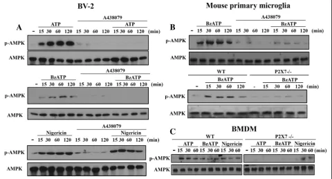
Fig. 1P2X7 induces AMPK activation in BV-2, primary microglia and BMDM. BV-2 cell line (a), mouse primary microglia (b) and BMDM (c) isolated from WT or P2X7−/−mice were treated with ATP (1 mM), BzATP (200μM), or nigericin (10μM), either with or without the pre-treatment of A438079
Statistical analysis
Data were expressed as mean ± S.E.M. Multiple groups were compared by one-way analysis of variance and Bonferroni post-test, making use of Graph pad software (Graph Pad Software, San Diego, CA, USA). Two groups were compared with an unpaired Student’s t test and two-tailpvalue. Results were considered statistically sig-nificant whenp< 0.05.
Results
P2X7 induces Ca/CaMKK- and ROS-dependent AMPK activation in microglia and macrophages
In order to link the energy-sensing kinase AMPK and P2X7 signaling, we first determined the effects of two P2X7 agonists, ATP and BzATP, on AMPK activation. In BV-2 microglia, we found a time-dependent and rapid AMPK phosphorylation upon agonist treatment.
A438079, the P2X7 competitive antagonist, can abolish ATP- and BzATP-induced AMPK phosphorylation, but not that induced by nigericin, which is a potassium ionophore and exerts some actions mimicking P2X7 such as NLRP3 inflammasome activation via K+ efflux (Fig. 1a). Similar to BV-2 cells, BzATP also activated AMPK in mouse primary microglial cells (Fig. 1b) and BMDM (Fig. 1c), and this effect of BzATP was also abolished by P2X7 knockout (P2X7-/-) (Fig. 1b and c) and/or A438079 (Fig. 1b). In contrast, nigericin-induced AMPK activation in BMDM was unaltered by P2X7 knockout (Fig. 1c). All blotting data were quantified and shown in the Additional file1: Figure S1.
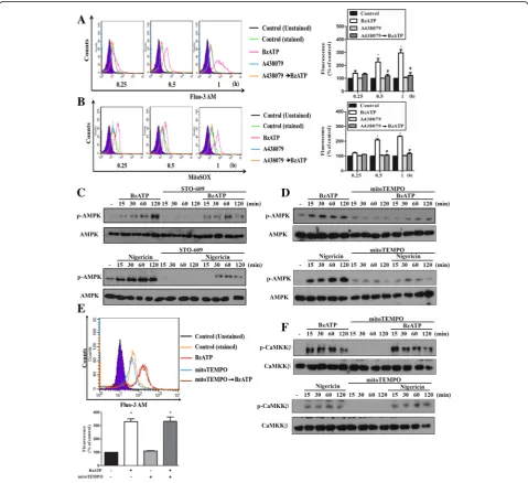
Next, we assessed the signaling events underlying P2X7-mediated AMPK activation. Since increases of intracellular calcium and ROS production upon P2X7
activation have been demonstrated in neurons [36] and microglial cells [37], we sought to determine whether Ca2+/calmodulin-dependent protein kinase kinase (CaMKK) and ROS involve in P2X7-mediated AMPK phosphorylation. Using Fluo-3 AM and mitoSOX, we found that [Ca2+]i and mitoROS levels were significantly and rapidly increased by BzATP within 60 min treat-ment, and both events were abolished by A438079 (10 μM) (Fig. 2a, b). We further used STO-609, a spe-cific inhibitor of CaMKK, to determine the involvement of CaMKK in AMPK signaling. As result shown in Fig. 2c, BzATP- and nigericin-induced AMPK phosphoryl-ation were inhibited in BV-2 cells pretreated with STO-609 (10μM). Likewise, we found that the presence of mitoTEMPO, an inhibitor of mitoROS, can abrogate AMPK phosphorylation caused by BzATP and nigericin
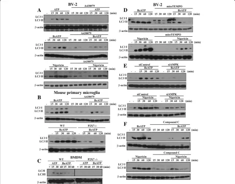
Fig. 3AMPK activation and mitoROS production mediate LC3II accumulation. BV-2 cells were pre-treated with A438079 (10μM,a), mitoTEMPO (250μM,d) or compound C (10μM,f) for 30 min, or siAMPK (100 nM,e) for 48 h. Then cells were treated with ATP (1 mM), BzATP (200μM), or nigericin (10μM) for the indicated time points. In some experiments, mouse primary microglia (b) and BMDM (c) isolated from WT and P2X7−/−
in BV-2 cells (Fig. 2d). To determine whether mitoROS might regulate Ca/CaMKK signaling, we treated cells with mitoTEMPO and found that there were no signifi-cant alterations on the increased [Ca2+]i (Fig. 2e) and the phosphorylation status of CaMKK (Fig. 2f ) induced by BzATP or nigericin. All these results suggest that CaMKK and mitoROS may contribute independently in P2X7-mediated AMPK activation. All blotting data of Fig. 2c, d and f were quantified and shown in the Additional file 2: Figure S2A, B and C, respectively.
P2X7 induces AMPK-dependent mitochondrial fission and mitophagy
To further reveal the consequence of P2X7-activated AMPK signaling on mitochondria function, we analyzed the mitochondrial dynamism and mitophagy processing. To assess mitophagy, we first determined LC3II, whose accumulation is an index of autophagy [38]. We found that both BzATP and ATP can increase LC3II accumula-tion in BV-2 (Fig. 3a), primary microglia (Fig. 3b) and BMDM (Fig.3c), and these effects were greatly attenuated by A438079 and/or P2X7 gene knockout. In contrast,
A438079 cannot abolish the LC3II accumulation triggered by nigericin (Fig. 3a). In addition, in the situation with mitoTEMPO treatment (Fig. 3d), AMPK silencing (Fig. 3e) or treatment with AMPK inhibitor compound C (Fig. 3f ), BzATP- and nigericin-induced LC3II increases in BV-2 cells were greatly inhibited. These findings further emphasize the AMPK-dependence of autophagy/mito-phagy induction. All blotting data of Fig.3were quantified and shown in the Additional file3: Figure S3.
Mitochondrial dynamics upon P2X7 activation was analyzed by confocal laser scanning microscopy with specific antibodies against Tom 20 and LC3. Tom-20 is a subunit of the translocase of the mitochondrial outer membrane (TOM) complex, and becomes a common marker of mitochondria. Its co-localization with LC3II indicates the formation of mitophagy. Our data revealed a higher mitochondrial fission upon BzATP or nigericin treatment, and the concomitant co-localization of LC3 with Tom 20 (Fig. 4a). These effects of BzATP and nigercin were markedly attenuated by AMPK silencing (Fig. 4b). Along with the changes of mitochondrial dy-namics, the fluorescence of Mitotracker green (an index
of mitochondrial mass) was reduced upon BzATP or nigericin treatment for 2 h (Fig.4c, d). Previously some studies concern the action specificity of higher concen-trations of Mitotracker green for its action might de-pend not only on mitochondrial mass but also on metabolic rate [39], thus we determined the response of FCCP, which is a mitochondrial uncoupler. As a result, in our working concentration of Mitotracker green (50 nM), FCCP (1μM) treatment for 2 h did not affect the fluorescence of mitotracker green (Fig. 4d). More-over, the action of BzATP was also blocked by A438079 and siAMPK, while that of nigericin was reversed by siAMPK only (Fig. 4d). All these data suggest that P2X7 activated AMPK signaling may mediate mito-chondrial fission.
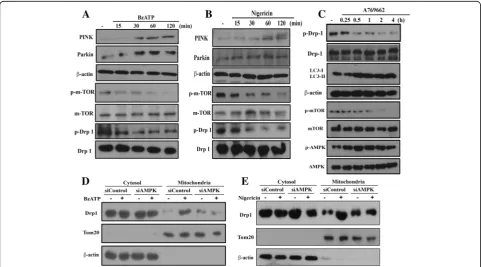
To further address the underlying basis for P2X7-induced mitophagy, we investigated some essential molecules contributing to mitophagy. As shown in Fig. 5a and b, we found that BzATP and nigericin increased the pro-tein levels of both PINK and Parkin, but inhibited the phosphorylation of mTOR and Drp-1 (Fig.5a, b). Because Drp-1 phosphorylation at S637 is a negative regulator of mitochondrial fission [40, 41], our findings of reduced Drp-1 phosphorylation support the P2X7-mediated mito-chondrial fission. Likewise, A769662 (an AMPK activator) can time-dependently induce AMPK phosphorylation and
LC3II accumulation, but decrease mTOR and Drp-1 phosphorylation (Fig.5c). Moreover, Drp-1 was translo-cated to mitochondria after BzATP or nigericin treat-ment for 1 h, and this effect was decreased in cells with AMPK silencing (Fig.5d, e). All these data together fur-ther support the mitophagy induced by BzATP and nigericin is mediated by AMPK activation. All blotting data of Fig.5were quantified and shown in the Additional file4: Figure S4.
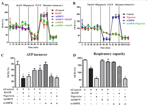
P2X7 inhibits mitochondrial respiration via a pathway independent of AMPK
To further investigate whether mitochondrial respiratory function is affected in parallel with mitochondrial fission and mitophagy triggered by P2X7, we measured the OCR with the Seahorse flux analysis. With sequential addition of several mitochondrial stressors such as oligomycin, FCCP, rotenone and antimycin, we determined ATP turnover and respiratory capacity (or named uncoupled respiration). Our results showed that within the 26 min incubation period, BzATP and nigericin had no signifi-cant effects on resting OCR, but both suppressed ATP turnover and respiratory capacity of mitochondria (Fig. 6a, b). Moreover, the effects of BzATP were abolished by the P2X7 antagonist A438079. Notably nigericin treatment led to an enormous decrease of
respiratory capacity compared to that stimulated by BzATP (Fig.6c, d). Next we determined the link between AMPK activation and respiratory dysfunction downstream P2X7 signaling. We found that AMPK silencing did not affect the BzATP- and nigericin-triggered suppression
of mitochondrial ATP turnover and respiratory
capacity (Fig. 6c, d). These findings indicate an AMPK-independent mechanism in P2X7 signaling to impair mitochondrial respiration.
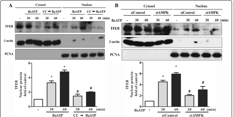
P2X7 induces lysosomal biogenesis via AMPK pathway Current understanding in regulation of lysosomal bio-genesis and lysosomal function is still limited. To reveal the involvement of P2X7 signaling in the process of lyso-somal biogenesis, we measured the TFEB expression and its cellular localization after BzATP treatment. TFEB is the major transcription factor for lysosomal biogenesis and its cellular localization is counter-regulated by
AMPK and mTOR [12]. As shown in Fig.7a, the cyto-solic level of TFEB was rapidly decreased along with an increase of TFEB in the nuclei after BzATP treatment. This nuclear translocation of TFEB was reduced by com-pound C pretreatment (Fig.7a) or siAMPK (Fig.7b).
Using Lysotracker Red staining as the index of lysosome stability, we found that BzATP did not change the Lyso-tracker Red intensity until 6 h, and the inhibitory effect of BzATP at 6 h was prevented by A438079 (Fig.8a, b) but not by AMPK silencing (Fig.8c). Accordingly, enzymatic activity of intracellular cathepsin B was increased after BzATP treatment for 6 h and this effect was blocked by A438079, P2X7 silencing or calcium-free media (Fig.8d). Previously cathepsin B was shown to mediate cell death [42] and negatively regulate TFEB translocation to nuclei [43]. To further understand the consequence of this event under P2X7 activation, we used cathepsin B inhibitor
CA-074Me. As shown in Fig. 8e, CA-074Me caused a
synergistic effect with BzATP to promote nuclear trans-location of TFEB. Moreover, our data showed that BzATP-induced decrease of BV-2 cell viability was re-versed by treatment with CA-074Me (Fig.8f ). In addition, because autophagy/mitophagy is a double edged sword on cell fate, we further used 3-MA to determine the role of mitophagy in P2X7-mediated cell death. 3-MA is an in-hibitor of class III PI3K and has been used as an inin-hibitor of autophagy/mitophagy. As a result, our data revealed the ability of 3-MA to reduce BzATP-induced cell death, and combination of CA-074Me and 3-MA had an additive ef-fect (Fig.8f ). These findings suggest the involvements of cathepsin B and mitophagy in P2X7-induced cell death.
Discussion
Failure of mitophagy is involved in the progression of several neurodegenerative disorders [44, 45]. Therefore, upregulation of mitophagy holds potential for the devel-opment of therapeutic interventions towards confronting neurodegenerative diseases in humans. Recently, a few studies demonstrate that P2X7 can modulate autophagic flux in microglia [27,28], and mitochondrial toxicity is a key event leading to cell death induced by P2X7 activa-tion [27, 46]. Furthermore, P2X7 induces autophagy in myoblasts and myotubes in a mouse model of Duchenne muscular dystrophy, and such autophagic cell death sub-sequently leads to muscular dystrophy [47]. Induction of autophagy in monocyte/macrophages by P2X7 activation
was found to contribute to the bacterial killing [48]. Moreover, P2X7 is suggested to be a key modulator in oxidative stress-mediated autophagy and inflammation in an experimental nonalcoholic steatohepatitis [49] and intestinal inflammation [50]. Of note, P2X7 also induces cellular stress events to impair lysosomal function and cause lysosomal alkalinization, which in turn negatively regulate autophagy [29]. To date the regulatory role of P2X7 activation in lysosome function is much less understood than that in mitochondria. The functional integration between mitochondria and lysosome in mito-phagy regulated by P2X7 activation requires further ex-ploration, in particular focusing on the action time course and mediated signaling pathways.
mitophagy, mitochondrial fission and TFEB nuclear translocation in microglia.
AMPK is crucial for mediating mitophagy and modulat-ing mitochondrial dynamics and biogenesis [51]. With re-gard to mitophagy, AMPK is a key signaling molecule to initiate autophagic flux. In addition, AMPK can regulate PINK (a mitochondrial kinase) and Parkin (an ubiquitin lig-ase) which are two key factors in mitophagy [52]. Accumu-lation of PINK on the mitochondrial outer membrane can regulate Parkin for mitophagy. Study further reveals the ability of AMPK to phosphorylate PINK1 at S495, leading
mitophagy and mitochondrial fission upon P2X7 stimula-tion of microglial cells. This is because AMPK silencing can reverse the effects of BzATP on LC3II accumulation and translocation to mitochondria, Drp-1 phosphorylation and mitochondrial translocation,and mitochondrial fission.
In addition to involvement in the degradation by forming autophagosome, lysosome is recently recog-nized as a signaling hub permitting the coordination of several homeostatic signaling pathways [11]. TFEB was recently reported as the major transcription factor to activate lysosome- and autophagy-related genes, thereby increasing the number of lysosomes and promoting autophagy-related cargo degradation [56]. TFEB activity is timely controlled antagonistically by mTOR and AMPK signaling pathways [11–13]. Accordingly, in our study AMPK silencing and compound C treatment can inhibit BzATP-induced TFEB translocation to nuclei (Fig.7a, b). Despite the enhanced TFEB activation by P2X7 signaling at early stage, longer incubation of BzATP led to an AMPK-independent lysosomal destabilization (Fig. 8c) and cathepsin B release (Fig.8d).Our data further suggest that cathepsin B release from lysosomes may contribute to cell death caused by P2X7 activated (Fig.8f ). Currently it remains unclear how the integrity of lysosomal membrane is altered, leading to the leakage of lysosomal enzymes to the cytoplasm and triggering cell death.
Although extracellular ATP and BzATP were shown to induce AMPK phosphorylation in MCA38 colon cancer cells [57] and HUVEC cells [58], the upstream signaling pathway remains to be elucidated. Our data revealed
that P2X7-induced mitoROS (Fig. 2d) and CaMKK acti-vation (Fig. 2c) are involved in the rapid activation of AMPK. Here we propose that both ROS and Ca2+ sig-nals are functionally independent, because mitoTEMPO which is a selective scavenger of mitoROS [59, 60] can-not alter the increased intracellular calcium and CaMKK activation (Fig. 2e, f ). Moreover, P2X7-mediated AMPK activation is accompanied by the inhibition of mTOR that is involved in protein translation. All these findings sup-port the notion that ROS and CaMKK function as two major upstream mediators of AMPK activation [61,62].
Besides timely regulating mitochondrial dynamics and mitophagy, AMPK activity also affects mitochondrial res-piration [63]. In our study, we found that P2X7-mediated inhibitions of ATP production and respiratory capacity are AMPK independent. In contrast to the reversal effects as we discussed above, AMPK silencing did not alter BzATP-induced inhibition of mitochondrial respiration. We speculate that this effect might be due to the changes of calcium and potassium homeostasis. Intracellular cal-cium overload and dramatic potassium efflux upon P2X7 activation might interrupt the mitochondrial membrane potential and cytosolic pH level that may play important roles in maintaining the electron transport efficiency and mitochondrial complex activities [64]. Supporting this no-tion is our finding that nigericin can induce a higher re-sponse than BzATP to decrease uncoupled respiration (Fig.6d). Thus we suggest that K+efflux induced by P2X7 and nigericin is the potential mediator to impair mito-chondrial respiration. In addition, based on the less
dependence of nigericin responses on AMPK as compared to BzATP, e.g. loss of mitochondrial mass (Fig.4d) and in-duction of Tom-20 and LC3 colocalization (Fig. 4b), K+ efflux might exert other direct actions independent of Ca/ CaMKK/AMPK to regulate mitochondrial dynamics and mitophagy. Moreover, we also observed the effects of nigericin to increase mitoROS and [Ca2+]i in BV-2 cells (data not shown). Therefore, it is suggested that K+efflux itself can indirectly change the intracellular calcium level and mitochondrial function.
In this study we used pharmacologically selective AMPK activator A769662 and genetic silencing ap-proach to demonstrate the role of AMPK in the P2X7 actions. A769662 is the relatively most selective AMPK agonist compared to others like metformin and AICAR. Nevertheless, some recent studies still question the selectivity of A769662 at concentrations above 50– 100 μM [63, 65, 66]. In our study, A769662 alone at our working concentration (i.e. 20 μM) did not alter intracellular calcium level in BV-2 cells (data not shown). Moreover, silencing AMPK exerted opposite actions of A769662, in terms of LC3II protein level and phosphoryl-ation of mTOR and Drp-1. Therefore, the involvement of AMPK in P2X7-mediated actions in BV-2 cells is strongly suggested.
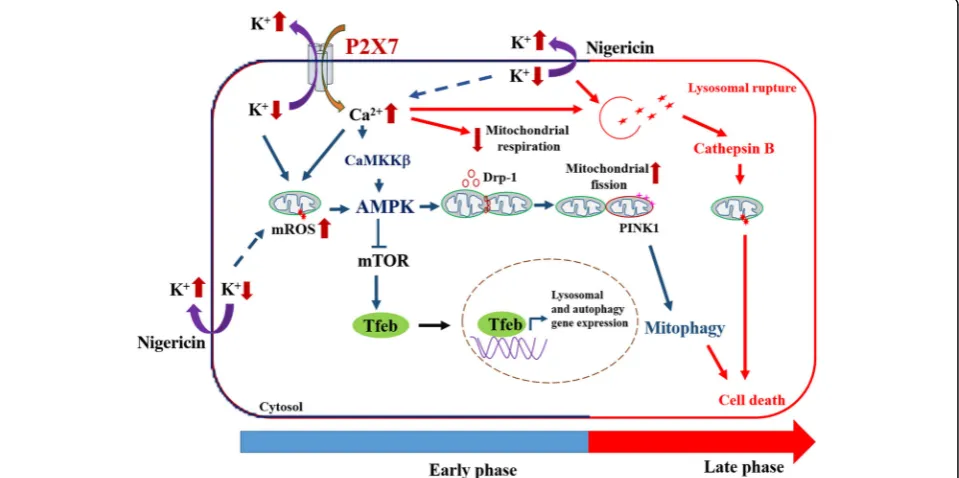
Conclusion
Taken together, our findings indicate the time-dependent actions of P2X7 in regulating the functions of mitochon-dria and lysosomes in microglial cells (Fig. 9). In early phase shortly after P2X7 activation, the Ca2+ influx and K+ efflux efficiently act as positive regulators for mito-phagy and lysosomal biogenesis, but a negative modulator for mitochondrial respiration. A prolonged action of P2X7 signaling would lead to lysosomal destabilization and cell death. We demonstrate for the first time that P2X7 activa-tion can rapidly trigger AMPK activaactiva-tion via ROS and CaMKKII pathways, which lead to mitochondrial fission, mitophagy induction and TFEB activation. However, in-hibition of mitochondrial respiration and induction of lysosomal destabilization caused by P2X7 activation is in-dependent of AMPK.
Additional files
Additional file 1:Figure S1.Immunoblotting data of main Fig.1a-c were quantified using Image J software. Data were the mean ± S.E.M. from 3 independent experiments. *p< 0.05, indicating the enhanced effects of ATP, BzATP and nigericin; #p< 0.05, indicating the antagonist effects of A438079 and/or P2X7−/−on the individual action of ATP and
BzATP. (TIF 3316 kb)
Additional file 2:Figure S2.Immunoblotting data of main Fig.2c, d and f were quantified using Image J software and shown in Additional file2: Figure S2A, 2B and 2C, respectively. Data were the mean ± S.E.M. from 3 independent experiments. *p< 0.05, indicating the significant
effects of BzATP and nigericin; #p< 0.05, indicating the antagonist effects of mitoTEMPO and STO-609 on the action of BzATP and nigericin. (TIF 2618 kb)
Additional file 3:Figure S3.Immunoblotting analysis of main Fig.3
were quantified using Image J software. Data were the mean ± S.E.M. from 3 independent experiments. *p< 0.05, indicating the significant effects of ATP, BzATP and nigericin; #p< 0.05, indicating the antagonist effects of A438079, mitoTEMPO, Compound C, siAMPK and P2X7−/−on
the individual action of ATP, BzATP and/or nigericin. (TIF 5179 kb)
Additional file 4:Figure S4.Immunoblotting analysis of main Fig.5
were quantified using Image J software. Data were the mean ± S.E.M. from 3 independent experiments. *p< 0.05, indicating the significant effects of BzATP, nigericin and A769662; #p< 0.05, indicating the antagonist effect of siAMPK on the individual action of BzATP and nigericin. (TIF 4890 kb)
Abbreviations
[Ca2+]i:Intracellular calcium; AMPK: 5’AMP-activated protein kinase;
ATP: Adenosine 5′-triphosphate; BMDM: Bone marrow-derived macrophages; BzATP: 2′(3′)-O-(4-Benzoylbenzoyl)adenosine 5′-triphosphate
triethylammonium salt; CC: Compound C; DAMP: Damage associated molecular pattern; DMEM: Dulbecco Modified Eagle Medium; Drp-1: Dynamin-related protein-1; FACS: Fluorescence-activated cell sorting; FBS: Fetal bovine serum; FCCP: Carbonyl
cyanide-p-trifluoromethoxyphenylhydrazone; OCR: Oxygen consumption rate; PBS: Phosphate-buffered saline; ROS: Reactive oxygen species
Funding
We like to thank the research funding from MOST (103–2320-B-002-069-MY3 and 106–2321-B-002-021), National Taiwan University Hospital (105F025–07) and Academia Sinica (IBMS-CRC106-P03 and AS-TP-106-L11–1).
Availability of data and materials Not applicable.
Authors’contributions
PS designed and performed the experiments, analyzed the data, and prepared and reviewed the manuscript. D-YH conducted the confocal microscopy experiment for mitophagy. S-FC involved in the discussion, experimental guidance and reviewed the manuscript. W-WL designed the experiments and prepared and reviewed the manuscript. All authors read and approved the final manuscript.
Ethics approval
All experimental procedures were approved by the National Taiwan University College of Medicine Ethics Committee in accordance with their guidelines for the care of animals (protocol no. 20160419).
Consent for publication Not applicable.
Competing interests
The authors declare that they have no competing interests.
Publisher’s Note
Springer Nature remains neutral with regard to jurisdictional claims in published maps and institutional affiliations.
Author details
1
Received: 8 August 2018 Accepted: 1 November 2018
References
1. Ransohoff RM. How neuroinflammation contributes to neurodegeneration. Science. 2016;353:777–83.
2. Lull ME, Block ML. Microglial activation and chronic neurodegeneration. Neurotherapeutics. 2010;7:354–65.
3. Cunningham C. Microglia and neurodegeneration: the role of systemic inflammation. Glia. 2013;61:71–90.
4. Exner N, Lutz AK, Haass C, Winklhofer KF. Mitochondrial dysfunction in Parkinson’s disease: molecular mechanisms and pathophysiological consequences. EMBO J. 2012;31:3038–62.
5. Kubli DA, Gustafsson AB. Mitochondria and mitophagy: the yin and yang of cell death control. Circ Res. 2012;111:1208–21.
6. Ni HM, Williams JA, Ding WX. Mitochondrial dynamics and mitochondrial quality control. Redox Biol. 2015;4:6–13.
7. Dorn GW 2nd, Kitsis RN. The mitochondrial dynamism-mitophagy-cell death interactome: multiple roles performed by members of a mitochondrial molecular ensemble. Circ Res. 2015;116:167–82.
8. Garcia D, Shaw RJ. AMPK: mechanisms of cellular energy sensing and restoration of metabolic balance. Mol Cell. 2017;66:789–800.
9. Toyama EQ, Herzig S, Courchet J, Lewis TL Jr, Loson OC, Hellberg K, Young NP, Chen H, Polleux F, Chan DC, Shaw RJ. Metabolism. AMP-activated protein kinase mediates mitochondrial fission in response to energy stress. Science. 2016;351:275–81.
10. Herzig S, Shaw RJ. AMPK: guardian of metabolism and mitochondrial homeostasis. Nat Rev Mol Cell Biol. 2018;19:121–35.
11. Carroll B, Dunlop EA. The lysosome: a crucial hub for AMPK and mTORC1 signalling. Biochem J. 2017;474:1453–66.
12. Hipolito VEB, Ospina-Escobar E, Botelho RJ. Lysosome remodelling and adaptation during phagocyte activation. Cell Microbiol. 2018;20.https://doi. org/10.1111/cmi.12824.
13. Lin SC, Hardie DG. AMPK: sensing glucose as well as cellular energy status. Cell Metab. 2018;27:299–313.
14. Young NP, Kamireddy A, Van Nostrand JL, Eichner LJ, Shokhirev MN, Dayn Y, Shaw RJ. AMPK governs lineage specification through Tfeb-dependent regulation of lysosomes. Genes Dev. 2016;30:535–52.
15. Sluyter R. The P2X7 receptor. Adv Exp Med Biol. 2017;1051:17–53. 16. Orioli E, De Marchi E, Giuliani AL, Adinolfi E. P2X7 receptor orchestrates
multiple Signalling pathways triggering inflammation, autophagy and metabolic/trophic responses. Curr Med Chem. 2017;24:2261–75. 17. Lin YC, Huang DY, Wang JS, Lin YL, Hsieh SL, Huang KC, Lin WW. Syk is
involved in NLRP3 inflammasome-mediated caspase-1 activation through adaptor ASC phosphorylation and enhanced oligomerization. J Leukoc Biol. 2015;97:825–35.
18. Adinolfi E, Giuliani AL, De Marchi E, Pegoraro A, Orioli E, Di Virgilio F. The P2X7 receptor: a main player in inflammation. Biochem Pharmacol. 2018; 151:234–44.
19. Savio LEB, de Andrade Mello P, da Silva CG, Coutinho-Silva R. The P2X7 receptor in inflammatory diseases: angel or demon? Front Pharmacol. 2018;9:52.
20. Sperlagh B, Illes P. P2X7 receptor: an emerging target in central nervous system diseases. Trends Pharmacol Sci. 2014;35:537–47.
21. Chen Z, He L, Li L, Chen L. The P2X7 purinergic receptor: an emerging therapeutic target in cardiovascular diseases. Clin Chim Acta. 2018;479: 196–207.
22. Bhattacharya A, Biber K. The microglial ATP-gated ion channel P2X7 as a CNS drug target. Glia. 2016;64:1772–87.
23. Bhattacharya A. Recent advances in CNS P2X7 physiology and pharmacology: focus on neuropsychiatric disorders. Front Pharmacol. 2018;9:30.
24. Beamer E, Fischer W, Engel T. The ATP-gated P2X7 receptor as a target for the treatment of drug-resistant epilepsy. Front Neurosci. 2017;11:21. 25. Burnstock G. Purinergic mechanisms and pain. Adv Pharmacol. 2016;75:
91–137.
26. McLarnon JG. Roles of purinergic P2X7 receptor in glioma and microglia in brain tumors. Cancer Lett. 2017;402:93–9.
27. Sekar P, Huang DY, Chang SF, Lin WW. Coordinate effects of P2X7 and extracellular acidification in microglial cells. Oncotarget. 2018;9:12718–31.
28. Fabbrizio P, Amadio S, Apolloni S, Volonte C. P2X7 receptor activation modulates autophagy in SOD1-G93A mouse microglia. Front Cell Neurosci. 2017;11:249.
29. Takenouchi T, Nakai M, Iwamaru Y, Sugama S, Tsukimoto M, Fujita M, Wei J, Sekigawa A, Sato M, Kojima S, et al. The activation of P2X7 receptor impairs lysosomal functions and stimulates the release of autophagolysosomes in microglial cells. J Immunol. 2009;182:2051–62.
30. Solle M, Labasi J, Perregaux DG, Stam E, Petrushova N, Koller BH, Griffiths RJ, Gabel CA. Altered cytokine production in mice lacking P2X(7) receptors. J Biol Chem. 2001;276:125–32.
31. Blasi E, Barluzzi R, Bocchini V, Mazzolla R, Bistoni F. Immortalization of murine microglial cells by a v-raf/v-myc carrying retrovirus. J Neuroimmunol. 1990;27: 229–37.
32. Mohanraj M, Sekar P, Liou HH, Chang SF, Lin WW. The mycobacterial adjuvant analogue TDB attenuates neuroinflammation via mincle-independent PLC-gamma1/PKC/ERK signaling and microglial polarization. Mol Neurobiol. 2018.https://doi.org/10.1007/s12035-018-1135-4. 33. Lin YC, Huang DY, Chu CL, Lin YL, Lin WW. The tyrosine kinase Syk
differentially regulates toll-like receptor signaling downstream of the adaptor molecules TRAF6 and TRAF3. Sci Signal. 2013;6:ra71.
34. Cottet-Rousselle C, Ronot X, Leverve X, Mayol JF. Cytometric assessment of mitochondria using fluorescent probes. Cytometry A. 2011;79:405–25. 35. Liao YH, Lin YC, Tsao ST, Lin YC, Yang AJ, Huang CT, Huang KC, Lin WW.
HMG-CoA reductase inhibitors activate caspase-1 in human monocytes depending on ATP release and P2X7 activation. J Leukoc Biol. 2013;93:289–99. 36. Nishida K, Nakatani T, Ohishi A, Okuda H, Higashi Y, Matsuo T, Fujimoto S,
Nagasawa K. Mitochondrial dysfunction is involved in P2X7 receptor-mediated neuronal cell death. J Neurochem. 2012;122:1118–28. 37. Bartlett R, Yerbury JJ, Sluyter R. P2X7 receptor activation induces reactive
oxygen species formation and cell death in murine EOC13 microglia. Mediat Inflamm. 2013;2013:271813.
38. Mizushima N, Yoshimori T. How to interpret LC3 immunoblotting. Autophagy. 2007;3:542–5.
39. Pendergrass W, Wolf N, Poot M. Efficacy of MitoTracker green and CMXrosamine to measure changes in mitochondrial membrane potentials in living cells and tissues. Cytometry A. 2004;61:162–9.
40. Cereghetti GM, Stangherlin A, Martins de Brito O, Chang CR, Blackstone C, Bernardi P, Scorrano L. Dephosphorylation by calcineurin regulates translocation of Drp1 to mitochondria. Proc Natl Acad Sci U S A. 2008;105: 15803–8.
41. Cribbs JT, Strack S. Reversible phosphorylation of Drp1 by cyclic AMP-dependent protein kinase and calcineurin regulates mitochondrial fission and cell death. EMBO Rep. 2007;8:939–44.
42. de Castro MA, Bunt G, Wouters FS. Cathepsin B launches an apoptotic exit effort upon cell death-associated disruption of lysosomes. Cell Death Discov. 2016;2:16012.
43. Qi X, Man SM, Malireddi RK, Karki R, Lupfer C, Gurung P, Neale G, Guy CS, Lamkanfi M, Kanneganti TD. Cathepsin B modulates lysosomal biogenesis and host defense against Francisella novicida infection. J Exp Med. 2016; 213:2081–97.
44. Metaxakis A, Ploumi C, Tavernarakis N. Autophagy in age-associated neurodegeneration. Cells. 2018;7.https://doi.org/10.3390/cells7050037. 45. Zhang J, Culp ML, Craver JG, Darley-Usmar V. Mitochondrial function and
autophagy: integrating proteotoxic, redox, and metabolic stress in Parkinson’s disease. J Neurochem. 2018;144:691–709.
46. Seeland S, Kettiger H, Murphy M, Treiber A, Giller J, Kiss A, Sube R, Krahenbuhl S, Hafner M, Huwyler J. ATP-induced cellular stress and mitochondrial toxicity in cells expressing purinergic P2X7 receptor. Pharmacol Res Perspect. 2015;3:e00123.
47. Young CN, Sinadinos A, Lefebvre A, Chan P, Arkle S, Vaudry D, Gorecki DC. A novel mechanism of autophagic cell death in dystrophic muscle regulated by P2RX7 receptor large-pore formation and HSP90. Autophagy. 2015;11:113–30.
48. Biswas D, Qureshi OS, Lee WY, Croudace JE, Mura M, Lammas DA. ATP-induced autophagy is associated with rapid killing of intracellular mycobacteria within human monocytes/macrophages. BMC Immunol. 2008;9:35.
50. Souza CO, Santoro GF, Figliuolo VR, Nanini HF, de Souza HS, Castelo-Branco MT, Abalo AA, Paiva MM, Coutinho CM, Coutinho-Silva R. Extracellular ATP induces cell death in human intestinal epithelial cells. Biochim Biophys Acta. 2012;1820:1867–78.
51. Shah SZA, Zhao D, Hussain T, Yang L. Role of the AMPK pathway in promoting autophagic flux via modulating mitochondrial dynamics in neurodegenerative diseases: insight into prion diseases. Ageing Res Rev. 2017;40:51–63.
52. Matsuda N. Phospho-ubiquitin: upending the PINK-Parkin-ubiquitin cascade. J Biochem. 2016;159:379–85.
53. Wang B, Nie J, Wu L, Hu Y, Wen Z, Dong L, Zou MH, Chen C, Wang DW. AMPKalpha2 protects against the development of heart failure by enhancing mitophagy via PINK1 phosphorylation. Circ Res. 2018;122:712–29. 54. Narendra D, Walker JE, Youle R. Mitochondrial quality control mediated by
PINK1 and Parkin: links to parkinsonism. Cold Spring Harb Perspect Biol. 2012;4.https://doi.org/10.1101/cshperspect.a011338.
55. Sakamoto A, Saotome M, Hasan P, Satoh T, Ohtani H, Urushida T, Katoh H, Satoh H, Hayashi H. Eicosapentaenoic acid ameliorates palmitate-induced lipotoxicity via the AMP kinase/dynamin-related protein-1 signaling pathway in differentiated H9c2 myocytes. Exp Cell Res. 2017;351:109–20.
56. Settembre C, Di Malta C, Polito VA, Garcia Arencibia M, Vetrini F, Erdin S, Erdin SU, Huynh T, Medina D, Colella P, et al. TFEB links autophagy to lysosomal biogenesis. Science. 2011;332:1429–33.
57. Bian S, Sun X, Bai A, Zhang C, Li L, Enjyoji K, Junger WG, Robson SC, Wu Y. P2X7 integrates PI3K/AKT and AMPK-PRAS40-mTOR signaling pathways to mediate tumor cell death. PLoS One. 2013;8:e60184.
58. da Silva CG, Jarzyna R, Specht A, Kaczmarek E. Extracellular nucleotides and adenosine independently activate AMP-activated protein kinase in endothelial cells: involvement of P2 receptors and adenosine transporters. Circ Res. 2006;98:e39–47.
59. Wu CA, Chao Y, Shiah SG, Lin WW. Nutrient deprivation induces the Warburg effect through ROS/AMPK-dependent activation of pyruvate dehydrogenase kinase. Biochim Biophys Acta. 2013;1833:1147–56. 60. Park J, Min JS, Kim B, Chae UB, Yun JW, Choi MS, Kong IK, Chang KT, Lee DS.
Mitochondrial ROS govern the LPS-induced pro-inflammatory response in microglia cells by regulating MAPK and NF-kappaB pathways. Neurosci Lett. 2015;584:191–6.
61. Sundararaman A, Amirtham U, Rangarajan A. Calcium-oxidant signaling network regulates AMP-activated protein kinase (AMPK) activation upon matrix deprivation. J Biol Chem. 2016;291:14410–29.
62. Mungai PT, Waypa GB, Jairaman A, Prakriya M, Dokic D, Ball MK, Schumacker PT. Hypoxia triggers AMPK activation through reactive oxygen species-mediated activation of calcium release-activated calcium channels. Mol Cell Biol. 2011;31:3531–45.
63. Vincent EE, Coelho PP, Blagih J, Griss T, Viollet B, Jones RG. Differential effects of AMPK agonists on cell growth and metabolism. Oncogene. 2015; 34:3627–39.
64. Berezhnov AV, Soutar MP, Fedotova EI, Frolova MS, Plun-Favreau H, Zinchenko VP, Abramov AY. Intracellular pH modulates autophagy and Mitophagy. J Biol Chem. 2016;291:8701–8.
65. Huang Y, Smith CA, Chen G, Sharma B, Miner AS, Barbee RW, Ratz PH. The AMP-dependent protein kinase (AMPK) activator A-769662 causes arterial relaxation by reducing cytosolic free calcium independently of an increase in AMPK phosphorylation. Front Pharmacol. 2017;8:756.