R E S E A R C H A R T I C L E
Open Access
Generation of polyclonal antibody with high
avidity to rosuvastatin and its use in
development of highly sensitive ELISA for
determination of rosuvastatin in plasma
Ibrahim A Darwish
*, Abdul-Rahman M Al-Obaid and Hamoud A Al-Malaq
Abstract
In this study, a polyclonal antibody with high avidity and specificity to the potent hypocholesterolaemic agent rosuvastatin (ROS) has been prepared and used in the development of highly sensitive enzyme-linked
immunosorbent assay (ELISA) for determination of ROS in plasma. ROS was coupled to keyhole limpt hemocyanin (KLH) and bovine serum albumin (BSA) using carbodiimide reagent. ROS-KLH conjugate was used for immunization of female 8-weeks old New Zealand white rabbits. The immune response of the rabbits was monitored by direct ELISA using ROS-BSA immobilized onto microwell plates as a solid phase. The rabbit that showed the highest antibody titer and avidity to ROS was scarified and its sera were collected. The IgG fraction was isolated and purified by avidity chromatography on protein A column. The purified antibody showed high avidity to ROS; IC50= 0.4 ng/ml. The specificity of the antibody for ROS was evaluated by indirect ELISA using various competitors from the ROS-structural analogues and the therapeutic agents used with ROS in a combination therapy. The proposed ELISA involved a competitive binding reaction between ROS, in plasma sample, and the immobilized ROS-BSA for the binding sites on a limited amount of the anti-ROS antibody. The bound anti-ROS antibody was quantified with horseradish peroxidase-labeled second anti-rabbit IgG antibody (HRP-IgG) and 3,3’,5,5’-tetramethylbenzidine (TMB) as a substrate for the peroxidase enzyme. The concentration of ROS in the sample was quantified by its ability to inhibit the binding of the anti-ROS antibody to the immobilized ROS-BSA and subsequently the color intensity in the assay wells. The assay enabled the determination of ROS in plasma at concentrations as low as 40 pg/ml.
Background
Rosuvastatin (ROS); (3R,5S,6E)-7-[4-((4-fluorophenyl)-6-(1-methylethyl)-2-[methyl (methylsulphonyl) amino]-5-pyrimidinyl]-3, 5-dihydroxy-6-heptenoic acid (Figure 1), is an effective 3-hydroxy-3-methylglutaryl-coenzyme A (HMG-CoA) reductase inhibitor and it is widely pre-scribed in the treatment of patients with hypercholester-olaemia [1]. ROS is available as Crestor in tablet form (5, 10, 20, or 40 mg) for oral administration. In clinical studies, ROS (1-80 mg) produced highly significant dose-dependent reductions in low-density lipoprotein cholesterol (up to 65%) and was well tolerated. The pharmacokinetics of ROS following single and
multiple-dose administration of the drug to healthy volunteers have been investigated. The absolute oral bioavailability is greater than 20%, and the elimination half-life is ~ 20 hrs. The reported ROS plasma therapeutic concentration was 19 ng/ml, after 40 mg-daily dosing. The elimination of ROS is primarily via the liver. In a clinical trial, ~ 90% of the orally administered dose of ROS is recovered in feces as unchanged drug [2].
Because of the clinical success of ROS, several meth-ods have been developed for its quantitative determina-tion in plasma samples. Almost all of these methods are liquid chromatography [3-9]. These methods involved tedious steps for the treatment of the samples, pre-derivatization with critical derivatizing reagents, and use of expensive detectors (e.g. tandem mass spectrometry) that are not available in most laboratories. For these * Correspondence: [email protected]
Department of Pharmaceutical Chemistry, College of Pharmacy, King Saud University, P.O. Box 2457, Riyadh 11451, Saudi Arabia
reasons, the development of new alternative analytical technology for determination of ROS in plasma with adequate sensitivity, improved simplicity, and lower cost was seriously needed.
Immunoassays have been widely used in pharmaceuti-cal and clinipharmaceuti-cal analysis because of their inherent specifi-city, applicability for a wide range of analytes, high-throughput, and low cost [10]. ELISA is the most versa-tile format of the immunoassays. ELISA is remarkably quick, easily performed, and also offers great sensitivity when appropriate enzyme labels are used. As well, ELISA is well suited for the screening of large number of samples, and the specificity for the analyte of interest even in multi-component complex sample matrix such as plasma [10]. The specificity of the antibody to the analyte of interest is the limiting factor in the validity of any immunoassay system. In order to establish a specific and sensitive ELISA for ROS, a specific antibody with high avidity for ROS was required. The present study describes, for the first time, the preparation of a polyclo-nal antibody that can specifically recognizes ROS with high avidity. The ELISA that has been developed using
this antibody is able to determine ROS in plasma sam-ples at concentrations as low as 40 pg/ml.
Experimental Apparatus
Elx808 microplate reader (Bio-Tek Instruments Inc., USA). Elx50 microplate washer (Bio-Tek Instruments Inc., USA). EM-36N microtube shaker (Taitec, Japan). Biofuge Pico centrifuge (Heraeus Instruments, Ger-many). Mini/18 incubator (Genlab Ltd., UK). Water pur-ification system (Milli-Q Labo, Millipore Ltd., Bedford, USA)
Materials
Rosuvastatin (ROS) was obtained from Biocon India Ltd., India. Horseradish peroxidase labeled goat anti-rabbit IgG (HRP-IgG), bovine serum albumin (BSA), 2,4,6-trinitrobenzene sulfonic acid, 1-ethyl-3-(3-dimethy-laminopropyl) carbodiimide hydrochloride (EDC), and tween-20 were purchased from Sigma Chemical Co. (St. Louis, MO, USA). Keyhole limpet hemocyanin (KLH) was purchased from Novabiochem Co. (La Jolla, CA, USA). 3,3’,5,5’-Tetramethylbenzidine (TMB) peroxidase substrate was obtained from Kirkegaard-Perry Labora-tories (Gaithersburg, MD, USA). ELISA high-binding microwell plates were a product of Corning/Costar, Inc. (Cambridge, MA, USA). Centricon-30 filter (Amicon, Inc., Beverly, MA, USA). BCA reagent for protein assay and protein A column were obtained from Pierce Bio-technology Inc. (Rockford, IL, USA).
Procedures
Preparation of ROS-protein conjugates
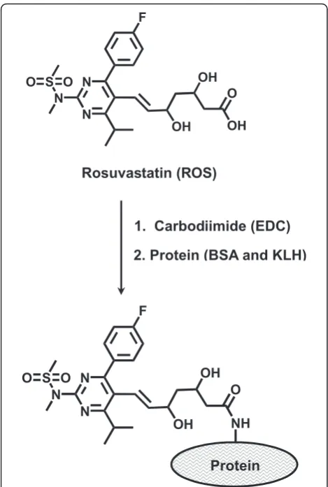
ROS was conjugated with keyhole limpet hemocyanin (KLH) and bovine serum albumin (BSA) according to the method described by Darwish et al [11]. Briefly, EDC (150 mg) was added to ROS solution (10-ml, 5 mg/ml) in 12.5 mM phosphate buffer (PB) of pH 5, and the pH of the reaction mixture was maintained at pH 5-5.5 using 0.01 M HCl for 5 min. Five ml of protein solu-tion (5 mg/ml, in 50 mM PB of pH 7.2) was added, and the pH was rapidly adjusted to pH 6.4 and maintained constant for 90 min. The reaction was left to proceed overnight in dark at 4°C. The uncojugated ROS was removed from the ROS-protein conjugates by buffer exchange using a Centricon-30 filter. Protein content of each conjugate was determined by BCA reagent kit and the extent of substitution of free amino groups on the protein was determined by estimation of free amino groups on equal amounts of the corresponding protein which underwent the same experimental treatment and on protein subjected to the conjugation by the proce-dure described by Habeeb [12]. The extent of conjuga-tion = (AROS-Protein/AProtein) × 100; where AROS-Protein
Rosuvastatin (ROS)
OH
OH OH
O N
N N S
F
O O
Protein
OH
OH NH
O N
N N S
F
O O
1. Carbodiimide (EDC)
2. Protein (BSA and KLH)
and AProtein were the absorbances obtained from the
reaction of 2,4,6-trinitrobenzene sulfonic acid with ROS-protein and ROS-protein, respectively. The extent of conjuga-tion was found to be 10.8 and 20.4% of the total amino group residues on KLH and BSA, respectively.
Immunization of animals and purification of antibody The immunogen used was ROS-KLH protein conjugate. Four female 8-weeks old New Zealand white rabbits were injected subcutaneously with 1 mg of ROS-KLH emulsified in Freund’s complete adjuvant, divided in dif-ferent sites for each rabbit. The same immunization pro-cedure was repeated 6 times with 2-weeks interval, however incomplete adjuvant was used instead. After 4-7 days from each immunization, blood samples (~500
μl) were collected and diluted tenfold with PBS. The diluted blood samples were centrifuged at 10,000 g at 4° C for 10 min, and the sera (supernatants) were col-lected. The antibody response in each rabbit was deter-mined by analysis of the collected antisera by direct enzyme immunoassay [11]. The rabbit whose diluted serum gave the highest avidity (lower IC50) to ROS was
selected as the most appropriate rabbit for collecting its total serum as as crude anti-ROS polyclonal antibody sample.
The serum (~20 ml) was kept overnight at 4°C and then centrifuged at 4°C for 10 min. To 5 ml of the supernatant, an equal volume of a saturated ammonium sulfate solution was gradually added and gently mixed. For complete precipitation of the IgG, the solution was kept over ice for 3 h. The precipitate was collected by centrifugation at 10,000 g at 4°C for 30 min. The preci-pitate was resuspended in 10 ml phosphate buffered sal-ine (PBS; 137 mM NaCl, 3 mM KCl, and 10 mM sodium phosphate, pH 7.4) followed by reprecipitation with ammonium sulfate. After repeating this step three times, the precipitate was dissolved in 10 ml of PBS. The produced antibody solution was purified by protein A column chromatography. One milliliter aliquot of the solution was mixed with an equal volume of the binding buffer (1.5 M glycine-NaOH containing 3 M NaCl, pH 8.7) and the mixture was applied to the protein A col-umn and the eluent was monitored for protein by mea-suring the absorbance of the eluted fractions at 280 nm. The column was washed with 50-60 ml of binding buf-fer, and the bound immunoglobulin was eluted with 0.1 M sodium citrate buffer (pH 3.0). The eluate was col-lected in 1.5 ml fractions into tubes containing 100μl of 1 M Tris-HCl buffer (pH 9.0), and mixed. The pooled fractions were dialyzed overnight against five changes of PBS (~ 4 h intervals). The protein content of the dialy-zate was determined by BCA reagent kit, and used as the pure anti-ROS antibody sample.
ELISA procedures and data analysis
Aliquot (50 μl) of either ROS-BSA conjugate or BSA protein solution (5μg/ml in PBS) was dispensed in each well of the microwell plate. The plates were incubated for 2 h at 37°C. The wells were washed with PBS con-taining 0.05% Tween-20 (PBS-T) and blocked with 200
μl of 3% BSA by incubation at 37°C for 1 h. A 50 μl of anti-ROS antibody sample (rabbit serum or purified IgG) was dispensed in each well and plate was incubated for 1.5 h at 37°C, then the plates were washed with PBS-T, and 50 μl of HRP-IgG (1/5,000 in PBS) was added to each well. After 1.5 h incubation, the plates were washed with PBS-T and the amount of the bound HRP-IgG was quantified using TMB microwell substrate.
In competitive ELISA, 50μl of ROS sample (standard ROS solution or plasma that have been tenfold diluted with PBS) was mixed with antibody solution and 50 μl of the mixture was dispensed into microplate wells that have been previously coated and blocked. After, the competition reaction, the signal was generated as above.
The data were acquisitioned by KC Junior software (Bio-Tek Instruments Inc., USA), and transformed to a four-parameter curve fitting using Slide Write software, version 5.011 (Advanced Graphics Software, Inc., USA). Values for IC50were those that gave the best fit to the
following equation:
A = A0− {(A0−A1)[ROS]/(IC50+ [ROS])}
Where A is the signal at a definite known concentra-tion of ROS, A0 is the signal in the absence of ROS, A1
is the signal at the saturating concentration of ROS, and IC50is the ROS concentration that produces a 50%
inhi-bition of the signal. The concentrations of ROS in the samples were then obtained by interpolation on the standard curve.
Results and discussion
Preparation and characterization of ROS-protein conjugates
to the carrier proteins (BSA and KLH) by carbodiimide reagent (Figure 1).
In order to ascertain the extent to which ROS was conjugated to the proteins, spectral analysis of the pro-teins and ROS-protein conjugates were conducted under the same pH conditions. The apparent molar absorptiv-ity of ROS-protein conjugates was higher than that of the unconjugated protein. This hyperchromic effect was evident for the successful conjugation of the chromo-phoric ROS molecule with both BSA and KLH. The extent of conjugation was determined employing BCA reagent kit for protein assay, and the spectrophotometric procedures described by Habeeb [12]. The percentages of ROS residues in ROS-BSA and ROS-KLH conjugates were found to be 20.4 and 10.8%, respectively.
Preparation and characterization of anti-ROS antibody Because of the high immunogenicity of KLH [14], ROS-KLH was selected as immunogen for immunization of animals and ROS-BSA was selected for immobilization onto the microwell plates in the ELISA. To monitor the progress of the immune response of the rabbits and confirm that they were sufficiently immunized, serum samples were collected from the rabbits on the fourth to seventh days after each immunization, and were ana-lyzed by direct ELISA [11]. As shown in Figure 2A, the titers of the antisera (indicated by the absorbance values) increased with the repetitive immunizations, however, rabbit No. 1 showed weak response and rabbit No. 4 has been lost after the third immunization. As
well, it was observed that the reactivity of the produced antibodies to the immobilized protein (BSA) was com-parable to that before immunization (Figure 2B). These data indicated the specificity of the raised antibodies to the ROS residues in the immobilized ROS-BSA, but not to BSA molecules. The measured small absorbance values were attributed to the nonspecific binding. Although, rabbits No. 2 and 3 responded equally to the repetitive immunizations, however in order to select the more appropriate one for scarifying and collecting the total sera, the avidity of their antisera were checked by competitive ELISA as described above. As shown in Figure 3A, the serum from rabbit No. 3 has the higher avidity (lower IC50) for ROS. Therefore this rabbit was
scarified and its total sera were collected as crude anti-ROS antibody, and it was purified by avidity chromato-graphy on protein A column.
Figure 2 Monitoring the immune response of rabbits immunized with ROS-KLH. Micowell plates were coated with ROS-BSA (panel A) and ROS-BSA (panel B). Antiserum samples after different consecutive immunizations were allowed to bind to the
immobilized antigen (BSA and ROS-BSA). Signals were generated as described in the Experimental section.
(A)
(B)
Figure 3Avidity of antisera from rabbits No. 2 (▲) and No. 3 (○) for ROS (panel A), and the calibration curve for
Optimum concentration of anti-ROS antibody
In order to determine the optimum limiting concentra-tion of the purified anti-ROS antibody for effective com-petition between free ROS (as competitor in the test sample) and immobilized ROS (ROS-BSA coated onto the plate wells), titration of anti-ROS versus the solid phase ROS-BSA was carried. The limited (not saturat-ing) antibody concentration that gave 1-1.5 absorbance unit (in the direct ELISA) was 2μg/ml (Figure 4). This concentration was used in assessment of the specificity of the anti-ROS antibody by the competitive ELISA.
Specificity of anti-ROS antibody
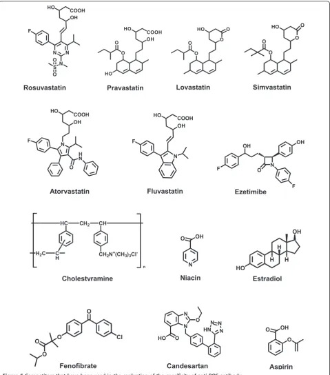
The specificity of the antibody was determined by carry-ing out the competitive assay uscarry-ing various competitors. These competitors were HMG-Co A reductase inhibi-tors (atorvastatin, pravastatin, lovastatin, and simvasta-tin), and various drugs that are administered in combined therapy with ROS. These drugs were ezeti-mibe [15], cholestyramine [16], niacin [17], estradiol [18], fenofibrate [19], candesartan [20], and aspirin [21]; the chemical structures of these competitors are given in Figure 5. The cross reactivity (CR) was calculated using the formula: IC50(ROS)/IC50 (competitor) × 100,
defining IC50as the concentration of the competitor
that causes 50% reduction of the maximum binding of anti-ROS antibody to the coating conjugates (i.e. 50% of the maximum color signal). The values of IC50that have
been obtained for the competitors, when possible, and their corresponding cross reactivities are given in Table 1. From these data, it was revealed that pravastatin, lovastatin and simvastatin, that share the 1,2,7,8,8a-hex-ahydronaphthyl moiety as a common backbone, did not cross react with the anti-ROS antibody. On the other hand, atorvastatin and fluvastatin exhibited small CR (4 and 2%, respectively) with ROS on the anti-ROS
antibody. This feature indicated that thep-fluorophenyl group (common moiety in all the drugs) is a recogniz-able epitope for the antibody. This assumption was also supported by the slight CR (0.8%) that has been exhib-ited by ezetimibe that has the same moiety in its struc-ture. The CR that has been exhibited by the HMG-CoA reductase inhibitors is not problematic in the analysis of ROS as these drugs are not co-administered simulta-neously. None of the other competitors (the drugs that are used in combination therapy with ROS; Figure 5) showed any CR with ROS in the proposed assay proving the high specificity of the antibody for ROS, and its pro-posed ELISA for ROS determination (Table 1).
Competitive ELISA for quantitation of ROS
The calibration curve of ROS using the proposed ELISA is shown in Figure 3B. This curve was generated using ROS at concentrations from 10 to 10000 pg/ml, pre-pared in PBS. The data showed good correlation coeffi-cient (r = 0.996) on the four-parameter curve fit. The limit of detection (LOD) of the proposed ELISA, defined as the lowest ROS concentration significantly different from zero concentration at 95% confidence limit (mean of zero ± 4.65 SD) was determined [22]. Based on the basis of 8 replicate measurements, the limit of detection in tenfold diluted plasma samples was found to be 40 pg/ml and the working range was 50-1000 pg/ml. This high sensitivity enables the determination of low thera-peutic concentrations of ROS in plasma.
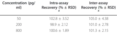
The intra-assay precision was assessed at three differ-ent concdiffer-entrations (low, middle, and high; Table 2) by analyzing 8 replicates of each sample in a single run and the inter-assay precision was assessed by analyzing the same sample, as duplicates, in 4 separate runs. Accord-ing to the recommendation of immunoassay validation [23], the assay gave satisfactory results as the values of relative standard deviations did not exceed 5% (Table 2). Studying the plasma matrix effect was required since the proposed assay was designed for quantitation of ROS in plasma. ROS-free plasma sample was serially diluted into PBS and each dilution was spiked with 200 pg/ml of ROS standard. The spiked samples were then analyzed by the proposed assay to investigate the feasi-bility of the assay. Acceptable recovery values [23] were obtained when plasma samples were diluted 10-fold with PBS. Therefore, plasma samples should be 10-fold diluted with PBS in order to avoid the possible false-positive analytical results. It is worth to mention that the high sensitivity of the assay (LOD was 40 pg/ml) allowed the high dilution of a clinical specimen (19 ng/ ml after 40 mg dosing) to attain the ROS concentrations in the working range of the assay.
The accuracy of the method was checked by recovery studies. ROS-free plasma samples (50 μl) were spiked
with 50 μl of varying concentrations of ROS, and the spiked samples were mixed with 400 μl of PBS to give final concentrations of 50-800 pg/ml. The samples were subjected to the analysis by the competitive ELISA, and the recovery values were calculated. The analytical recovery values were 98.9-105.0 ± 1.89-4.38% (Table 2).
This indicated the accuracy of the proposed method for determination of ROS in plasma samples, and absence of endogenous interfering substances in the plasma samples.
In order to compare the proposed ELISA with HPLC, plasma samples were spiked with ROS at known
O O HO
O O
Simvastatin
COOH HO
O O
HO
OH
Pravastatin
HO
O
O O
O
Lovastatin Rosuvastatin
HO
OH
N N
N S F
O
O
COOH
HC CH2 CH
CH2N+(CH3)3Cl -C
H H2C
n
Cholestyramine
H
H H HO
OH
Estradiol N
O OH
Niacin
N
OH
F O
OH
F
Ezetimibe Fluvastatin
COOH HO
OH
N F
Atorvastatin
N
COOH HO
OH
F
O H N
O
Cl O
O
O
Fenofibrate
O O OH
Aspirin
N N N HN N
N O
HO O
Candesartan
concentrations and analyzed by a reported HPLC method. These concentrations were 0.001-0.05 μg/ml. These concentrations were selected based on the sensi-tivity of the HPLC method. As the proposed ELISA has higher sensitivity, the same samples were diluted with PBS to make their concentrations within the working range of the proposed ELISA method. The concentra-tions measured by HPLC were plotted versus their cor-responding values that have been determined by the proposed ELISA. Regression analysis of the results was performed, and the results revealed the good agreement between the two methods: EIA = 0.00198 + 0.8768 HPLC (r = 0.988).
Conclusions
The present study described the preparation of a highly specific polyclonal antibody against ROS. The antibody recognizes ROS with high avidity. The high specificity and avidity of the produced antibody enabled the devel-opment of highly specific and sensitive ELISA for the accurate determination of ROS in plasma without pre-treatment at concentrations as low as 40 pg/ml. The assay produces a colored read-out, thus only a colori-metric plate reader is required. The entire protocol of the present assay is very easy to perform in a 96-well plate and permits an operator to analyze a batch of 200
samples per day when uses a pre-coated plates facilitat-ing the processfacilitat-ing of large number of samples. The pro-posed ELISA is expected to contribute to the pharmacokinetic studies of ROS as an effective alterna-tive to the existing cosalterna-tive and instrument-intensive chromatographic technologies.
Abbreviations
HMG-CoA: 3-hydroxy-3-methylglutaryl-coenzyme A; ROS: rosuvastatin; ELISA: enzyme-linked immunosorbent assay; KLH: keyhole limpt hemocyanin; BSA: bovine serum albumin; TMB: 3,3’,5,5’-tetramethylbenzidine; HRP-IgG: horseradish peroxidase-labeled second anti-rabbit IgG antibody; EDC: 1-ethyl-3-(3-dimethylaminopropyl) carbodiimide hydrochloride; PB: phosphate buffer; PBS: phosphate buffered saline; PBS-T: phosphate buffered saline containing 0.05% Tween-20; IC50: the concentration of the drug that causes
50% inhibition from the maximum binding of the antibody to the immobilized ROS-BSA conjugate; CR: cross reactivity; LOD: limit of detection; SD: standard deviation; pg: picogram.
Acknowledgements
The authors extend their appreciation to the Deanship of Scientific Research at King Saud University for funding the work through the research group No. RGP-VPP-065.
Authors’contributions
ID designed the study and supervised HA in conducting all the experiments, discussed the results and prepared the manuscript. AA participated in preparation of protein conjugates and discussion of the results. HA prepared the antibody and conducted the assay development.
Competing interests
The authors declare that they have no competing interests.
Received: 22 May 2011 Accepted: 5 July 2011 Published: 5 July 2011 References
1. Antonopoulos S, Mikros S, Mylonopoulou M, Kokkoris S, Giannoulis G:
Rosuvastatin as a novel treatment of non-alcoholic fatty liver disease in hyperlipidemic patients.Atherosclerosis2006,184:233-234.
2. Cooper KJ, Martin PD, Dane AL, Warwick MJ, Raza A, Schneck DW:Lack of effect of ketoconazole on the pharmacokinetics of rosuvastatin in healthy subjects.Br J Clin Pharmacol2003,55:94-99.
3. Lan K, Jiang X, Li Y, Wang L, Zhou J, Jiang Q, Ye L:Quantitative determination of rosuvastatin in human plasma by ion pair liquid-liquid extraction using liquid chromatography with electrospray ionization tandem mass spectrometry.J Pharm Biomed Anal2007,44:540-546. 4. Dong-Hang X, Zou-Rong R, Quan Z, Hong Y, Bo J:Quantitative
determination of rosuvastatin in human plasma by liquid chromatography with electrospray ionization tandem mass spectrometry.Rapid Commun Mass Spectrom2006,20:2369-2375. 5. Gao J, Zhong D, Duan X, Chen XJ:Liquid chromatography/negative ion
electrospray tandem mass spectrometry method for the quantification of rosuvastatin in human plasma: Application to a pharmacokinetic study.Chromatogr B Biomed Sci Appl2007,856:35-40.
6. Vittal S, Shitut NR, Kumar TR, Vinu MCA, Mullangi R, Srinivas NR:
Simultaneous quantitation of rosuvastatin and gemfibrozil in human plasma by high-performance liquid chromatography and its application to a pharmacokinetic study.Biomed Chromatogr2006,20:1252-1259. 7. Kumar TR, Shitut NR, Kumar PK, Vinu MCA, Kumar VVP, Mullangi R,
Srinivas NR:Determination of rosuvastatin in rat plasma by HPLC: validation and its application to pharmacokinetic studies.Biomed Chromatogr2006,20:881-887.
8. Pasha MK, Muzeeb S, Basha SJS, Shashikumar D, Mullangi R, Srinivas NR:
Analysis of five HMG-CoA reductase inhibitors: atorvastatin, lovastatin, pravastatin, rosuvastatin and simvastatin: pharmacological,
pharmacokinetic and analytical overview and development of a new method for use in pharmaceutical formulations analysis and in vitro metabolism studies.Biomed Chromatogr2006,20:282-293.
Table 1 Specificity of anti-ROS antibody
Competitor IC50(ng/ml) Cross reactivity (%)
Rosuvastatin 0.4 100
Pravastatin > 1000 Not determined
Lovastatin > 1000 Not determined
Simvastatin > 1000 Not determined
Atorvastatin 10 4
Fluvastatin 20 2
Ezetimibe 50 0.8
Cholestyramine > 1000 Not determined
Niacin > 1000 Not determined
Estradiol > 1000 Not determined
Fenofibrate > 1000 Not determined Candesartan > 1000 Not determined
Aspirin > 1000 Not determined
Table 2 Precisions and recovery studies of the proposed ELISA for determination of ROS at three different concentration levels
Concentration (pg/ ml)
Intra-assay Recovery (% ± RSD)
a
Inter-assay Recovery (% ± RSD)
a
50 102.8 ± 3.52 105.0 ± 4.38
200 98.9 ± 2.12 101.0 ± 2.78
800 100.6 ± 1.89 101.3 ± 2.15
a
9. Nirogi R, Mudigonda K, Kandikere V:Chromatography-mass spectrometry methods for the quantitation of statins in biological samples.J Pharm Biomed Anal2007,44:379-387.
10. Darwish IA:Immunoassay methods and their applications in pharmaceutical analysis: basic methodology and recent advances.Int J Biomed Sci2006,2:217-235.
11. Darwish IA, Al-Obaid AM, Al-Malaq HA:New highly sensitive enzyme immunoassay for the determination of pravastatin in human plasma.
Talanta2009,79:1478-1483.
12. Habeeb AF:Determination of free amino groups in proteins by trinitrobenzenesulfonic acid.Anal Biochem1966,14:328-336. 13. Weeks I:Chemiluminescence Immunoassay.InComprehensive Analytical
Chemistry. Volume XXIX.Edited by: Sevehla G. Elsevier, Amsterdam; 1992. 14. Mastronicolis S, Kapoulas V, Kröger H:The isolation of specific antibodies
for 5-methylcytidine–bovine serum albumin.Z Natureforsch1981,
36:459-463.
15. Ballantyne CM, Weiss R, Moccetti T, Vogt A, Eber B, Sosef F, Duffield E:
Efficacy and safety of rosuvastatin 40 mg alone or in combination with ezetimibe in patients at high risk of cardiovascular disease (results from the EXPLORER study).Am J Cardiol2007,99:673-680.
16. Christie BM, Elinor M, Rohini C:Efficacy and safety of rosuvastatin alone and in combination with cholestyramine in patients with severe hypercholesterolemia: A randomized, open-label, multicenter trial.Clin Ther2004,26:1855-1864.
17. David CM, John MM, Robert WJ, Rohini CR, Howard HG, Michael CD:
Beneficial effects ofrosuvastatinalone and in combination with extended-releaseniacinin patients with a combined hyperlipidemia and low high-density lipoprotein cholesterol levels.Am J Cardiol2003,
91:1304-1310.
18. Mueck AO, Seeger H, Deuringer FU, Wallwiener D:Effect of an estrogen/ statin combination on biochemical markers of endothelial function in human coronary artery cell cultures.Menopause2001,8:216-221. 19. Jones PH, Davidson MH, Kashyap MI, Kelly MT, Buttler SM, Setze CM,
Sleep DJ, Stolzenbach JC:Efficacy and safety of ABT-335 (fenofibric acid) in combination with rosuvastatin in patients with mixed dyslipidemia: A phase 3 study.Atherosclerosis2009,204:208-215.
20. Calkin AC, Giunti S, Sheehy KJ, Chew C, Boolell V, Rajaram YS, Cooper ME, Jandeleit-Dahm KA:The HMG-CoA reductase inhibitor rosuvastatin and the angiotensin receptor antagonist candesartan attenuate
atherosclerosis in an apolipoprotein E-deficient mouse model of diabetes via effects on advanced glycation, oxidative stress and inflammation.Diabetologia2008,51:1731-1740.
21. Chapman MJ:From pathophysiology to targeted therapy for atherothrombosis: A role for the combination of statin and aspirin in secondary prevention.Pharmacol Ther2007,113:184-196.
22. Anderson DJ:Determination of the lower limit of detection.Clin Chem 1989,35:2152-2153.
23. Findlay JWA, Smith WC, Lee JW, Nordblom GD, Das I, DeSilva BS, Khan MN, Bowsher RR:Validation of immunoassays for bioanalysis: a
pharmaceutical industry perspective.J Pharm Biomed Anal2000,
21:1249-1273.
doi:10.1186/1752-153X-5-38
Cite this article as:Darwishet al.:Generation of polyclonal antibody
with high avidity to rosuvastatin and its use in development of highly sensitive ELISA for determination of rosuvastatin in plasma.Chemistry Central Journal20115:38.
Open access provides opportunities to our colleagues in other parts of the globe, by allowing
anyone to view the content free of charge.
Publish with
Chemistry
Central and every
scientist can read your work free of charge
W. Jeffery Hurst, The Hershey Company.
available free of charge to the entire scientific community peer reviewed and published immediately upon acceptance cited in PubMed and archived on PubMed Central yours you keep the copyright
Submit your manuscript here: