R E S E A R C H
Open Access
Somatostatin receptor 2A protein
expression characterizes anaplastic
oligodendrogliomas with favorable
outcome
Romain Appay
1,3, Emeline Tabouret
2,3, Mehdi Touat
4,5, Catherine Carpentier
5, Carole Colin
3, François Ducray
6,7,
Ahmed Idbaih
4,5, Karima Mokhtari
5,8, Emmanuelle Uro-Coste
9,10, Caroline Dehais
4,
Dominique Figarella-Branger
1,3*and the POLA network
Abstract
Diffuse gliomas are classified according to the 2016 WHO Classification of Tumors of the Central Nervous System, which now defines entities by both histology and molecular features. Somatostatin receptor subtype 2A (SSTR2A) expression has been reported in various solid tumors as associated with favorable outcomes. Its expression has been reported in gliomas with uncertain results regarding its prognostic value. The objective of this study was to assess the prognostic impact of SSTR2A protein expression in a large cohort of grade III and IV gliomas classified according to the updated 2016 WHO classification. We further validated our result with an independent cohort of low grade glioma using dataset generated by The Cancer Genome Atlas (TCGA) Research Network.
We analyzed clinical and molecular data from 575 patients. SSTR2A protein expression was evaluated using immunohistochemistry on tissue microarrays. High expression of SSTR2A protein associated with the anaplastic oligodendrogliomaIDH-mutant and 1p/19q-codeleted subgroup (p< 0.001). Among these tumors, SSTR2A protein expression was significantly associated with a lower proliferative index, the absence of microvascular proliferation and the absence of necrosis (p< 0.001). Furthermore SSTR2A protein expression associated with better overall survival (p= 0.007) and progression-free survival (p= 0.01) in both univariate and multivariate analysis when adjusted by the age, the presence of necrosis and the mitotic index. Similar results were obtained regarding
SSTR2 mRNA expression in the TCGA low grade glioma, subtypeIDH-mutant and 1p/19q-codeleted, dataset.
SSTR2A might represent an attractive biomarker and therapeutic target in anaplastic oligodendrogliomaIDH-mutant and 1p/19q-codeleted specific subgroup. Understanding the implicated molecular pathways may represent a step forward to improve therapeutic approaches.
Keywords:Somatostatin receptor subtype 2A (SSTR2A), Glioma, Biomarker, Therapeutic target
Introduction
Diffuse gliomas are the most common primary brain cancers. They are classified according to the 2016 WHO (World Health Organization) Classification of Tumors of the Central Nervous System (CNS), which combines for the first time histological and molecular
features in an “integrated diagnosis” [20]. This novel classification system incorporates mutations in the isocitrate deshydrogenase 1 and 2 genes (IDH1 and
IDH2) and the whole-arm chromosomal loss of 1p and 19q (1p/19q-codeletion), which are both required to be present for confirming the diagnosis of oligo-dendroglioma. IDH mutations are the key genetic alterations characterizing grade II and III gliomas and glioblastomas with favorable outcome [37]. Diagnostic strategy and therapeutic management depend on each subtype and the identification of distinct prognostic * Correspondence:dominique.figarella-branger@univ-amu.fr
1APHM, Hôpital de la Timone, Service d’Anatomie Pathologique et de
Neuropathologie, Marseille, France
3Aix-Marseille Univ, CNRS, INP, Inst Neurophysiopathol, Marseille, France
Full list of author information is available at the end of the article
subgroups among gliomas belonging to the same histo-molecular category is crucial to open perspec-tives of therapeutic development.
Somatostatin (SST), also known as growth hormone-inhibiting hormone (GHIH), was first described in 1968 as a hormone secretion [18]. The effects of SST are mediated through its interaction with somatostatin receptors (SSTR), a family of G protein-coupled receptors consisting of 6 different subtypes (SSTR1, 2A, 2B, 3, 4 and 5) [26, 32].
SSTR2A is the predominant subtype. Its expression has been reported in various solid tumors as associated with favorable outcomes [1, 19, 23, 25, 28]. SSTRs are commonly expressed on neuroendocrine tumors (NETs). In NETs, the expression of SSTR2A by tumor cells is of interest for both diagnostic and therapeutic strategy. Indeed, SSTR2A is a target for radiolabeled imaging (OCTREOSCAN, PET 68Ga-DOTATOC) as well as ther-apy using SST analogs labelled with β-emitting isotopes (90Y-DOTATOC and 177Lu-DOTATATE) [2, 5, 29]. In addition, SST analogs (Octreotide and Lanreotide) are used to inhibit the release of hormones and control secretory symptoms [1, 13, 14, 16, 26]. Interestingly, recent studies demonstrated that SST analogs can also inhibit growth of SSTRs-dependent tumors by regulating intracellular signaling pathways, including dephosphoryla-tion of actors implicated in the mitogen-activated protein kinase (MAPK) signaling and induction of apoptosis [13, 26, 32].
Few studies have previously reported the expression of SSTR2A in gliomas with discrepant results regard-ing their association with grade [11, 17, 21, 26]. In a recent study, Kiviniemi et al. [17] reported high expression of SSTR2A protein predominant in oligo-dendrogliomas in a cohort of 184 gliomas classified according to the specific molecular signatures of the updated WHO classification. Furthermore, they reported a survival benefit in gliomas with high ex-pression of SSTR2A protein. However, this difference might be related to the association between SSTR2A and the oligodendroglioma subtype and it is not clear whether the level of SSTR2A expression has prognostic significance among the oligodendroglioma subgroup.
In France, since 2008, the POLA network provides a centralized review and molecular analysis of de novo adult high-grade glioma with an oligodendroglial component. Using the tissue samples and dataset provided by this network, our objective was to assess the prognostic impact of the SSTR2A protein expres-sion in a large cohort of grade III and IV gliomas. We further validated our result with an independent cohort using dataset generated by the TCGA Research Network [8].
Materials and methods
Study population
A total number of 575 patients from the French nation-wide POLA cohort were included in this study. Inclusion criteria were the written consent of the pa-tient for clinical data collection and genetic analysis according to national and POLA network policies, sufficient tissue material for molecular studies allow-ing classification accordallow-ing to the WHO 2016 (i.e. evaluation of the IDH mutation and 1p/19q-codele-tion status) and an established diagnosis of high grade glioma (WHO grade III or IV).
IDH mutation status was evaluated using automated immunohistochemistry (IHC) and direct sequencing using the Sanger method as previously described [30]. The genomic profile and assessment of the 1p/19q-code-letion status was determined based on single nucleotide polymorphism (SNP) arrays, comparative genomic hybridization (CGH) arrays, or microsatellite marker analysis as previously described [30].
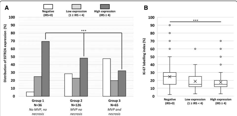
Anaplastic oligodendroglioma, IDH-mutant, 1p19q-codeleted were classified into 3 pathological subgroups based on mitotic index, microvascular proliferation (MVP), and necrosis [12]. Group 1, involved cases with more than 5 mitoses per 10-high power field (HPF), no MVP, and no necrosis, group 2 displayed MVP but no necrosis, and group 3 showed MVP and necrosis.
Proliferative index was evaluated using Ki67 antibody (clone Mib1; 1:100; Dako) and scored as percentage by counting the immunostained nuclei of 400 cells in the most positive area.
Clinical data collected included gender, age at surgery, extend of surgical resection, preoperative Karnofsky performance scale (KPS) and post-operative treatment. Clinical information regarding the outcome was recorded as follow: progression-free survival (PFS) was defined as the time from the date of surgery to recur-rence or death from any cause, censored at the date of the last documented disease evaluation. Overall survival (OS) was defined as the time from the date of surgery to death from any cause, censored at the date of last contact.
Tissue microarray (TMA) design
SSTR2A immunohistochemistry and evaluation on TMA
Immunohistochemical detection of SSTR2A protein ex-pression was performed on 5 μm thick TMA sections with Ventana Benchmark XT. The monoclonal SSTR2A antibody (clone UMB1) was purchased from Abcam and used at 1/4000 dilution. A Benchmark Ventana autostai-ner (Ventana Medical Systems SA, Illkirch, France) was used for detection and TMA slides were simultaneously immunostained to avoid inter-manipulation variability.
Immunostaining was scored for 185 triplicates by two independent pathologists (RA & DFB). In almost all cases the staining intensity was roughly similar among the different cores of the triplicate, thus the core demon-strating the stronger immunoreaction was analyzed to establish an immunoreactive score (IRS) as previously described by Casar-Borota et al. [9]. Staining intensity was scored as 0 (no immunostaining), 1 (weak), 2 (mod-erate), or 3 (strong). The percentage of immunoreactive cells was scored as 0 (none), 1 (< 10%), 2 (10–50%), 3 (51–80%), or 4 (> 80%). Multiplication of the staining intensity score and the percent immunoreactivity score resulted in an IRS score, which ranged from 0 to 12. Because the results were highly concordant among the two pathologists, the 390 remaining triplicates were scored by a single pathologist (RA).
The median IRS score based on analysis of all cases in the cohort was equal to 1 and corresponded to a cutoff between positive (IRS≥1) and negative expres-sion (IRS = 0). The third quartile (IRS = 4) was used as the cutoff to define positive cases with high expression (IRS≥4) versus positive cases with low expression (1≤IRS < 4). Representative images of different IRS are shown in Fig. 1.
Public data acquisition
Informed consent and ethical approval was obtained when The National Cancer Institute (NCI) and National Human Genome Research Institute (NHGRI) had col-lected tissue for TCGA [8]. All sequencing or clinical
datasets used in the present study are publicly available to researchers without restrictions or limitations accord-ing to TCGA policy. Therefore, the results shown here are in whole based upon data generated by the TCGA Research Network: http://cancergenome.nih.gov/. We used GlioVis data portal for visualization and analysis of brain tumor expression datasets [6] ( http://gliovis.-bioinfo.cnio.es/), we limited our analysis to data from adult patients with LGG.
Statistical analysis
For statistical analysis, the Chi-square test was used to compare qualitative variables when they were scored as positive or negative. Continuous variables were com-pared using the Mann–Whitney U test. The Kaplan– Meier method was used to estimate survival distribu-tions. Log-rank tests were used for univariate compari-sons. Age at diagnosis, KPS, presence of necrosis and proliferative index were used to build the multivariate Cox proportional hazard backward models. All statistical tests were two-sided, and the threshold for statistical significance was p= 0.05. Statistical analysis was done using IBM SPSS statistics software version 23 (IBM SPSS Inc., Chicago, IL, USA).
Results
Clinicopathologic characteristics
We evaluated 227 anaplastic oligodendroglioma IDH --mutant and 1p/19q-codeleted. In addition, we ana-lyzed 86 anaplastic astrocytoma (80 IDH-mutant and 6 IDH-wildtype) and 262 glioblastomas (124 IDH -mu-tant and 138 IDH-wildtype). Overall 575 patients were included in this study. Patient characteristics are pre-sented in Table 1.
Scoring of SSTR2A immunohistochemistry and its association with tumor entity
Expression (any level; IRS≥1) of SSTR2A was detected in 59% (337/575) of gliomas. The distribution of
Fig. 1Immunohistochemical staining of SSTR2A protein in anaplastic oligodendrogliomaIDH-mutant and 1p/19q-codeleted(a)Negative staining (IRS = 0)(b)Weak staining intensity (score = 1) in 20% of the cells (score = 2) corresponding to positive cases with low expression (IRS = 3)(c)
SSTR2A protein expression according to gliomas sub-type is shown in Fig.2. SSTR2A protein expression was significantly associated with IDH mutation (66% of
IDH-mutant tumors were positive for SSTR2A expres-sion versus 39% of IDH-wild type, p< 0.001). High expression of SSTR2A (IRS score≥4) was detected in
31% (180/575) of gliomas. High expression of
SSTR2A was significantly associated with anaplastic oligodendroglioma, IDH-mutant and 1p/19q-codeleted and was found in approximatively half of the studied samples whereas it was uncommon in astrocytoma and glioblastoma independently of the presence ofIDH -muta-tion (p< 0.001).
Association between expression of SSTR2A protein and the anaplastic oligodendrogliomaIDH-mutant and 1p/19q-codeleted pathological subgroups
In the anaplastic oligodendroglioma subgroup, expres-sion levels of SSTR2A protein varied depending on the pathological subgroup as shown in Fig. 3a (p< 0.001). High SSTR2A expression was detected in the majority
(70%) of anaplastic oligodendroglioma belonging to the pathological group 1. It was less frequent in group 2 (50%) and only one-third of the anaplastic oligodendroglioma of group 3 presented with high SSTR2A expression. In accordance with the previous result, the absence of SSTR2A expression was uncom-mon auncom-mong the patients of the group 1 (5%) whereas approximately half of the group 3 presented with no SSTR2A expression.
We selected six representative cases (i.e. two negative for SSTR2A expression, IRS = 0; two positive with low expression, IRS = 3; and two positive with high ex-pression, IRS = 9) from anaplastic oligodendroglioma,
IDH-mutant and 1p/19q-codeleted belonging to the group 3 and evaluated intratumoral heterogeneity of SSTR2A expression on whole tumor sample. For each selected case, the percentage and intensity of the immunostaining was in agreement with the result obtained on TMA. One of the two cases evaluated with an IRS score equal to 0 showed focal positivity away from the area selected for the TMA (this area
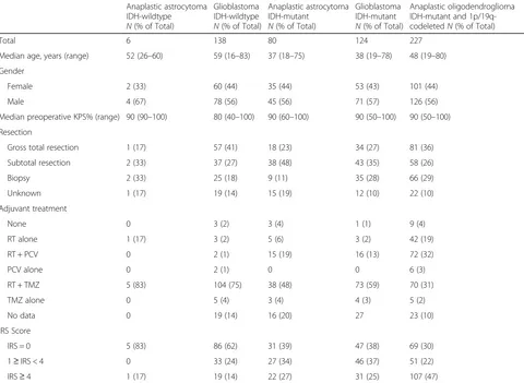
Table 1Characteristics of the patient population (N= 583)
Anaplastic astrocytoma IDH-wildtype
N(% of Total)
Glioblastoma IDH-wildtype
N(% of Total)
Anaplastic astrocytoma IDH-mutant
N(% of Total)
Glioblastoma IDH-mutant
N(% of Total)
Anaplastic oligodendroglioma IDH-mutant and 1p/19q-codeletedN(% of Total)
Total 6 138 80 124 227
Median age, years (range) 52 (26–60) 59 (16–83) 37 (18–75) 38 (19–78) 48 (19–80)
Gender
Female 2 (33) 60 (44) 35 (44) 53 (43) 101 (44)
Male 4 (67) 78 (56) 45 (56) 71 (57) 126 (56)
Median preoperative KPS% (range) 90 (90–100) 80 (40–100) 90 (60–100) 90 (50–100) 90 (50–100)
Resection
Gross total resection 1 (17) 57 (41) 18 (23) 34 (27) 81 (36)
Subtotal resection 2 (33) 37 (27) 38 (48) 43 (35) 58 (26)
Biopsy 2 (33) 25 (18) 9 (11) 35 (28) 66 (29)
Unknown 1 (17) 19 (14) 15 (19) 12 (10) 22 (10)
Adjuvant treatment
None 0 3 (2) 3 (4) 1 (1) 9 (4)
RT alone 1 (17) 3 (2) 5 (6) 3 (2) 42 (19)
RT + PCV 0 2 (1) 15 (19) 16 (13) 72 (32)
PCV alone 0 2 (1) 0 0 6 (3)
RT + TMZ 5 (83) 104 (75) 38 (48) 73 (59) 70 (31)
TMZ alone 0 5 (4) 3 (4) 4 (3) 5 (2)
No data 0 19 (14) 16 (20) 27 23 (10)
IRS Score
IRS = 0 5 (83) 86 (62) 31 (39) 47 (38) 69 (30)
1≥IRS < 4 0 33 (24) 27 (34) 46 (37) 51 (22)
IRS≥4 1 (17) 19 (14) 22 (27) 31 (25) 107 (47)
corresponded to a much less aggressive part of the tumor) but the vast majority of the sample was nega-tive, the second one was completely negative. In the other four cases, the SSTR2A protein expression was in keeping with results evaluated on TMA and always absent in tumor cells located in the vicinity of foci of
palisading necrosis (data not shown). Therefore, the low SSTR2A expression in group 3 is not related to necrosis per se but likely represent the acquisition of a more aggressive phenotype. It should be noted that SSTR2A protein expression was also observed in neu-rons within the peripheral cortex.
Fig. 2Distribution of SSTR2A protein expression according to tumor subtypeAbbreviations: AIII IDHwt, Anaplastic astrocytomaIDH-wildtype; GB IDHwt, GlioblastomaIDH-wildtype; AIII IDHmut, Anaplastic astrocytomaIDH-mutant; GB IDHmut, GlioblastomaIDH-mutant; OIII, Anaplastic oligodendroglioma,IDH-mutant and 1p/19q-codeleted. *** =p< 0.001
Association between expression of SSTR2A protein and proliferative index in anaplastic oligodendroglioma, IDH-mutant and 1p/19q-codeleted
To examine whether SSTR2A protein expression was associated with differences in the proliferative index we compared quantitative evaluation of Ki-67 (MIB-1) la-belling index using immunohistochemistry. As presented in Fig. 3b, anaplastic oligodendroglioma, IDH-mutant and 1p/19q-codeleted with high or low SSTR2A protein expression presented with a lower Ki-67 labelling index when compared to SSTR2A negative gliomas (median Ki-67 expression = 15% in SSTR2A positive gliomas versus 26% in SSTR2A negative,p< 0.001).
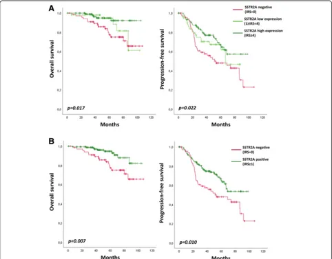
Association between SSTR2A protein expression and survival in anaplastic oligodendroglioma,IDH-mutant and 1p/19q-codeleted
We further analyzed the prognostic significance of SSTR2A expression in gliomas. Among the anaplastic oligodendroglioma, IDH-mutant and 1p/19q-codeleted, SSTR2A protein expression is prognostic for PFS and OS (Fig. 4a). Both low and high SSTR2A expressive anaplastic oligodendroglioma presented with better OS (p= 0.022) and PFS (p= 0.017) when compared to nega-tive gliomas. No significant prognostic difference was observed between low expression and high expression in terms of PFS (p= 0.293) and OS (p= 0.280).
Accordingly, expression of SSTR2A protein (any level, IRS≥1) was significantly associated with longer PFS (p= 0.010) and OS (p= 0.007) among the sub-group of anaplastic oligodendroglioma, IDH-mutant and 1p/19q-codeleted (Fig. 4b).
In multivariate analysis, expression of SSTR2A (any level, IRS≥1) was also significantly associated with better OS when adjusted by the age (HR = 0.414; 95% CI, 0.185–0.929; p= 0.033), by the presence of necrosis (HR = 0.391; 95% CI, 0.174–0,877; p= 0.023) or by the proliferative index (HR = 0.411; 95% CI, 0.176–0.959;
p= 0.04). When adjusted by the preoperative KPS, the result did not reach statistical significance (HR = 0.413; 95% CI, 0.165–1.034; p= 0.059), which might be attributed to an insufficient number of collected data. The extend of surgical resection and postopera-tive treatment did not reached a p-value > 0.2 in univariate analysis thus have not been integrated in the multivariate analysis.
No association between survival and SSTR2A protein expression was observed in patients with other gliomas subtypes (data not shown).
TCGA low-grade glioma RNA-seq data as confirmation dataset
As presented in Fig.5a, among the independent TCGA low-grade glioma dataset we observed a higher SSTR2
mRNA expression in IDH-mutant glioma when
compared to IDH-wild type (p< 0.001). Furthermore,
IDH-mutant and 1p/19q-codeleted gliomas presented with the highest level of SSTR2 mRNA expression (p< 0.001).
When categorized into two groups according to the median score, we observed a better overall survival in glioma with high SSTR2 mRNA expression (p= 0.056) among the low grade glioma IDH-mutant and 1p/ 19q-codeleted subgroup (Fig.5b).
No association between survival and SSTR2 mRNA expression was observed in patient with IDH-mutant without 1p/19q-codeletion (p= 0.478) and IDH-wildtype gliomas (p= 0.301) (data not shown).
Discussion
Gliomas are the most common primary CNS tumors. Updated WHO classification of CNS tumors combines for the first time histological and molecular features for an integrated diagnosis. IDH-mutant gliomas display a more favorable outcome than theIDH-wildtype counter-part. However, despite aggressive treatment,IDH-mutant gliomas are characterized by a malignant transformation over time with a median survival of approximately 10 years. Therefore, the identification of distinct prognostic groups amongIDH-mutant gliomas might be of interest to better stratified the patients and improve therapeutic approaches.
In the present study, we have evaluated the protein ex-pression of SSTR2A by immunohistochemistry in a large cohort of gliomas classified according to the WHO 2016 classification. Because of the inclusion criteria defined by the POLA network (i.e. high-grade glioma with oligodendroglial component) it is worth noticing that the percentage of each category in our study does not reflect the normal distribution of gliomas.
obtained regarding SSTR2 mRNA expression in an inde-pendent cohort using the low grade gliomas TCGA dataset.
Thus, our results indicate that the immunohistochemi-cal expression of SSTR2A protein could serve as a prog-nostic biomarker among anaplastic oligodendroglioma,
IDH-mutant and 1p/19q-codeleted.
SSTR2A is strongly expressed in neuroendocrine tumors and also in normal neurons according to the brain transcriptome database [7]. The high expression of SSTR2A in IDH-mutant adult high grade gliomas in comparison to IDH-wildtype gliomas is in accordance with the proneural subtype of IDH-mutant gliomas reported by the Cancer genome Atlas [33]. Importantly, among this group, the highest SSTR2A expression is recorded in anaplastic oligodendrogliomas IDH-mutant and 1p/19q-codeleted. This is in keeping with the
neuronal differentiation of these tumors highlighted by ultrastructural and transcriptional studies [4, 10, 35]. Indeed, Ducray et al. [10] demonstrated that there is a strong correlation between 1p/19q-codeletion and the expression of proneural genes in malignant gliomas. Interestingly, in their cohort, SSTR2 was significantly overexpressed (p= 0.0001) in the 1p/19q-codeleted group when compared to the EGFR amplified high grade gliomas. Furthermore, Bielle et al. [4] reported the oc-currence of neuronal intermediate progenitors (NIP) markers in a subset of anaplastic oligodendroglioma
IDH-mutant and 1p/19q-codeleted especially in cases associated with necrosis (p= 0.0034). It is possible that NIP-high subgroup could result from tumor dedifferenti-ation. In the same line, we can postulate that loss of SSTR2A expression among anaplastic oligodendrogliomas
IDH-mutant and 1p/19q-codeleted is correlated to the
Fig. 4Overall survival and Progression-free survival according to SSTR2A protein expression in anaplastic oligodendroglioma,IDH-mutant and 1p/19q-codeleted.aNo SSTR2A expression (IRS = 0) versus low SSTR2A expression (1≤IRS < 4) versus high SSTR2A expression (IRS≥4).
acquisition of a more immature neuronal phenotype. We could hypothesize that SSTR2A, through its inhibition of the MAPK-pathway and induction of apoptosis, may constitute a brake against in the acquisition of markers of aggressiveness.
Surgery, when possible, is the first line treatment for
IDH-mutant and 1p/19q-codeleted oligodendroglioma and “watch-and-wait” strategies may be justified in patients with macroscopically complete resection or in young patients (aged < 40 years) with incomplete resec-tion if the tumor is not symptomatic [36]. If further treatment beyond surgery is considered necessary the standard treatment is radiotherapy followed by procar-bazine, lomustine and vincristine chemotherapy (PCV).
“Watch-and-wait” strategies might be considered even with WHO grade III tumors [36]. According to our re-sults, SSTR2A protein expression is an independent bio-marker of good prognosis in both uni- and multivariate analysis that might guide the decision of a postsurgical treatment among the subgroup of anaplastic oligo-dendrogliomas,IDH-mutant and 1p/19q-codeleted.
Moreover, SSTR2A is an attractive therapeutic target. An inhibitory effect of SST analogs on cell migration and invasion has been demonstrated in various tumors [3,24,31,34]. Moreover, therapy using radiolabeled SST analogs is commonly used as a therapeutic option for the treatment of metastatic or inoperable neuroendo-crine tumors [5, 29]. Interestingly, encouraging results have been reported in some studies evaluating locoregio-nal delivery of targeted radionuclide therapy in gliomas [15,22,27]. Our results suggest the identification of the specific oligodendroglioma subgroup as a possible target of therapies using SST analogs.
Conclusion
Our results demonstrate that high expression of SSTR2A is commonly associated with anaplastic oligodendrogli-omas. Moreover, among the anaplastic oligodendroglioma subgroup SSTR2A expression is associated with a better OS. Therefore, SSTR2A might represent an attractive biomarker and target for these tumors.
Abbreviations
CNS:Central Nervous System;; HPF: High power field (HPF); IDH 1 & 2: Isocitrate deshydrogenase 1 and 2 genes; IRS: Immunoreactive score; KPS: Karnofsky performance status scale; MVP: Microvascular proliferation; NIP: Neuronal Intermediate Progenitor; OS: Overall survival; PCV: Procarbazine + Lomustine + Vincristine chemotherapy; PFS: Progression-free survival; RT: Radiotherapy; SST: Somatostatin; SSTR2A: Somatostatin receptor subtype 2A; TCGA: The Cancer Genome Atlas; TMA: Tissue microarray;
TMZ: Temozolomide
Acknowledgements
We thank the ARTC-Sud patients’s association (Association pour le Recherche sur les Tumeurs Cérébrales) and the Cancéropôle PACA.
POLA Network :
Amiens (C. Desenclos, H. Sevestre), Angers (P. Menei, A. Rousseau), Annecy (T. Cruel, S. Lopez), Besançon (M-I Mihai, A. Petit), Bicêtre (C. Adam, F. Parker), Brest (P. Dam-Hieu, I. Quintin-Roué), Bordeaux (S. Eimer, H. Loiseau), Caen (L. Bekaert, F. Chapon), Clamart (D. Ricard) Clermont-Ferrand (C. Godfraind, T. Khallil), Clichy (D. Cazals-Hatem, T. Faillot), Colmar (C. Gaultier, MC. Tortel), Cornebarrieu (I. Carpiuc, P. Richard), Créteil (W. Lahiani), Dijon (H. Aubriot-Lorton, F. Ghiringhelli), Lille (CA. Maurage, C. Ramirez), Limoges (EM. Gueye, F. Labrousse), Marseille (O Chinot), Montpellier (L. Bauchet, V. Rigau), Nancy (P. Beauchesne, G. Gauchotte), Nantes (M. Campone, D. Loussouarn), Nice (D. Fontaine, F. Vandenbos-Burel) , Nimes (A. Le Floch, P. Roger) Orléans (C. Blechet, M. Fesneau), Paris (A.Carpentier, JY Delattre (POLA Network National coordinator), S. Elouadhani-Hamdi, M. Polivka), Poitiers
(D. Larrieu-Ciron,S. Milin), Reims (P. Colin, MD. Diebold), Rennes (D. Chiforeanu, E. Vauleon), Rouen (O. Langlois, A. Laquerriere), Saint-Etienne (F. Forest, MJ. Motso-Fotso), Saint-Pierre de la Réunion (M. Andraud, G. Runavot), Strasbourg (B. Lhermitte, G. Noel), Suresnes (S. Gaillard, C. Villa), Toulon (N. Desse), Tours (C. Rousselot-Denis, I. Zemmoura), Toulouse (E. Cohen-Moyal, E. Uro-Coste), Villejuif (F. Dhermain)
Funding
This work was funded by the French Institut National du Cancer (INCa, POLA network) and is part of the national program Cartes d′Identité des Tumeurs (CIT) (http://cit.ligue-cancer.net/), which is funded and developed by the Ligue nationale contre le cancer.
Availability of data and materials
The datasets used and/or analysed during the current study available from the corresponding author on reasonable request.
Authors’contributions
RA wrote the manuscript, performed the SSTR2A immunohistochemical examination & all the statistical analysis. ET, MT, AI, CC, AI & DFB were major contributors in writing the manuscript. All authors (RA, ET, MT, CC, CC, FD, AI, KM, EUC, CD, DFB) contributed to the data collection. All authors read and approved the final manuscript.
Ethics approval and consent to participate
Patients included prospectively in the POLA network have provided their written consent for clinical data collection and genetic analysis according to national and POLA network policies.
Consent for publication
Not applicable.
Competing interests
The authors declare that they have no competing interests.
Publisher’s Note
Springer Nature remains neutral with regard to jurisdictional claims in published maps and institutional affiliations.
Author details
1
APHM, Hôpital de la Timone, Service d’Anatomie Pathologique et de Neuropathologie, Marseille, France.2APHM, Hôpital de la Timone, Service de
Neurooncologie, Marseille, France.3Aix-Marseille Univ, CNRS, INP, Inst Neurophysiopathol, Marseille, France.4AP-HP, Hôpitaux Universitaires La Pitié
Salpêtrière-Charles Foix, Service de Neurologie 2–Mazarin, Paris, France.
5Sorbonne Université, Inserm, CNRS, UMR S 1127, Institut du Cerveau et de la
Moelle épinière, ICM, APHP, Hôpitaux Universitaires La Pitié Salpêtrière -Charles Foix, Service de Neurologie 2-Mazarin, F-75013 Paris, France.
6
Hospices Civils de Lyon, Hôpital Pierre Wertheimer, Service de
Neuro-oncologie, Bron, France.7Department of Cancer Cell Plasticity, Cancer
Research Centre of Lyon, Inserm U1052, CNRS UMR5286, Lyon, France.
8AP-HP, Hôpitaux Universitaires La Pitié Salpêtrière-Charles Foix, Service de
Neuropathologie Raymond Escourolle, Paris, France.9CHU Toulouse, Hôpital Rangueil, Service d’Anatomie Pathologique et Histologie-Cytologie, Toulouse, France.10Inserm U1037, Centre de Recherche en Cancérologie de Toulouse, Université de Toulouse, Toulouse, France.
Received: 10 August 2018 Accepted: 2 September 2018
References
1. Abdel-Rahman O, Lamarca A, Valle JW, Hubner RA (2014) Somatostatin receptor expression in hepatocellular carcinoma: prognostic and therapeutic considerations. Endocr Relat Cancer 21:R485–R493
2. Ambrosini V, Fani M, Fanti S, Forrer F, Maecke HR (2011) Radiopeptide imaging and therapy in Europe. J Nucl Med 52(Suppl 2):42S–55S 3. Annunziata M, Luque RM, Durán-Prado M, Baragli A, Grande C, Volante M et
al (2012) Somatostatin and somatostatin analogues reduce PDGF-induced endometrial cell proliferation and motility. Hum Reprod 27:2117–2129 4. Bielle F, Ducray F, Mokhtari K, Dehais C, Adle-Biassette H, Carpentier C et al
(2017) Tumor cells with neuronal intermediate progenitor features define a subgroup of 1p/19q co-deleted anaplastic gliomas. Brain Pathol 27:567–579 5. Bodei L, Mueller-Brand J, Baum RP, Pavel ME, Hörsch D, O’Dorisio MS et al
(2013) The joint IAEA, EANM, and SNMMI practical guidance on peptide receptor radionuclide therapy (PRRNT) in neuroendocrine tumours. Eur J Nucl Med Mol Imaging 40:800–816
6. Bowman RL, Wang Q, Carro A, Verhaak RGW, Squatrito M (2017) GlioVis data portal for visualization and analysis of brain tumor expression datasets. Neuro-Oncology 19:139–141
7. Cahoy JD, Emery B, Kaushal A, Foo LC, Zamanian JL, Christopherson KS et al (2008) A transcriptome database for astrocytes, neurons, and
oligodendrocytes: a new resource for understanding brain development and function. J Neurosci 28:264–278
8. Cancer Genome Atlas Research Network, Brat DJ, RGW V, Aldape KD, WKA Y, Salama SR et al (2015) Comprehensive, Integrative Genomic Analysis of Diffuse Lower-Grade Gliomas. N Engl J Med 372:2481–2498
9. Casar-Borota O, Heck A, Schulz S, Nesland JM, Ramm-Pettersen J, Lekva T et al (2013) Expression of SSTR2a, but not of SSTRs 1, 3, or 5 in somatotroph adenomas assessed by monoclonal antibodies was reduced by octreotide and correlated with the acute and long-term effects of octreotide. J Clin Endocrinol Metab 98:E1730–E1739
10. Ducray F, Idbaih A, de Reyniès A, Bièche I, Thillet J, Mokhtari K et al (2008) Anaplastic oligodendrogliomas with 1p19q codeletion have a proneural gene expression profile. Mol Cancer 7:41
11. Dutour A, Kumar U, Panetta R, Ouafik L, Fina F, Sasi R et al (1998) Expression of somatostatin receptor subtypes in human brain tumors. Int J Cancer 76:620–627
12. Figarella-Branger D, Mokhtari K, Dehais C, Jouvet A, Uro-Coste E, Colin C et al (2014) Mitotic index, microvascular proliferation, and necrosis define 3 groups of 1p/19q codeleted anaplastic oligodendrogliomas associated with different genomic alterations. Neuro-Oncology 16:1244–1254
13. Florio T (2008) Molecular mechanisms of the antiproliferative activity of somatostatin receptors (SSTRs) in neuroendocrine tumors. Front Biosci 13:822–840
14. Gatto F, Hofland LJ (2011) The role of somatostatin and dopamine D2 receptors in endocrine tumors. Endocr Relat Cancer 18:R233–R251 15. Heute D, Kostron H, von Guggenberg E, Ingorokva S, Gabriel M,
Dobrozemsky G et al (2010) Response of recurrent high-grade glioma to treatment with 90Y-DOTATOC. J Nucl Med 51:397–400
16. Kim HS, Lee HS, Kim WH (2011) Clinical significance of protein expression of cyclooxygenase-2 and somatostatin receptors in gastroenteropancreatic neuroendocrine tumors. Cancer Res Treat 43:181–188
17. Kiviniemi A, Gardberg M, Kivinen K, Posti JP, Vuorinen V, Sipilä J et al (2017) Somatostatin receptor 2A in gliomas: association with oligodendrogliomas and favourable outcome. Oncotarget 8:49123
18. Krulich L, Dhariwal AP, McCann SM (1968) Stimulatory and inhibitory effects of purified hypothalamic extracts on growth hormone release from rat pituitary in vitro. Endocrinology 83:783–790
19. Liu Y, Jiang L, Mu Y (2013) Somatostatin receptor subtypes 2 and 5 are associated with better survival in operable hepatitis B-related hepatocellular carcinoma following octreotide long-acting release treatment.
Oncol Lett 6:821–828
20. Louis DN, Perry A, Reifenberger G, von Deimling A, Figarella-Branger D, Cavenee WK et al (2016) The 2016 World Health Organization classification of tumors of the central nervous system: a summary. Acta Neuropathol 131:803–820
21. Mawrin C, Schulz S, Pauli SU, Treuheit T, Diete S, Dietzmann K et al (2004) Differential expression of sst1, sst2A, and sst3 somatostatin receptor proteins in low-grade and high-grade astrocytomas. J Neuropathol Exp Neurol 63:13–19
22. Merlo A, Hausmann O, Wasner M, Steiner P, Otte A, Jermann E et al (1999) Locoregional regulatory peptide receptor targeting with the diffusible somatostatin analogue 90Y-labeled DOTA0-D-Phe1-Tyr3-octreotide (DOTATOC): a pilot study in human gliomas. Clin Cancer Res 5:1025–1033
23. Okuwaki K, Kida M, Mikami T, Yamauchi H, Imaizumi H, Miyazawa S et al (2013) Clinicopathologic characteristics of pancreatic neuroendocrine tumors and relation of somatostatin receptor type 2A to outcomes. Cancer 119:4094–4102
24. Pola S, Cattaneo MG, Vicentini LM (2003) Anti-migratory and anti-invasive effect of somatostatin in human neuroblastoma cells: involvement of Rac and MAP kinase activity. J Biol Chem 278:40601–40606
25. Raggi CC, Maggi M, Renzi D, Calabrò A, Bagnoni ML, Scaruffi P et al (2000) Quantitative determination of sst2 gene expression in neuroblastoma tumor predicts patient outcome. J Clin Endocrinol Metab 85:3866–3873
27. Schumacher T, Hofer S, Eichhorn K, Wasner M, Zimmerer S, Freitag P et al (2002) Local injection of the 90Y-labelled peptidic vector DOTATOC to control gliomas of WHO grades II and III: an extended pilot study. Eur J Nucl Med Mol Imaging 29:486–493
28. Song KB, Kim SC, Kim JH, Seo D-W, Hong S-M, Park K-M et al (2016) Prognostic value of somatostatin receptor subtypes in pancreatic neuroendocrine tumors. Pancreas 45:187–192
29. Strosberg J, El-Haddad G, Wolin E, Hendifar A, Yao J, Chasen B et al (2017) Phase 3 trial of 177Lu-Dotatate for midgut neuroendocrine tumors. N Engl J Med 376:125–135
30. Tabouret E, Nguyen AT, Dehais C, Carpentier C, Ducray F, Idbaih A et al (2016) Prognostic impact of the 2016 WHO classification of diffuse gliomas in the French POLA cohort. Acta Neuropathol 132:625–634
31. Tang X, Takekoshi S, Itoh J, Umemura S, Shoji S, Terachi T et al (2010) Somatostatin analogue inhibits the mobility of prostate carcinoma cells: a new therapeutic method for advanced prostate carcinoma. Int J Oncol 37:1077–1083
32. Theodoropoulou M, Stalla GK (2013) Somatostatin receptors: from signaling to clinical practice. Front Neuroendocrinol 34:228–252
33. Verhaak RGW, Hoadley KA, Purdom E, Wang V, Qi Y, Wilkerson MD et al (2010) Integrated genomic analysis identifies clinically relevant subtypes of glioblastoma characterized by abnormalities in PDGFRA, IDH1, EGFR, and NF1. Cancer Cell 17:98–110
34. Vitali E, Cambiaghi V, Zerbi A, Carnaghi C, Colombo P, Peverelli E et al (2016) Filamin-a is required to mediate SST2 effects in pancreatic neuroendocrine tumours. Endocr Relat Cancer 23:181–190 35. Vyberg M, Ulhøi BP, Teglbjaerg PS (2007) Neuronal features of
oligodendrogliomas--an ultrastructural and immunohistochemical study. Histopathology 50:887–896
36. Weller M, van den Bent M, Tonn JC, Stupp R, Preusser M, Cohen-Jonathan-Moyal E et al (2017) European Association for Neuro-Oncology (EANO) guideline on the diagnosis and treatment of adult astrocytic and oligodendroglial gliomas. Lancet Oncol 18:e315–e329