product accelerates brain remyelination
Arjun Saha, … , Joanne Kurtzberg, Andrew E. Balber
JCI Insight.
2016;
1(13)
:e86667.
https://doi.org/10.1172/jci.insight.86667
.
Microglia and monocytes play important roles in regulating brain remyelination. We
developed DUOC-01, a cell therapy product intended for treatment of demyelinating
diseases, from banked human umbilical cord blood (CB) mononuclear cells.
Immunodepletion and selection studies demonstrated that DUOC-01 cells are derived from
CB CD14
+monocytes. We compared the ability of freshly isolated CB CD14
+monocytes
and DUOC-01 cells to accelerate remyelination of the brains of NOD/SCID/IL2R
g
nullmice
following cuprizone feeding–mediated demyelination. The corpus callosum of mice
intracranially injected with DUOC-01 showed enhanced myelination, a higher proportion of
fully myelinated axons, decreased gliosis and cellular infiltration, and more proliferating
oligodendrocyte lineage cells than those of mice receiving excipient. Uncultured CB CD14
+monocytes also accelerated remyelination, but to a significantly lesser extent than
DUOC-01 cells. Microarray analysis, quantitative PCR studies, Western blotting, and flow cytometry
demonstrated that expression of factors that promote remyelination including PDGF-AA,
stem cell factor, IGF1, MMP9, MMP12, and triggering receptor expressed on myeloid cells 2
were upregulated in DUOC-01 compared to CB CD14
+monocytes. Collectively, our results
show that DUOC-01 accelerates brain remyelination by multiple mechanisms and could be
beneficial in treating demyelinating conditions.
Research Article
Neuroscience
Therapeutics
Find the latest version:
R E S E A R C H A R T I C L E
Conflict of interest: The authors have declared that no conflict of interest exists.
Submitted: February 3, 2016 Accepted: July 15, 2016 Published: August 18, 2016 Reference information:
JCI Insight. 2016;1(13):e86667. doi:10.1172/jci.insight.86667.
A cord blood monocyte–derived cell
therapy product accelerates brain
remyelination
Arjun Saha,1 Susan Buntz,1 Paula Scotland,1 Li Xu,1 Pamela Noeldner,1 Sachit Patel,1 Amy Wollish,1
Aruni Gunaratne,1 Tracy Gentry,1 Jesse Troy,1 Glenn K. Matsushima,2 Joanne Kurtzberg,1
and Andrew E. Balber1
1Robertson Clinical and Translational Cell Therapy Program, Duke Translational Medicine Institute, Duke University
Medical Center, Durham, North Carolina, USA. 2Department of Microbiology and Immunology, UNC Neuroscience Center,
Integrative Program for Biological and Genome Sciences, University of North Carolina, Chapel Hill, North Carolina, USA.
Introduction
Microglia play critical but incompletely understood roles in propagation and resolution of central nervous system (CNS) injuries. These cells modulate neuroinflammation, produce factors that regulate activities of astrocytes, oligodendrocytes, and neurons, and clear debris to provide an environment for oligodendrocytes to begin to remyelinate neurons (1). In mice, microglia arise from a unique pool of replicating precursors in the brain that is originally derived from the extraembryonic yolk sac early in fetal development (2). Bone marrow–derived, circulating blood monocytes constitute another potential source of infiltrating phagocytic cells that can exacerbate or ameliorate CNS damage (3). Although a pathway for circulation of monocytes between lymph and brain parenchyma has recently been described (4), large numbers of circulating mono-cytes do not enter the uninjured, adult mouse brain but may infiltrate the CNS following insult such as brain irradiation (5, 6), chemotherapy or injury (7), demyelinating conditions (8), or chronic stress (9, 10). In some models, these infiltrating blood monocytes may activate inflammation and participate in demyelin-ating events (11, 12). In others, blood monocytes may facilitate remyelination (13, 14).
Limited information is available concerning the role of human blood monocytes in the dynamics of repair of brain injury. Circulating human monocytes include subpopulations that differ in their ability to migrate to tissues, proliferate, and form inflammatory or reparative macrophages at sites of injury (15). Based on experiments in rodents, several groups have proposed that cell products composed of human monocytes could be considered as candidates for the treatment of injury-induced CNS demyelination (16, 17). CD14+ monocytes present in human umbilical cord blood (CB) are among these candidates. CB
Microglia and monocytes play important roles in regulating brain remyelination. We developed DUOC-01, a cell therapy product intended for treatment of demyelinating diseases, from banked human umbilical cord blood (CB) mononuclear cells. Immunodepletion and selection studies demonstrated that DUOC-01 cells are derived from CB CD14+ monocytes. We compared the ability
of freshly isolated CB CD14+ monocytes and DUOC-01 cells to accelerate remyelination of the
brains of NOD/SCID/IL2Rγnull mice following cuprizone feeding–mediated demyelination. The
corpus callosum of mice intracranially injected with DUOC-01 showed enhanced myelination, a higher proportion of fully myelinated axons, decreased gliosis and cellular infiltration, and more proliferating oligodendrocyte lineage cells than those of mice receiving excipient. Uncultured CB CD14+ monocytes also accelerated remyelination, but to a significantly lesser extent than
DUOC-01 cells. Microarray analysis, quantitative PCR studies, Western blotting, and flow cytometry demonstrated that expression of factors that promote remyelination including PDGF-AA, stem cell factor, IGF1, MMP9, MMP12, and triggering receptor expressed on myeloid cells 2 were upregulated in DUOC-01 compared to CB CD14+ monocytes. Collectively, our results show
mononuclear cells are protective in several in vitro culture and animal models of CNS injury (reviewed in ref. 18), and CB CD14+ cells are essential for the protective ability of intravenously injected CB mono-nuclear cells in the rat middle cerebral artery occlusion model of stroke (19).
We have recently developed DUOC-01, a cell therapy product composed of cells with characteristics of macrophages and microglia that is intended for use in the treatment of demyelinating CNS diseases. DUOC-01 is manufactured by culturing banked CB-derived mononuclear cells (MNCs). The motile, phagocytic cells in DUOC-01 express CD45, CD11b, CD14, CD16, CD206, ionized calcium binding adaptor molecule 1 (Iba1), HLA-DR, and iNOS, secrete IL-10 and IL-6, and upregulate the secretion of cytokines in response to TNF-α and IFN-γ (20). DUOC-01 cells derived from genetically normal donors also secrete a battery of lysosomal hydrolases that are missing in children with leukodystrophies, and the initial DUOC-01 clinical trial (NCT02254863) is evaluating the safety and feasibility of treating pediat-ric leukodystrophy patients with the product in the setting of systemic allogeneic CB transplantation. The trial was designed so that DUOC-01, administered intrathecally, can provide cross-correcting normal enzyme to slow neurodegeneration before definitive engraftment by wild-type enzyme–producing cells from the systemic CB transplant. Studies of the biological activities of DUOC-01 suggest that it may modulate ongoing disease in other ways that could expand the potential therapeutic use of DUOC-01 to other demyelinating conditions (20).
The studies described in this report were designed to provide proof of concept for the use of DUOC-01 in treatment of demyelinating diseases that do not arise from enzyme deficiency. To accomplish this, we assessed the ability of DUOC-01 to promote remyelination of mouse brain after cuprizone-induced (CPZ-induced) demyelination, a model that has been widely used to study the mechanisms and cellular dynamics of remyelination in the corpus callosum (CC) region (21–26), and also to test the effects of vari-ous interventions, including cell therapy agents (27–30). CPZ is a Cu++-chelating agent that is highly toxic to oligodendrocytes (26, 31–34), and CPZ feeding results in demyelination that can be assessed in the CC where abundant neural fiber bundles become disorganized as myelin degrades. When CPZ is removed from the diet, newly differentiated oligodendrocytes remyelinate the CC over a period of weeks. Astrocytes (35), microglia (11, 34, 36, 37), and infiltrating peripheral monocytes (11, 38, 39) have been shown to participate in the remyelination process in this model. In this study, we used the immunodeficient NOD/ SCID/IL2Rγnull (NSG) mice that lack functional T cells, B cells, and NK cells and readily accept human tissue grafts (40). We showed, to the best of our knowledge for the first time, that CPZ feeding in NSG mice results in reversible demyelination in the CC with a time course similar to the process in immune-competent mouse strains, and that this model can be used to assess the activity of human cell therapy prod-ucts in promoting brain remyelination. Using this model, we demonstrate that the DUOC-01 cell product accelerates brain remyelination following CPZ feeding. We also show that uncultured CD14+ CB cells that give rise to DUOC-01 also accelerate remyelination, but significantly less actively than DUOC-01 cells. A comparison of whole-genome expression arrays of CB CD14+ monocytes and DUOC-01 revealed large differences in gene expression, and helped identify candidate molecules that may participate in remyelin-ation. We subsequently confirmed that cells in the DUOC-01 product express and secrete several factors that promote myelination by several mechanisms.
Results
CB CD14+ monocytes are essential for the production of DUOC-01 cells. To test the hypothesis that the DUOC-01
R E S E A R C H A R T I C L E
To test the possibility that CD34+ hematopoietic progenitor cells could give rise to DUOC-01 cells during manufacturing, similar experiments were carried out using immunomagnetically selected CD34+ CB cells and CD34+-depleted populations. In 6 experiments using fresh and 3 using cryopreserved CB, CD34+cells survived poorly, and no cells resembling DUOC-01 arose in culture (data not shown). In con-trast, CD34+-depleted cell populations gave rise to normal numbers of DUOC-01 cells.
used for treatment in this xenogeneic model, we chose to use the immune-incompetent NSG mice for our CPZ-mediated demyelination and remyelination studies.
CC region of NSG mice was severely demyelinated and disorganized following CPZ feeding. Because different
mouse strains may respond to CPZ feeding in significantly different ways (23) and because NSG mice have not previously been used in this model, we first studied the process of demyelination and remyelination of the CC in NSG animals in the absence of cell therapy. Very similar results were obtained in each of 4 experiments. Similar to C57BL/6 mice fed 0.2% CPZ (38) for 5 weeks, NSG mice weighed 12%–16% less than mice on nor-mal diets (Supplemental Figure 2) and gained weight similarly to the control strain when they were returned to the normal laboratory chow after 5 weeks of CPZ feeding. Neither CPZ feeding nor injection of any of the 3 cell populations induced any obvious changes in overall behavior or general health of the animals.
R E S E A R C H A R T I C L E
After 5 weeks of CPZ feeding, the extent of myelination in the CC was examined by staining coronal brain sections with Luxol fast blue-periodic acid Schiff (LFB-PAS). The CC region of NSG mice was severely demyelinated, with gliosis following CPZ feeding compared with control mice on standard chow (Figure 1A). Figure 1A also shows that peripheral regions of the CC were less affected. Thus, NSG mice exposed to CPZ demyelinate in the CC, similarly to what has been reported for C57BL/6 mice (41).
Immunohistochemical analysis confirmed morphological and cellular effects of CPZ feeding. The mid-line CC region of the brains of mice fed CPZ for 5 weeks showed very little or no staining for myelin basic protein (MBP) compared with uniform MBP staining in control animals (Figure 1B). Astrocytes positive for glial fibrillary acidic protein (GFAP) and microglia positive for Iba1 were much more profuse in the CC region of CPZ-fed animals than those of controls (Figure 1C), indicative of severe gliosis. Expression of both GFAP and Iba1 per unit surface area in the CC region was significantly greater in the CPZ-treated mice than in the control mice (Figure 1D).
Electron microscopic analysis of the CC also confirmed that CPZ caused severe demyelination and showed additional disruptions of axonal structure in the region (Supplemental Figure 3A). Spontaneous remyelination kinetics of NSG mouse brains after CPZ withdrawal was evaluated by LFB-PAS staining. We found that, 1 week after CPZ withdrawal, most of the midline CC area of NSG mouse brains remained severely demyelinated (Supplemental Figure 4). However, 2 weeks after CPZ withdrawal, the midline CC area of the brain was significantly remyelinated (Supplemental Figure 4). Thus, the effects of CPZ feeding on the CC region of NSG mice are generally similar to the effects in the more commonly used C57BL/6 mouse strain, and we used this model to explore the effects of DUOC-01 treatment on the kinetics of remy-elination once CPZ feeding was terminated.
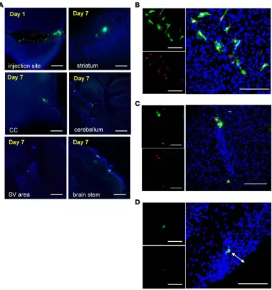
DUOC-01 cells disseminated from the injection site and persisted in the brain for up to 1 week after intracranial injection. To trace human cells in the brain
following stereotactic injection in the midline CC area, NSG mice that had been fed CPZ for 5 weeks were injected intracranially with 1.0 × 10⁵ CFSE-labeled DUOC-01 cells. CFSE stains the cells fluorescent green, and the dye is stable for weeks in vivo (42). CFSE-labeled cells were found at the injection site as well as in the striatum, CC, cerebellum, brain stem, and subventricular area up to 7 days after injection (Figure 2A). To further confirm that the CFSE-positive cells observed in brain sections were injected DUOC-01 cells, we performed immunostaining with an antibody that specifically detects human nuclei (anti-HuN). Mouse cells in the brain sections were not positive for this anti-HuN antibody. In contrast, the CFSE-positive cells costained with anti-HuN (Figure 2B), confirming that the CFSE-stained cells were not mouse brain cells that might have taken up CFSE released by DUOC-01 or stained human cell debris. CSFE- and HuN-costained DUOC-01 cells were detected deep in the brain parenchyma and as far from the CC injection site as the frontal cortex, and persisted for up to 1 week until assessment of myelination (Figure 2C). We also found CFSE-stained CD14+ cells in various parts of the brain even after 7 days after intracranial injections (Supplemental Figure 5). Thus, DUOC-01 cells disseminated bilaterally from the injec-tion site and persisted in the brain during the 1-week period between cell injection and harvesting brains for assessment of myelination status.
DUOC-01 treatment accelerates remyelination after CPZ feeding in the CC region of NSG mice. As noted above, LFB-PAS staining showed that NSG
mice spontaneously remyelinated the CC region during 2 weeks follow-ing termination of CPZ feedfollow-ing (Supplemental Figure 4). In all 4 exper-iments, the CC of CPZ-fed mice treated with Ringer’s remained severe-ly demyelinated 1 week after diet change and injection (Figure 3A). In contrast, LFB-PAS staining showed extensive myelin fiber formation in the CC 1 week after treatment with DUOC-01 (Figure 3A). Myelination scores of the CC of DUOC-01–treated mice were significantly higher than those of the Ringer’s-injected group (Figure 3B). CD14+ cell–treated mice also showed an increased amount of remyelination compared with the Ringer’s control group, but significantly less than the DUOC-01–treated group (Figure 3, A and B). We examined the effects of DUOC-01 treatment in remyelination in more detail.
Immunohistochemical analysis with an anti-MBP antibody confirmed that DUOC-01–treated mice remyelinated more extensively than Ringer’s control animals during the week after diet change and treat-ment (Figure 4A). Analysis of higher magnification confocal images revealed a higher density and level of organization of MBP-containing fibers in the CC of DUOC-01–treated mice (Figure 4B), and MBP appeared to colocalize with neurofilament-H (NFH) (Figure 4B), indicative of myelin wrapping along the axonal fibers.
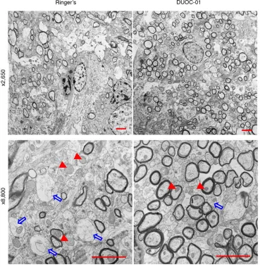
Electron microscopic analysis revealed that the newly synthesized myelin detected by immuno-histochemistry in the CC of DUOC-01–treated mice was organized into myelin sheaths on axons (Figure 5). Morphometric analysis revealed that the CC of DUOC-01–treated mice had significantly more myelinated axons than the CC of animals treated with Ringer’s (Figure 6A). To further assess
R E S E A R C H A R T I C L E
the organization of the myelin sheath, we counted the number of turns of myelin sheath wrapped around the axons. The DUOC-01–treated group had approximately 2 additional turns of myelin sheath per axon compared with the control group (Figure 6B). We also found that the g-ratio (ratio of the inner axonal diameter to the total outer [including myelin wrap] diameter) value was lower in DUOC-01–treated compared with the Ringer’s-treated mice, indicating increased myelin thick-ness in DUOC-01–treated mice (Figure 6C). Supplemental Figure 6A shows that axonal diameters displayed a similar distribution of higher and lower g-ratios across various axon diameters both in Ringer’s- and DUOC-01–treated groups. We also explored whether cell treatment had an impact on axonal density in the CC area. Axonal density, measured as the number of axons present per microscopic field (×8,800 magnification) of electron micrographs, was not significantly different (P < 0.075) in the DUOC-01–treated and the Ringer’s-treated samples (Supplemental Figure 6B). Taken together, these data show that relative to Ringer’s treatment, administration of DUOC-01 cells increased the number of remyelinated axons and augmented the myelin thickness and organization in the CC in the 7 days following treatment.
Morphometric analysis also showed that treatment with DUOC-01 accelerated the reversal of mega-mitochondria formation (Figure 5 and Supplemental Figure 3B). One week after DUOC-01 cell treat-ment, the average size of mitochondria in the brain cells of Ringer’s-treated mice was significantly larger
Figure 5. Electron microscopic analysis of remyelination status upon DUOC-01 treatment. Representative ×2,650
than in cells of the DUOC-01–treated group (Figure 6D). Electron micrographs (Supplemental Figure 3B) show that mitochondria of DUOC-01–treated brains were similar in size to those in unmyelinated con-trol brains. In the Ringer’s-treated group, enlarged mitochondria were present in both axons as well as in other cells, possibly in oligodendrocytes (Supplemental Figure 3B). Brains from DUOC-01–treated mice had a greater number of mitochondria per electron microscopic field than brains from Ringer’s-treated animals (Figure 6E). This reduction in mitochondrial size coupled with the observed increase in mega-mitochondria formation and larger numbers of mega-mitochondria suggests that DUOC-01 cells helped restore mitochondrial activity during remyelination.
R E S E A R C H A R T I C L E
Cellularity scoring of LFB-PAS stains of CC sections showed that DUOC-01 treatment also signifi-cantly reduced cellular accumulation and gliosis in the CC region 1 week following diet change (Figure 7A). Reduced glial accumulation was also evident in brain sections stained with antibodies against GFAP to detect astrocytes and Iba1 to detect microglia (Figure 7B). We performed quantitative analysis of areas covered by Iba1- and GFAP-positive cells, indicative of their numbers, along the CC. Both the numbers of Iba1-positive (microglia) and GFAP-positive (astrocytes) cells were significantly lower in the CC area of the DUOC-01–treated animal brains (Figure 7C). The cellularity score also was decreased in the CD14+ cell–injected group (Supplemental Figure 7) compared to the Ringer’s-injected control, but not as markedly as the DUOC-01–treated group.
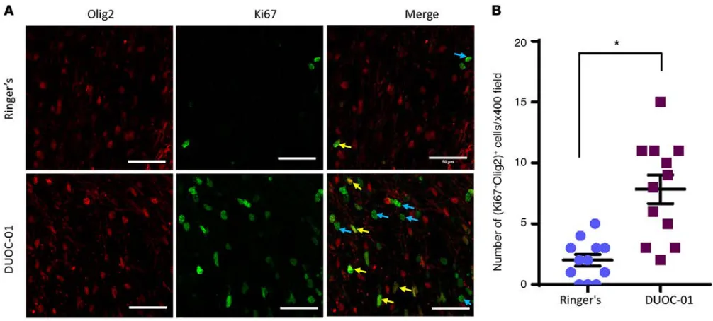
DUOC-01 cell treatment promotes oligodendrocyte progenitor proliferation. We next determined whether
DUOC-01 treatment increased the number of proliferating oligodendrocyte progenitor cells in the CC area following cessation of CPZ feeding (Figure 8). In adult brains, the oligodendrocyte lineage transcrip-tion factor 2 (Olig2) is present in the nuclei of oligodendrocyte progenitors and mature oligodendrocytes (43, 44). Ki67 is only present in proliferating cells (45). Thus, we used the combination of anti-Olig2 and anti-Ki67 antibodies to detect newly generated cells in the oligodendrocyte lineage. The number of proliferating Olig2+Ki67+ oligodendrocytes (Figure 8B) present in the CC region was significantly higher in brain sections from DUOC-01–treated animals than in controls that did not receive cell therapy. There was no significant increase in the number of proliferating Olig2+Ki67+ oligodendrocytes in CB CD14+– treated brains as compared with the Ringer’s-injected group (data not shown). Thus, DUOC-01 treatment promotes oligodendrogenesis, which in turn could facilitate remyelination.
Identification of gene products expressed by DUOC-01 that may promote remyelination. We used
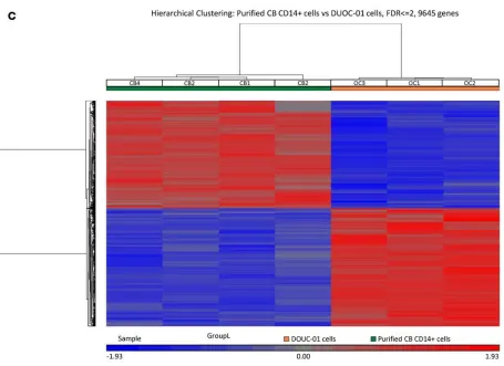
more robustly than CB CD14+ monocytes, our initial strategy was to identify differentially expressed tran-scripts that were more abundant in DUOC-01 than in CB CD14+ monocytes. We performed whole-genome microarray analysis on 4 highly purified, flow cytometry–sorted, CB CD14+ monocytes and 3 DUOC-01 cell products. Complete expression data have been deposited in the NCBI’s Gene Expression Omnibus (GEO GSE76803).
We used stringent MAS5 analysis to identify expressed genes. In order for a transcript correspond-ing to a probe to be scored as “present,” we required that all 4 CB CD14+ cell samples or all 3 DUOC-01 samples on the chip showed expression with a specific probe set (Supplemental File 1). A Venn diagram displaying the findings from microarray analysis showing the number of genes only expressed in purified fresh CD14+ or DUOC-01 cells as well as genes expressed by both cell types is shown in Figure 9A. For less stringent analysis, transcripts that were not detected in at least 1 sample, but detected in others were scored as “mixed,” and transcripts absent in all samples were scored as “absent.” The 2 cell populations differed considerably in gene expression. Thus, 1,184 probe sets detected transcripts in all DUOC-01 samples that were absent in all CB CD14+ monocyte samples and, conversely, 1,017 transcripts were present in all CB CD14+ monocytes and absent in all DUOC-01 samples. In addition, 3,189 probe sets detected transcripts in 1 or 2 of the 3 DUOC-01 lots but none of the 4 CB CD14+ preparations. Conversely, 3,496 probes detected transcripts in 1, 2, or 3 of the 4 CB CD14+ preparations but none of the DUOC-01 lots. Additional differ-ences in expression were observed when requirements were less stringent.
R E S E A R C H A R T I C L E
Pearson’s correlation coefficients among the samples (Supple-mental Figure 8) showed a lower correlation (0.87–0.90) among the CB CD14+ and DUOC-01 groups, although the correlation coefficient was much higher (0.97–0.99) between the samples within the same group. Differentially expressed transcripts are listed in GEO GSE76803. The heat map presented in Figure 9C also demonstrates that DUOC-01 and CB CD14+ cells fall into discrete populations defined by a large number of differentially expressed transcripts.
To annotate the function of genes that were differentially expressed, we eliminated uncharacterized transcripts, pseu-dogenes, and non–protein-coding transcripts from further analysis. The resulting gene lists were examined using the tools aggregated at the DAVID website (46, 47). Supplemen-tal File 2 shows the functional cluster analysis for the genes that are more highly expressed in DUOC-01 cells than CB CD14+ monocytes. This list is enriched for genes involved in all aspects of cell division and mitosis. Pathway analysis also shows an abundance of genes involved in cell division and in lyso-somal activity and trafficking of intracellular vesicles. Of interest, genes encoding factors previously shown to be secreted and/or increased (e.g., IL-10, TGF-β, and galactocerebrosidase [GALC]) during manufacturing of DUOC-01 (20), were identified in the DUOC-01 upregulated gene list. In addition, transcripts for several other lysosomal enzymes that are secreted by DUOC-01 (20) are more abundant in the cell product than in CB CD14+ cells, and pathway analysis shows a very high probability of enrichment for genes encoding proteins in the lysosomal lumen in DUOC-01 (Supplemental File 2). A variety of transcripts that can influence myelination are highly overexpressed in DUOC-01 cells rela-tive to CD14+ cells. Some of these are listed in Supplemental File 2. In contrast, Supplemental File 2 shows that the list of genes more highly expressed in CB CD14+ cells is enriched in transcription fac-tors and signaling molecules, particularly in repressors of transcription. Genes active in hematopoiesis and myeloid cell differentiation are also more common. Genes active in mitosis and cell cycle entry are much less common than in the list derived from DUOC-01.
We selected some of the transcripts overexpressed in DUOC-01 that are known to be important in pro-moting remyelination and confirmed their level of expression in CB CD14+ and DUOC-01 cells by quan-titative PCR methods. Expression of these candidate molecules is presented in Table 1. Platelet-derived growth factor subunit A (PDGFA), KIT-ligand (KITLG, also known as stem cell factor [SCF]), insulin-like growth factor-1 (IGF1), triggering receptor expressed on myeloid cells 2 (TREM2), matrix metalloproteinase-9 (MMP9), and MMP12 were highly upregulated in DUOC-01 cells compared with CB CD14+ monocytes; bioplex analysis also demonstrated that DUOC-1 cells secrete MMP9, MMP12, and other matrix proteases into culture supernatants (unpublished observation). IL10 transcript levels were also enriched in DUOC-01 cells (Supplemental File 2), confirming our previous studies (20). Western blot analysis confirmed enrichment of TREM2, SCF, MMP9, and MMP12 proteins in DUOC-01 relative to CB CD14+ monocyte homogenates (Supplemental Figure 9). Higher expression of TREM2 on the DUOC-01 cell surface was also verified by flow cytometry (Supplemental Figure 9B). However, the relative abundance of IGF1 and PDGF-AA protein detected by Western blotting did not replicate transcript abundance (Supplemental Figure 9A). IGF1 and PDGF-AA proteins were detected in CD14+ CB monocytes but not in DUOC-01 homogenates. We hypoth-esized that our failure to detect IGF1 and PDGF-AA in DUOC-01 homogenates might result from their rapid secretion from the cells, as we have previously demonstrated that DUOC-01 cells secrete several proteins (20). To test this idea, DUOC-01 cells were allowed to adhere to glass slides and then incubated for 5 hours with brefeldin A (BFA). BFA treatment rapidly inhibits transport of secretory proteins from the ER to the Golgi, resulting in accumulation of proteins within the ER (48). Western blot analysis of DUOC-01 cells after BFA treatment showed higher intracellular concentrations of both IGF1 and PDGF-AA (Supplemental Figure 9C), indicating that these proteins are rapidly secreted by DUOC-01 cells. Immunocytochemical analysis of BFA-treated DUOC-01 cells using IGF1 and PDGF-AA antibodies also showed higher levels of staining after BFA treatment compared to cells without any BFA pretreatment (Supplemental Figure 9D). These data sug-gest that DUOC-01 cells express and secrete factors known to promote remyelination by several mechanisms and enhance oligodendrocyte precursor proliferation and differentiation.
Table 1. Quantitative PCR determination of abundance of transcripts of factors that promote oligodendrogenesis and myelination in DUOC-01 and CB CD14+ monocytes.
Gene name Fold change (Mean ± SEM, n > 3) P value
PDGFA 32.3 ± 8.3 ≤0.01
KITLG/SCF 26.7 ± 4.8 ≤0.033
IGF1 799 ± 294 ≤0.05
MMP9 632 ± 109 ≤0.002
MMP12 2057 ± 460 ≤0.006
R E S E A R C H A R T I C L E
Discussion
As we have discussed in detail elsewhere (20), the current NCT02254863 clinical trial using DUOC-01 to treat genetic metabolic diseases was predicated on the mechanistic assumption that intrathecally injected cell product can provide wild-type lysosomal enzyme replacement therapy to immunoablated CB trans-plant recipients. However, studies of the biological activities of DUOC-01 raised the possibility that the cell product has other types of potential benefits in treatment of demyelinating diseases (20). The studies pre-sented here confirm that the DUOC-01 cell product strongly promotes remyelination in an animal model that does not depend on enzyme replacement: CPZ-induced demyelination of the CC. Injecting DUOC-01 cells into the CC area 1 day after CPZ-fed NSG mice were returned to normal diets dramatically accelerat-ed reversal of all the pathological manifestations of CPZ feaccelerat-eding during the following week. The course of demyelination, astrogliosis, microgliosis, cellular accumulation, and the reversal of these processes follow-ing cessation of CPZ feedfollow-ing in NSG mice closely resembled what has been reported in C57BL/6 mice (23, 25). Using CFSE-labeled DUOC-01 cells, we found that DUOC-01 cells reached brain regions remote from the injection site and that cells could be detected in the brain throughout the 1-week experiment. Storms et al. (49) have reported that a low percentage of intrathecally injected DUOC-01 cells persist in the brain of neonatal NSG mice for up to 56 days. Immunohistochemical and LFB-PAS staining demonstrated that DUOC-01 treatment accelerated remyelination of the CC, and electron microscopy revealed that adminis-tration of DUOC-01 cells increased the proportion of remyelinated axons and the thickness and organiza-tion of the myelin sheath following treatment. DUOC-01 treatment also significantly resolved gliosis in the CC induced by CPZ feeding. Finally, electron microscopic analysis also showed that DUOC-01 treatment reduced the number of megamitochondria in the CC region, suggesting that DUOC-01 treatment reverses metabolic stress (41, 50–52), abnormality of mitochondrial fission (53, 54), or oxidative stress induced by CPZ treatment. Thus, DUOC-01 cells can deploy within the cerebral cortex following intracerebral injection and accelerate remyelination after cessation of CPZ feeding. During manufacture of DUOC-01 from CD14+ CB monocytes, expression of many factors that can influence remyelination by a variety of mechanisms is upregulated. These studies provide proof of concept for the clinical use of DUOC-01 in the treatment of demyelinating diseases.
Freshly isolated CB CD14+ monocytes also accelerated remyelination and reduced cellular infiltration in the CC following discontinuation of CPZ feeding, but did so significantly less markedly than DUOC-01 cells. Because we are primarily interested in delineating the mechanism of action of the DUOC-01 cell product that is in the clinic, we have not studied the biodistribution or persistence of these cells following injection or performed detailed immunohistochemical or morphometric analysis of the effects of CD14+ CB cells in the CPZ system. We do not know if both cell populations use the same mechanisms of action to accelerate remyelination. We note in this context that DUOC-01 treatment increased proliferation of oligo-dendrocyte lineage cells, but treatment with CD14+ CB monocytes did not.
changes in injured tissue. Thus, the gene expression data provide a starting point to elucidate the mecha-nisms by which DUOC-01 promotes remyelination.
Several of the proteins that are expressed by DUOC-01 cells are known to regulate the number or activ-ity of oligodendrocyte progenitor cells (OPCs). PDGFs regulate the OPC numbers in the adult CNS and their activity following CNS demyelination (55–57), and PDGFA transcript expression was upregulated 32-fold in the DUOC-01 compared with CB CD14+ monocytes. SCF has been implicated in the mainte-nance, migration, and survival of the OPC population (58, 59), and its transcript was expressed at a 26-fold higher level in DUOC-01 cells than in CB CD14+ cells. Similarly, expression of IGF1 transcripts was almost 800-fold higher in DUOC-01 compared to the CD14+ cells. IGF1 has been shown to induce myelination in vitro and in vivo and also protects mature oligodendrocytes from a pathological insult (34, 60, 61). Fur-thermore, IGF1 promotes the long-term survival of mature oligodendrocytes in culture and inhibits mature oligodendrocyte apoptosis in vitro (61, 62). Brefeldin A–mediated inhibition of secretory proteins demon-strated that PDGF-AA and IGF1 are both rapidly secreted from DUOC-01 cells. These factors, then, could directly drive the large increase in proliferating oligodendrocyte lineage cells that we observed in the CC of DUOC-01–treated animals compared with controls receiving no cell therapy.
TREM2 is another molecule expressed by DUOC-01 cells that may play an important role in remy-elination. CD14+ monocytes do not express TREM2 transcripts or protein. This surface receptor senses lipid debris and regulates signaling by glial cells that modulate myelination (63, 64). It also functions in clearance of cellular and myelin debris, an important early step for recovery and remyelination follow-ing CNS injury (12, 65). DUOC-01 cells are highly phagocytic (20) and could play a significant role in myelin clearance and intercellular signaling through the TREM2 receptor. DAVID analysis of differentially expressed genes showed that the lysosomal/intracellular vesicular pathway and Fcγ-mediated phagocytosis were among the most highly upregulated group of genes in DUOC-01 compared with CD14+ CB mono-cytes (Supplemental File 2).
DUOC-01 cells express many other proteins that could participate more indirectly in promoting remy-elination and in resolving cellular accumulation in the CC. Cytokine-activated microglia can stimulate the differentiation of oligodendrocytes from neural progenitor cells (66). While oligodendrocytes affect the remyelination of nerve fibers, other cell types are important for this repair process (33). Astrocytes provide trophic factors for oligodendrocytes and also for microglia (35). Microglia also provide trophic factors and remove myelin debris that inhibit remyelination by oligodendrocytes (11, 34, 36, 37). The microarray data indicate that several chemokines and other regulators of neuroinflammation are upregulated in DUOC-01 cells. We previously reported that DUOC-01 cells secrete IL-10 and TNF-α in culture (20). Yang et al. have demonstrated that neuronal stem cells producing IL-10 not only effectively suppress CNS inflammation but also promote remyelination and neuron/oligodendrocyte repopulation in a mouse model of experimental autoimmune encephalomyelitis (67). Furthermore, IL-10 promotes survival of neurons and oligodendro-cytes by protecting them from inflammation-induced damage (68–70). It has been shown that TNF-α plays an important reparative role in the demyelinating brain. Lack of TNF-α led to a reduction in the pool of proliferating OPCs and subsequent significant delay in remyelination in CPZ-mediated demyelinated brain (71). The microarray data indicate that several other factors including chemokines and other regulators of neuroinflammation are upregulated in DUOC-01 cells.
DUOC-01 cells also overexpress proteases that can regulate remyelination through modification of the extracellular matrix. We confirmed upregulation of MMP9 and MMP12 by PCR and Western blotting. CD14+ CB monocytes did not detectably express either protease. MMP9 activity is required to clear NG2 chondroitin sulfate proteoglycan deposition and overcome the negative impact of NG2 on oligodendrocyte maturation and remyelination (72). High expression of MMP12, which is required for proteolysis and matrix invasion by macrophages in mice, might facilitate the migration of DUOC-01 from the injection site in the CC to other regions of the brain that we observed using CFSE-labeled cells. MMPs also play a role in angiogenesis, in the release of growth factors sequestered by the extracellular matrix (73), and in processing of cell-cell recognition molecules that allow repair (74).
R E S E A R C H A R T I C L E
In the model presented in this report, we used intracerebral injection to maximize localization of cells adja-cent to the injured area. This is a plausible, but not an optimal, route of clinical administration. Second, the question of whether there is persistence of cell product in an immune-competent patient will need to be explored. While this is not an issue in the studies presented here using immune-incompetent NSG mice or in the ongoing NCT02254863 clinical trial, as the patients are myelo- and immunoablated and the CB unit used for transplant is the same as the unit used to manufacture DUOC-01 (20), immunogenicity is likely to be important in other clinical settings. In the current clinical trial, systemic engraftment of the donor CB unit used to manufacture DUOC-01 must occur before DUOC-01 is administered intrathecally. Thus, the recipient is tolerized to the DUOC-01 donor. In other indications, systemic tolerization is unlikely to be included in the therapeutic approach, rather DUOC-01 will be administered as a stand-alone, partially HLA-matched, patient-directed cell therapy product. DUOC-01 is moderately immunogenic in mixed lym-phocyte assays, although less so than peripheral blood MNCs (20), and the impact of this is not known but can be further explored in the CPZ model since we have found that the kinetics of de- and remyelination appear to be the same in immune-competent and NSG animals. Also, the gene expression data presented here provide the basis for continuing studies of the mechanism of action of the DUOC-01 cell product and assays of product potency and release. The biological activity of DUOC-01 reported here suggests that it has potential for the treatment of disorders of myelination in the clinic and that these preclinical studies should be considered as part of the clinical development program of this interesting and potentially novel CB-derived cell therapy product.
Methods
Animals and animal welfare. Male NSG mice (NOD/SCID/IL2Rγnull ) were supplied by NSG Breeding Core
of Duke Division of Laboratory Animal Resources and were maintained under specific pathogen–free conditions.
Manufacture of DUOC-01. DUOC-01 cells were prepared from cryopreserved CB units and formulated in
Ringer’s solution using the protocols developed to make products for clinical use (20). These methods have been described in detail (20). For convenience, this material is reproduced in the Supplemental Materials.
Separation of specific cell populations from CB. CD14+ populations from cryopreserved CB were
immuno-magnetically selected using Whole Blood CD14 Microbeads as described by the manufacturer (Miltenyi Biotec). Cells that did not adhere to the anti-CD14 antibody columns comprised the CD14+-depleted popu-lation. Some experiments were carried out with cells from CD14+ cells from freshly collected CB. MNC populations depleted of erythrocytes were prepared from fresh CB either by centrifugation on Ficoll or in SepMate tubes (STEMCELL Technologies) as described by the manufacturer. CD14+ cells were immu-nomagnetically purified from MNC preparations using the CD14 Microbeads. Similar experiments were carried out with CB cell populations enriched for or depleted of CD34-expressing cells using anti-CD34 Microbeads (Miltenyi Biotec).
To prepare CD14+ cell RNA for microarray analysis, freshly collected CB was centrifuged on Ficoll to prepare MNC fractions. These fractions were treated with 0.15 M NH4Cl to lyse erythrocytes, washed in PBS, and then incubated on ice with PeCy7-mouse anti–human CD14 (catalog 562698), FITC-mouse anti–human CD3 (catalog 555339), and FITC-mouse anti–human CD235a (catalog 559943) antibodies (all from BD). Cells were then sorted twice by flow cytometry to yield CD14+CD235a–CD3– popula-tions. The first enrichment sort was followed by a second purity sort. Cells were maintained at 0°C–4°C during all procedures, including flow sorting. The purity of selected populations and the extent of CD14+ cell depletion were determined by flow cytometry as previously described (20).
CPZ demyelination in NSG mice. Eight-week-old male NSG mice were acclimated to milled standard
PBS and then with 4% paraformaldehyde. Paraffin-embedded coronal sections were prepared for analysis of myelination status, the organization of neural fibers, and persistence of injected human cells by LFB-PAS staining, immunohistochemistry, and electron microscopy as described below. Cohorts of 5 or 6 mice were analyzed under each set of experimental conditions.
Myelination, cellular infiltration, and gliosis were assessed by LFB-PAS staining of the CC region, (approximately at the level of the bregma –0.2 to –0.9 mm) (75). We used 5.0-μm-thick paraffin-embedded coronal sections of the CC region. LFB stains the myelin blue, and PAS stains demyelinated axons pink. Three independent, blinded readers scored coded LFB-PAS–stained sections between 0 and 3. A score of 3 is equivalent to the myelin status of a brain not treated with CPZ; 0 is equivalent to a completely demy-elinated brain area. A score of 1 or 2 corresponds to one-third or two-third fiber myelination, respectively. Similarly, a quantitative cellularity score was obtained by counting the number of nuclei in the CC region of LFB-stained brain slices on a scale of 0 to 3, by blinded readers.
Immunohistochemistry. Detailed protocols are presented in the Supplemental Methods. Brain slices from
3 animals in each treatment group were analyzed. Primary antibodies used were: rat anti-MBP (1:1,000, Abcam, catalog ab7349); chicken anti-NFH (1:100,000, EnCor Biotech, catalog CPCA-NF-H); mouse anti-HuN (1:250, Millipore, catalog MAB1281); chicken anti-GFAP (1:500, Abcam, catalog ab4674); goat Iba1 (1:200, Abcam, catalog ab5076); rabbit Ki67 (1:300, Abcam, catalog ab15580); and goat anti-Olig2 (1:50, R&D Systems, catalog AF2418). Secondary antibodies used were: Alexa-488 donkey anti-rat, Alexa-647 donkey anti-chicken, Alexa-568 donkey anti-mouse (1:500, Molecular Probes). Confocal micro-graphs were obtained using constant settings including laser power, stack thickness, and camera resolution. The number of stained cells per microscopic field in the CC region and the average area covered by cells stained with each antibody were quantified by ImageJ software (NIH).
Electron microscopy. Preparation of brains for electron microscopy is described in detail in the
Supple-mental Methods. Images were then analyzed using ImageJ software. For analysis, g-ratio analysis was modified such that the inner diameter of compact myelin (instead of the axon diameter) was divided by the outer diameter of the myelin sheath. Diameters were calculated from enclosed areas. Fibers with promi-nent outfoldings in the plane of section were excluded. We implemented a plugin for the ImageJ software (http://rsbweb.nih.gov/ij), which allowed for semiautomated analysis of randomly selected sets of fibers (76). Plugin and source code are available online (http://gratio.efil.de). A minimum of 100 fibers/mouse, 3 mice/time point/treatment, were analyzed. The number of mitochondria in all cells in the CC area was counted in all the electron micrographs, and average mitochondria present per ×8,800-magnified field was calculated. To determine the size of the mitochondria, electron microscopic images were analyzed with ImageJ, using the area analysis function. For area measurement, the mitochondria were circled by the lasso tool, and then the areas of the circles were calculated and converted to their actual values using the scale bar. At least 10 images were analyzed per sample in a blinded fashion.
Tracking DUOC-01 cells in the brain. DUOC-01 cells were stained with 5 μM Vybrant CFDA SE Cell
Tracer dye (CFSE, V12883, green fluorescence, Life Technologies) and injected into the CC as described above. One, four, and seven days later, brains were harvested, sliced, and processed for confocal microscopy.
Expression analysis by microarrays. RNA for microarray analysis was prepared from 4 flow-sorted CD14+
CB and 3 DUOC-01 products using the QIAGEN RNeasy Mini Kit as described by the manufacturer. These samples were used for whole-genome microarray analysis on 1 chip. Microarray analysis was performed by the Microarray Shared Resource in the Duke Center for Genomic and Computational Biology using Affymetrix GeneChip Human Transcriptome Array 2.0 microarrays. Partek Genomics Suite 6.6 (Partek Inc.) was used to perform data analysis. Robust multichip analysis (RMA) normalization was performed on the entire dataset. Multi-way ANOVA and analysis of the fold change were performed to select target genes that were differentially expressed. Hierarchical clustering was performed on differentially expressed genes based on average linkage with Pearson’s dissimilarity.
RNA isolation and quantitative real-time PCR. Quantitative real-time RT-PCR was used to measure levels
R E S E A R C H A R T I C L E
(sense 5′-TGTACCGCTATGGTTACACTCG-3′, antisense 5′- GGCAGGGACAGTTGCTTCT-3′);
IGF1 (sense 5′-GCCTCCTTAGATCACAGCTC-3′, antisense 5′-GATGCTCTTCAGTTCGTGTGT-3′);
IL10 (sense 5′-GCGCTGTCATCGATTTCTTC-3′, antisense 5′-TCACTCATGGCTTTGTAGATGC-3′);
MMP12 (sense 5′-CAAAACTCAAATTGGGGTCACAG-3′, antisense 5′
-CTCTCTGCTGATGACATAC-GTG-3′), KITLG (sense 5′-AGCTGAAGATAAATGCAAGTGAG-3′, antisense 5′ -CAGAACAGCTA-AACGGAGTCG-3′), and TREM2 (sense 5′-TCATAGGGGCAAGACACCT-3′, antisense 5′ -GCTGCT-CATCTTACTCTTTGTC-3′). Values were normalized to GAPDH expression.
Statistics. In most cases statistical comparisons were conducted with 2-tailed Student’s t tests with
unequal variance. For comparing LFB and cellularity scores we used Wilcoxon rank-sum tests. Statistical comparisons were performed using the Wilcoxon rank-sum test for clustered data using the clusrank pack-age in R. Mean differences were considered significant if P values were less than 0.05.
Study approval. All animal experimentation protocols were approved by the Duke University IACUC and
were conducted in accordance with the United States Public Health Service Policy on Human Care and Use of Laboratory Animals.
Author contributions
All authors reviewed the manuscript. In addition, JK conceived and directed the DUOC-01 development program and edited the manuscript; AEB designed and directed the research, analyzed data, and edited the manuscript; AS designed, directed, and performed experiments, and drafted the manuscript; SB man-aged the research, performed experiments, and analyzed the data; LX, PS, PN, AW, SP, and AG performed experiments; GM advised on CPZ experiments, analyzed related data, and critically reviewed the manu-script; TG developed and directed cGMP manufacturing and designed experiments.
Acknowledgments
The authors are grateful to the staff at the Carolinas Cord Blood Bank for providing cord blood units for the experiments described, Lynn Cheatham for help with graphics, Marcia Bentz and Roderick Franczak for helping in tissue preparation and staining, Neil Medvitz of Duke Pathology for performing electron micros-copy, April Ozamiz, Benjamin Rusche, Norin Meadows, and Frankie Shaw of the GMP lab for helping with DUOC-01 cells preparation, David Snyder of Department of Surgery for IC injections, Michael Cook at the Duke Cancer Institute Core Flow Cytometry Facility for assistance with cell sorting, Zhengzheng Wei at the Duke Institute for Genomic Sciences Microarray Core Facility for performing and helping analyze microar-ray data, and to Benjamin Carlson at the Light Microscopy Core Facility at Duke University for assistance with image analysis. Amy Wollish was supported by a National Cancer Institute of the National Institutes of Health Award (grant 5T32CA074736, Research Training in Neuro-Oncology). This work was supported by the Julian Robertson Foundation and by the Marcus Foundation.
Address correspondence to: Arjun Saha, Room 0104, Research Park 2, PO Box 103455, Robertson Clinical and Translational Cell Therapy Program, Duke Translational Medicine Institute, Duke University Medical Center, Durham, North Carolina 27710, USA. Phone: 919.684.3934; E-mail:arjun.saha@duke.edu.
AW’s present address is: PPD Inc., Wilmington, North Carolina, USA.
1. Nayak D, Roth TL, McGavern DB. Microglia development and function. Annu Rev Immunol. 2014;32:367–402. 2. Ginhoux F, Lim S, Hoeffel G, Low D, Huber T. Origin and differentiation of microglia. Front Cell Neurosci. 2013;7:45. 3. Matsushima GK, et al. Absence of MHC class II molecules reduces CNS demyelination, microglial/macrophage infiltration,
and twitching in murine globoid cell leukodystrophy. Cell. 1994;78(4):645–656.
4. Louveau A, et al. Structural and functional features of central nervous system lymphatic vessels. Nature. 2015;523(7560):337–341. 5. Ginhoux F, et al. Fate mapping analysis reveals that adult microglia derive from primitive macrophages. Science.
2010;330(6005):841–845.
6. Mildner A, et al. Microglia in the adult brain arise from Ly-6ChiCCR2+ monocytes only under defined host conditions. Nat
Neu-rosci. 2007;10(12):1544–1553.
7. Lampron A, Pimentel-Coelho PM, Rivest S. Migration of bone marrow-derived cells into the central nervous system in models of neurodegeneration. J Comp Neurol. 2013;521(17):3863–3876.
8. McMahon EJ, Suzuki K, Matsushima GK. Peripheral macrophage recruitment in cuprizone-induced CNS demyelination despite an intact blood-brain barrier. J Neuroimmunol. 2002;130(1–2):32–45.
of immune-to-brain communication that influences mood and behavior. Front Neurosci. 2014;8:447.
10. Reader BF, Jarrett BL, McKim DB, Wohleb ES, Godbout JP, Sheridan JF. Peripheral and central effects of repeated social defeat stress: monocyte trafficking, microglial activation, and anxiety. Neuroscience. 2015;289:429–442.
11. Miron VE, Franklin RJ. Macrophages and CNS remyelination. J Neurochem. 2014;130(2):165–171.
12. Lampron A, et al. Inefficient clearance of myelin debris by microglia impairs remyelinating processes. J Exp Med. 2015;212(4):481–495.
13. Shechter R, et al. Recruitment of beneficial M2 macrophages to injured spinal cord is orchestrated by remote brain choroid plexus. Immunity. 2013;38(3):555–569.
14. Ruckh JM, et al. Rejuvenation of regeneration in the aging central nervous system. Cell Stem Cell. 2012;10(1):96–103. 15. Shi C, Pamer EG. Monocyte recruitment during infection and inflammation. Nat Rev Immunol. 2011;11(11):762–774. 16. Shechter R, Schwartz M. Harnessing monocyte-derived macrophages to control central nervous system pathologies: no longer
‘if ’ but ‘how’. J Pathol. 2013;229(2):332–346.
17. Sanberg PR, et al. Monocyte transplantation for neural and cardiovascular ischemia repair. J Cell Mol Med. 2010;14(3):553–563. 18. Sun JM, Kurtzberg J. Cord blood for brain injury. Cytotherapy. 2015;17(6):775–785.
19. Womble TA, et al. Monocytes are essential for the neuroprotective effect of human cord blood cells following middle cerebral artery occlusion in rat. Mol Cell Neurosci. 2014;59:76–84.
20. Kurtzberg J, et al. Preclinical characterization of DUOC-01, a cell therapy product derived from banked umbilical cord blood for use as an adjuvant to umbilical cord blood transplantation for treatment of inherited metabolic diseases. Cytotherapy. 2015;17(6):803–815.
21. Zendedel A, Beyer C, Kipp M. Cuprizone-induced demyelination as a tool to study remyelination and axonal protection. J Mol
Neurosci. 2013;51(2):567–572.
22. Skripuletz T, Gudi V, Hackstette D, Stangel M. De- and remyelination in the CNS white and grey matter induced by cuprizone: the old, the new, and the unexpected. Histol Histopathol. 2011;26(12):1585–1597.
23. Kipp M, Clarner T, Dang J, Copray S, Beyer C. The cuprizone animal model: new insights into an old story. Acta Neuropathol. 2009;118(6):723–736.
24. Torkildsen O, Brunborg LA, Myhr KM, Bø L. The cuprizone model for demyelination. Acta Neurol Scand, Suppl. 2008;188:72–76. 25. Matsushima GK, Morell P. The neurotoxicant, cuprizone, as a model to study demyelination and remyelination in the central
nervous system. Brain Pathol. 2001;11(1):107–116.
26. Bénardais K, et al. Cuprizone [bis(cyclohexylidenehydrazide)] is selectively toxic for mature oligodendrocytes. Neurotox Res. 2013;24(2):244–250.
27. Einstein O, Friedman-Levi Y, Grigoriadis N, Ben-Hur T. Transplanted neural precursors enhance host brain-derived myelin regeneration. J Neurosci. 2009;29(50):15694–15702.
28. Crocker SJ, et al. Intravenous administration of human embryonic stem cell-derived neural precursor cells attenuates cuprizone-induced central nervous system (CNS) demyelination. Neuropathol Appl Neurobiol. 2011;37(6):643–653.
29. Hedayatpour A, et al. Promotion of remyelination by adipose mesenchymal stem cell transplantation in a cuprizone model of multiple sclerosis. Cell J. 2013;15(2):142–151.
30. Nessler J, et al. Effects of murine and human bone marrow-derived mesenchymal stem cells on cuprizone induced demyelin-ation. PLoS One. 2013;8(7):e69795.
31. Arnett HA, et al. bHLH transcription factor Olig1 is required to repair demyelinated lesions in the CNS. Science. 2004;306(5704):2111–2115.
32. Mason JL, et al. Oligodendrocytes and progenitors become progressively depleted within chronically demyelinated lesions. Am
J Pathol. 2004;164(5):1673–1682.
33. Gudi V, Gingele S, Skripuletz T, Stangel M. Glial response during cuprizone-induced de- and remyelination in the CNS: lessons learned. Front Cell Neurosci. 2014;8:73.
34. Mason JL, Ye P, Suzuki K, D’Ercole AJ, Matsushima GK. Insulin-like growth factor-1 inhibits mature oligodendrocyte apopto-sis during primary demyelination. J Neurosci. 2000;20(15):5703–5708.
35. Biancotti JC, Kumar S, de Vellis J. Activation of inflammatory response by a combination of growth factors in cuprizone-induced demyelinated brain leads to myelin repair. Neurochem Res. 2008;33(12):2615–2628.
36. Skripuletz T, et al. Astrocytes regulate myelin clearance through recruitment of microglia during cuprizone-induced demyelin-ation. Brain. 2013;136(Pt 1):147–167.
37. Clarner T, et al. Myelin debris regulates inflammatory responses in an experimental demyelination animal model and multiple sclerosis lesions. Glia. 2012;60(10):1468–1480.
38. Hiremath MM, Saito Y, Knapp GW, Ting JP, Suzuki K, Matsushima GK. Microglial/macrophage accumulation during cupri-zone-induced demyelination in C57BL/6 mice. J Neuroimmunol. 1998;92(1–2):38–49.
39. Hibbits N, Yoshino J, Le TQ, Armstrong RC. Astrogliosis during acute and chronic cuprizone demyelination and implications for remyelination. ASN Neuro. 2012;4(6):393–408.
40. Shultz LD, et al. Human lymphoid and myeloid cell development in NOD/LtSz-scid IL2R gamma null mice engrafted with mobilized human hemopoietic stem cells. J Immunol. 2005;174(10):6477–6489.
41. Praet J, Guglielmetti C, Berneman Z, Van der Linden A, Ponsaerts P. Cellular and molecular neuropathology of the cuprizone mouse model: clinical relevance for multiple sclerosis. Neurosci Biobehav Rev. 2014;47:485–505.
42. Karrer FM, Reitz BL, Hao L, Lafferty KJ. Fluorescein labeling of murine hepatocytes for identification after intrahepatic trans-plantation. Transplant Proc. 1992;24(6):2820–2821.
43. Dimou L, Simon C, Kirchhoff F, Takebayashi H, Götz M. Progeny of Olig2-expressing progenitors in the gray and white mat-ter of the adult mouse cerebral cortex. J Neurosci. 2008;28(41):10434–10442.
44. Fancy SP, Chan JR, Baranzini SE, Franklin RJ, Rowitch DH. Myelin regeneration: a recapitulation of development? Annu Rev
Neurosci. 2011;34:21–43.
45. Scholzen T, Gerdes J. The Ki-67 protein: from the known and the unknown. J Cell Physiol. 2000;182(3):311–322.
R E S E A R C H A R T I C L E
resources. Nat Protoc. 2009;4(1):44–57.
47. Huang da W, Sherman BT, Lempicki RA. Bioinformatics enrichment tools: paths toward the comprehensive functional analysis of large gene lists. Nucleic Acids Res. 2009;37(1):1–13.
48. Misumi Y, Misumi Y, Miki K, Takatsuki A, Tamura G, Ikehara Y. Novel blockade by brefeldin A of intracellular transport of secretory proteins in cultured rat hepatocytes. J Biol Chem. 1986;261(24):11398–11403.
49. Storms R, et al. Tissue distribution of a cord blood-derived cell product following intrathecal transplantation. Cytotherapy. 2014;16(4):S63.
50. Kashani IR, et al. Protective effects of melatonin against mitochondrial injury in a mouse model of multiple sclerosis. Exp Brain
Res. 2014;232(9):2835–2846.
51. Tandler B, Hoppel CL. Division of giant mitochondria during recovery from cuprizone intoxication. J Cell Biol. 1973;56(1):266– 272.
52. Acs P, Komoly S. Selective ultrastructural vulnerability in the cuprizone-induced experimental demyelination. Ideggyogy Sz. 2012;65(7–8):266–270.
53. Flatmark T, Kryvi H, Tangerås A. Induction of megamitochondria by cuprizone(biscyclohexanone oxaldihydrazone). Evidence for an inhibition of the mitochondrial division process. Eur J Cell Biol. 1980;23(1):141–148.
54. Asano M, Wakabayashi T, Ishikawa K, Kishimoto H. Mechanism of the formation of megamitochondria by copper-chelating agents. IV. Role of fusion phenomenon in the cuprizone-induced megamitochondrial formation. Acta Pathol Jpn. 1978;28(2):205–213.
55. Woodruff RH, Fruttiger M, Richardson WD, Franklin RJ. Platelet-derived growth factor regulates oligodendrocyte progenitor numbers in adult CNS and their response following CNS demyelination. Mol Cell Neurosci. 2004;25(2):252–262.
56. Murtie JC, Zhou YX, Le TQ, Vana AC, Armstrong RC. PDGF and FGF2 pathways regulate distinct oligodendrocyte lineage responses in experimental demyelination with spontaneous remyelination. Neurobiol Dis. 2005;19(1–2):171–182.
57. Vana AC, Flint NC, Harwood NE, Le TQ, Fruttiger M, Armstrong RC. Platelet-derived growth factor promotes repair of chronically demyelinated white matter. J Neuropathol Exp Neurol. 2007;66(11):975–988.
58. Ida JA, Dubois-Dalcq M, McKinnon RD. Expression of the receptor tyrosine kinase c-kit in oligodendrocyte progenitor cells. J
Neurosci Res. 1993;36(5):596–606.
59. Erlandsson A, Larsson J, Forsberg-Nilsson K. Stem cell factor is a chemoattractant and a survival factor for CNS stem cells. Exp
Cell Res. 2004;301(2):201–210.
60. D’Ercole AJ, Ye P, Calikoglu AS, Gutierrez-Ospina G. The role of the insulin-like growth factors in the central nervous system.
Mol Neurobiol. 1996;13(3):227–255.
61. García-Segura LM, et al. Interaction of the signalling pathways of insulin-like growth factor-I and sex steroids in the neuroen-docrine hypothalamus. Horm Res. 1996;46(4–5):160–164.
62. Cho KH, Kim MW, Kim SU. Tissue culture model of Krabbe’s disease: psychosine cytotoxicity in rat oligodendrocyte culture.
Dev Neurosci. 1997;19(4):321–327.
63. Wang Y, et al. TREM2 lipid sensing sustains the microglial response in an Alzheimer’s disease model. Cell. 2015;160(6):1061–1071. 64. Cantoni C, et al. TREM2 regulates microglial cell activation in response to demyelination in vivo. Acta Neuropathol.
2015;129(3):429–447.
65. Tsiperson V, Li X, Schwartz GJ, Raine CS, Shafit-Zagardo B. GAS6 enhances repair following cuprizone-induced demyelin-ation. PLoS One. 2010;5(12):e15748.
66. Butovsky O, et al. Microglia activated by IL-4 or IFN-gamma differentially induce neurogenesis and oligodendrogenesis from adult stem/progenitor cells. Mol Cell Neurosci. 2006;31(1):149–160.
67. Yang J, et al. Adult neural stem cells expressing IL-10 confer potent immunomodulation and remyelination in experimental autoimmune encephalitis. J Clin Invest. 2009;119(12):3678–3691.
68. Strle K, et al. Interleukin-10 in the brain. Crit Rev Immunol. 2001;21(5):427–449.
69. Molina-Holgado F, Grencis R, Rothwell NJ. Actions of exogenous and endogenous IL-10 on glial responses to bacterial LPS/ cytokines. Glia. 2001;33(2):97–106.
70. Boyd ZS, Kriatchko A, Yang J, Agarwal N, Wax MB, Patil RV. Interleukin-10 receptor signaling through STAT-3 regulates the apoptosis of retinal ganglion cells in response to stress. Invest Ophthalmol Vis Sci. 2003;44(12):5206–5211.
71. Arnett HA, Mason J, Marino M, Suzuki K, Matsushima GK, Ting JP. TNF alpha promotes proliferation of oligodendrocyte progenitors and remyelination. Nat Neurosci. 2001;4(11):1116–1122.
72. Larsen PH, Wells JE, Stallcup WB, Opdenakker G, Yong VW. Matrix metalloproteinase-9 facilitates remyelination in part by processing the inhibitory NG2 proteoglycan. J Neurosci. 2003;23(35):11127–11135.
73. Whitelock JM, Murdoch AD, Iozzo RV, Underwood PA. The degradation of human endothelial cell-derived perlecan and release of bound basic fibroblast growth factor by stromelysin, collagenase, plasmin, and heparanases. J Biol Chem.
1996;271(17):10079–10086.
74. Yong VW, Power C, Forsyth P, Edwards DR. Metalloproteinases in biology and pathology of the nervous system. Nat Rev
Neu-rosci. 2001;2(7):502–511.
75. Doan V, Kleindienst AM, McMahon EJ, Long BR, Matsushima GK, Taylor LC. Abbreviated exposure to cuprizone is suffi-cient to induce demyelination and oligodendrocyte loss. J Neurosci Res. 2013;91(3):363–373.