Piezo1 incorporates mechanical force signals
into the genetic program that governs lymphatic
valve development and maintenance
Dongwon Choi, … , Il-Taeg Cho, Young-Kwon Hong
JCI Insight.
2019;
4(5)
:e125068.
https://doi.org/10.1172/jci.insight.125068
.
The lymphatic system plays crucial roles in tissue homeostasis, lipid absorption, and
immune cell trafficking. Although lymphatic valves ensure unidirectional lymph flows, the
flow itself controls lymphatic valve formation. Here, we demonstrate that a mechanically
activated ion channel Piezo1 senses oscillating shear stress (OSS) and incorporates the
signal into the genetic program controlling lymphatic valve development and maintenance.
Time-controlled deletion of
Piezo1
using a pan-endothelial Cre driver (
Cdh5[PAC]-CreER
T2) or lymphatic-specific Cre driver (
Prox1-CreER
T2) equally inhibited lymphatic
valve formation in newborn mice. Furthermore,
Piezo1
deletion in adult lymphatics caused
substantial lymphatic valve degeneration.
Piezo1
knockdown in cultured lymphatic
endothelial cells (LECs) largely abrogated the OSS-induced upregulation of the lymphatic
valve signature genes. Conversely, ectopic Piezo1 overexpression upregulated the
lymphatic valve genes in the absence of OSS. Remarkably, activation of Piezo1 using
chemical agonist Yoda1 not only accelerated lymphatic valve formation in animals, but also
triggered upregulation of some lymphatic valve genes in cultured LECs without exposure to
OSS. In summary, our studies together demonstrate that Piezo1 is the force sensor in the
mechanotransduction pathway controlling lymphatic valve development and maintenance,
and Piezo1 activation is a potentially novel therapeutic strategy for congenital and
surgery-associated lymphedema.
Research Article
Vascular biology
Conflict of interest: The authors have declared that no conflict of interest exists.
License: Copyright 2019, American Society for Clinical Investigation.
Submitted: September 21, 2018
Accepted: January 17, 2019
Published: March 7, 2019
Reference information:
JCI Insight. 2019;4(5):e125068. https://doi.org/10.1172/jci. insight.125068.
Piezo1 incorporates mechanical force
signals into the genetic program that
governs lymphatic valve development
and maintenance
Dongwon Choi,1,2 Eunkyung Park,1,2 Eunson Jung,1,2 Boksik Cha,3 Somin Lee,4 James Yu,4 Paul M. Kim,1,2 Sunju Lee,1,2 Yeo Jin Hong,1,2 Chester J. Koh,5 Chang-Won Cho,1,6 Yifan Wu,1,2 Noo Li Jeon,4 Alex K. Wong,1 Laura Shin,1 S. Ram Kumar,1 Ivan Bermejo-Moreno,7
R. Sathish Srinivasan,3 Il-Taeg Cho,1 and Young-Kwon Hong1,2
1Department of Surgery, and 2Department of Biochemistry and Molecular Medicine, Norris Comprehensive Cancer Center,
Keck School of Medicine, University of Southern California, Los Angeles, California, USA. 3Cardiovascular Biology Research
Program, Oklahoma Medical Research Foundation, Oklahoma City, Oklahoma, USA. 4Department of Mechanical and
Aerospace Engineering, Seoul National University, Seoul, South Korea. 5Division of Pediatric Urology, Texas Children’s
Hospital, Baylor Collexge of Medicine, Houston, Texas, USA. 6Traditional Food Research Group, Korea Food Research
Institute, Wanju-gun, Jeollabuk-do, South Korea. 7Department of Aerospace and Mechanical Engineering, University of
Southern California, Los Angeles, California, USA.
Introduction
The lymphatic system controls tissue fluid homeostasis, immune cell trafficking, and lipid absorption. Lym-phatic valves ensure a unidirectional flow of lymph fluid in lymLym-phatic vessels. Dysfunctional or malformed lymphatic valves significantly impair fluid drainage, immune responses, and lipid uptake in the gut (1–4). Flu-id flow–generated mechanical force regulates mechanotransduction pathways that incorporate the flow-in-duced shear stress signals into genetic programs that govern development and function of the vascular system (5–8). Previous studies have dissected mechanotransduction pathways that control different aspects of vascu-lar pathophysiology (9–14). In particuvascu-lar, recent work has elegantly identified and characterized important molecular constituents in lymphatic valve formation (15–22). Notably, these molecular players, intricately controlled by fluid flow–generated oscillating shear stress (OSS), cooperatively orchestrate the genetic and epigenetic programs responsible for the development, function, and maintenance of lymphatic valves.
Piezo proteins, encoded by Piezo1 and Piezo2, were originally identified as pore-forming subunits of mechanically activated ion channels (23, 24). Piezo1 has subsequently been demonstrated to be a cell stretch
The lymphatic system plays crucial roles in tissue homeostasis, lipid absorption, and immune cell trafficking. Although lymphatic valves ensure unidirectional lymph flows, the flow itself controls lymphatic valve formation. Here, we demonstrate that a mechanically activated ion channel Piezo1 senses oscillating shear stress (OSS) and incorporates the signal into the genetic program
controlling lymphatic valve development and maintenance. Time-controlled deletion of Piezo1
using a pan-endothelial Cre driver (Cdh5[PAC]-CreERT2) or lymphatic-specific Cre driver (
Prox1-CreERT2) equally inhibited lymphatic valve formation in newborn mice. Furthermore, Piezo1 deletion
R E S E A R C H A R T I C L E
sensor that integrates physiological force into vascular architecture, functioning as a critical molecular player for vascular development and function (23–27). Patient-based studies have recently associated mutations in the PIEZO1 gene with generalized lymphatic dysplasia and dysfunction (28–30). Despite the strong clinical associations, the role of Piezo1 in a mechanotransduction that controls lymphatic development, maintenance, and function is largely unknown. In this paper, we aimed to elucidate the function of Piezo1 in embryonic and postnatal lymphatic growth and valve development. Based on the data presented here, we propose that Piezo1 functions as a force sensor of the mechanotransduction that delivers hemodynamic signals from oscil-lating fluid flow to a genetic program that regulates lymphatic valve formation and maintenance.
Results
Piezo1 is essential for lymphatic valve development. We set out to investigate the effect of Piezo1 knockout (KO) on lymphatic valve formation by inducing conditional deletion of Piezo1 in lymphatic endothelial cells (LECs). For this goal, we produced mouse pups harboring a combination of Prox1-CreERT2 (19),
Prox1-td-Tomato (31), and/or Piezo1fl/fl (32) alleles. Lymphatic Piezo1 deletion (Piezo1ΔLEC) was then induced in these
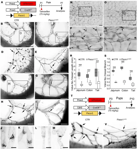
pups at postnatal day 1 (P1) by tamoxifen injection, and subsequently, lymphatic valve formation in the mesentery and tail skin was analyzed at P7 (Figure 1A). Mesenteric lymphatic valves, clearly marked with strong tdTomato expression because of high expression of Prox1 in lymphatic valve leaflet LECs, were normally formed at vascular branch points of the control animals. In the lymphatic Piezo1-KO pups, how-ever, not only were mesenteric lymphatic vessels scarce in terms of their density, but they also lacked mature valves at the branching points (Figure 1, B–I). Accordingly, the total numbers of valves (matured and immatured) were significantly reduced by Piezo1 deletion in both jejunum and colon (Figure 1, R and S). Moreover, high-powered images identified that, while all the control valves appeared to be fully mature, Piezo1-deleted mice demonstrated immature valves that were arrested at different stages of valve develop-ment (ref. 16 and Figure 1, J–M). We could detect the absence of lymphatic valve–forming activity at the expected valve branching points (Figure 1, E and I), constriction of vessels (Figure 1L), and immature valve leaflets (Figure 1M). This observation raised a possibility that Piezo1 may be essential for many stages of lymphatic valve development. Furthermore, we could also detect defective lymphatic valve formation in the tail skin after Piezo1 deletion (Figure 1, N–S). Notably, these conditional lymphatic Piezo1-KO pups exhib-ited a substantially retarded body growth (Supplemental Figure 1; supplemental material available online with this article; https://doi.org/10.1172/jci.insight.125068DS1). To confirm these phenotypes using an independent Cre allele, we next used the Cdh5(PAC)-CreERT2 line (33). Because Cdh5/vascular endothelial
cadherin is expressed in nearly all ECs (34), this Cre allele was expected to induce Piezo1 deletion in both blood and LECs. Using this driver line, Piezo1 was deleted at P3, and its consequence on lymphatic valve formation was studied at P10 (Figure 1T). Indeed, Piezo1 ablation by the Cdh5(PAC)-CreERT2 allele inhibited
lymphatic valve formation (Figure 1, U and V, and Supplemental Figure 2).
As mentioned above, Piezo1 deletion also reduced lymphatic vessel density, possibly by suppressing lym-phatic vessel growth or sprouting (Figure 1, B–I, and Supplemental Figure 3). We thus hypothesized that Piezo1 may be expressed not only in lymphatic valves (valvular LECs), but also in lymphatic vessels (luminal LECs). To date, the expression pattern of Piezo1 in the lymphatics in vivo has not been clearly documented. We therefore investigated the expression pattern of Piezo1 in valvular and luminal LECs using a Piezo1 reporter mouse, Piezo1P1−tdT, which encodes a C-terminus fusion protein of Piezo1 with tdTomato protein
(26). This reporter mouse, combined with immunofluorescence assays, showed that both valvular and luminal LECs express Piezo1 with much stronger expression in the valvular leaflet LECs (Figure 2, A–D).
We next asked whether lymphatic valve formation might have been prevented by defective lymphangiogene-sis in Piezo1-KO animals. Although a lymphatic valve–specific Cre line would be the most desirable tool to delete Piezo1 exclusively in valvular LECs, but not in luminal LECs, no animal model has been established offering such selectivity. Therefore, we set out to deal with this technical limitation by significantly reducing tamoxifen dosage: Because Prox1 expression is 5–10 times higher in valvular LECs than luminal LECs (35, 36), CreERT2
protein encoded by the Prox1-CreERT2 allele is much more abundant in valvular LECs than luminal LECs. We
thus speculate that, at low-dose tamoxifen, Piezo1 deletion by Prox1-CreERT2 allele would occur more readily in
Figure 1. Piezo1 is essential for lymphatic valve development. (A–S) Effect of lymphatic deletion of Piezo1 on lymphatic valve development. (A) Experimental design. Control pups (CTR) harbor Prox1-tdTomato and Prox1-CreERT2, but lack the Piezo1fl/fl allele, while lymphatic Piezo1-KO pups (Piezo1ΔLEC) have all
R E S E A R C H A R T I C L E
Figure 2. Piezo1 deletion in lymphatic valves by low-dose tamoxifen. (A–D) Dermal lymphatics in the ear of Piezo1-tdTomato adult reporter mouse Piezo1P1−tdT (26) were immunostained for tdTomato protein (to amplify the reporter signal) and Prox1 (to detect LECs). Images were captured by an (A and
B) epifluorescence or (C and D) confocal microscope. Note an enriched expression of Piezo1-tdTomato fusion protein in Prox1hi valve leaflet LECs. (E–N)
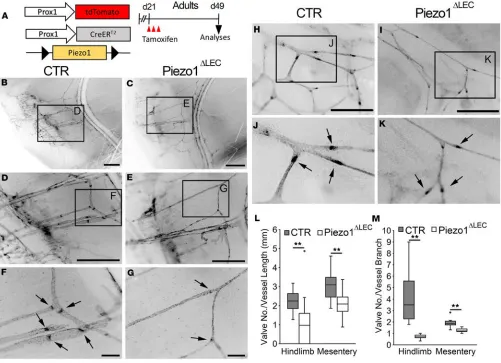
Piezo1 deletion leads to degeneration of lymphatic valves in adults. We next asked whether Piezo1 function is continuously needed to maintain lymphatic valves after development. Lymphatic Piezo1 deletion was induced at day 21 (juvenile), and the integrity of lymphatic valves in the skin and mesentery was investi-gated at day 49 (adult) (Figure 3A). We first examined matured lymphatic collectors that run along with the saphenous vein in the hind limb of the control and Piezo1-mutant mice. Notably, postdevelopmental lymphatic Piezo1 deletion caused significant degeneration of both lymphatic valves and lymphatic vessels in the skin (Figure 3, B–G, L, M). These lymphatic atrophies were also detected in the mesentery, where the lymphatics of Piezo1-deficient mice were much thinner and scarce, with fewer valves, compared with those of the control mice (Figure 3, H–M). Together, these data suggest that Piezo1 function is continuously necessary to maintain the integrity of lymphatic vessels and valves in adults.
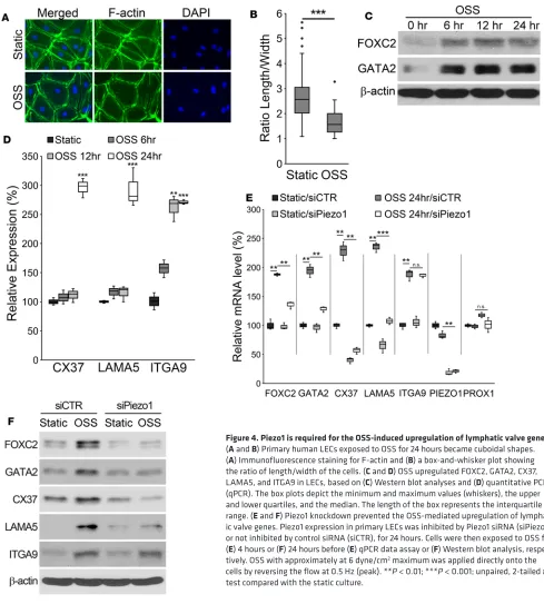
Piezo1 is required for the OSS-mediated upregulation of lymphatic valve genes. The in vivo lymphatic pheno-types of Piezo1-KO animals led us to investigate the mechanotransduction mechanism for the lymphatic valve formation. Fluid flow is known to play a key role in vascular development and morphogenesis (5). In particular, OSS has been reported to upregulate the lymphatic valve–associated genes, such as FOXC2, GATA2, CX37, LAMA5, and ITGA9, and to induce cellular morphology change (15–22). We thus inves-tigated how the mechanosensor Piezo1 affects the mechanotransduction process by performing in vitro loss-of-function studies. Consistent with the previous reports (15–20), OSS changed the morphology of cul-tured primary human LECs to more cuboidal shapes (Figure 4, A and B, and Supplemental Figure 5) and upregulated the lymphatic valve–associated genes mentioned above (Figure 4, C and D, and Supplemen-tal Figure 6). Remarkably, the OSS-induced upregulation of these genes was largely abrogated by Piezo1 knockdown (Figure 4, E and F, and Supplemental Figure 7). Notably, upregulation of ITGA9 by OSS was not affected by Piezo1 knockdown. Together, these loss-of-function studies suggest that Piezo1 plays a key role in the OSS-activated mechanotransduction that regulates lymphatic valve gene expression.
Ectopic expression of Piezo1 triggers lymphatic valve gene expression profile. Conversely, we next asked whether Piezo1 gain of function could recapitulate the molecular phenotypes caused by OSS. We transfected primary LECs with a plasmid bicistronically encoding mouse Piezo1 and EGFP and then evaluated the expression of the lymphatic valve–associated genes. EGFP+, thus overexpressing Piezo1,
upregulated FOXC2, GATA2, CX37, and LAMA5 expression by 3- to 4-fold, in the absence of OSS (arrowheads, Figure 5, A and B). However, neighboring EGFP– (untransfected) LECs did not
upregu-late these genes (arrows, Figure 5, A and B). Notably, ITGA9 was not upreguupregu-lated by Piezo1 expres-sion. To confirm these data, LECs were transfected with a control or Piezo1-expressing vector and further cultured in the absence of OSS. Indeed, quantitative PCR (qPCR) and Western blot analyses revealed that overexpression of Piezo1 upregulated the expression of mRNA and protein of FOXC2, GATA2, CX37, and LAMA5 (Figure 5, C and D). ITGA9 expression was consistently unaltered. Together, these gain-of-function studies demonstrate that ectopic expression of Piezo1 was able to partly recapitulate the lymphatic valve gene profile in the absence of OSS.
Activation of Piezo1 accelerates lymphatic valve formation in a mouse model. The outcome of the in vitro gain-of-function study led us to investigate whether activation of Piezo1 promotes lymphatic valve formation in animals. Yoda1, identified as the first small chemical agonist of Piezo1, delays the inactivation phase of transient currents and sensitizes Piezo1 response to external mechanical stimuli (37). Thus, we asked whether Yoda1 treatment enhances lymphatic valve formation in vivo. Toward this goal, Yoda1, or vehicle alone, was injected into Prox1-EGFP mice at E18.5 (into pregnant females) and again at P0 (into new-borns). The next day at P1, lymphatic valve formation was analyzed in the mesenteries of the jejunum and colon and in the tail skin of the control or Yoda1-treated pups (Figure 6A). While valve formation activity was not yet detected at the proximal regions of the mesentery lymphatics of the vehicle-treated jejunum, a number of lymphatic valves were already visible in the Yoda1-treated jejunum mesentery (Figure 6B). Similarly, Yoda1 accelerated lymphatic valve formation in the colon and tail skin: A higher number of lymphatic valves could be found in the Yoda1-treated pups (Figure 6, C and D). Yoda1 treatment signifi-cantly enhanced lymphatic valve formation in all the tissues tested (Figure 6, E and F). Together, we con-cluded that chemical activation of Piezo1 was sufficient to augment lymphatic valve development, possibly through sensitization of Piezo1 to lymphatic fluid flow in vivo.
R E S E A R C H A R T I C L E
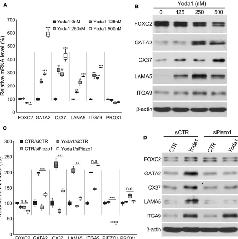
different doses of Yoda1 under the standard static condition. Indeed, Yoda1 treatment of LECs led to upregulation of both mRNA and protein of GATA2, CX37, LAMA5, and ITGA9 (Figure 7, A and B). Interesting, FOXC2 was rather slightly downregulated by increasing doses of Yoda1. Prox1 expression was not altered. Despite the reported EC50 of Yoda1 activation of Piezo1 in transfected HEK293 cells being about 20–25 μM (37), primary human LECs treated with Yoda1 higher than 1 μM for 24 hours revealed several signs of significant cytotoxicity, including cell detachment, cell-cell junction disruption, and cell contraction and spindle formation (data not shown). Moreover, we investigated whether the Yoda1-me-diated upregulation of these lymphatic valve genes was meYoda1-me-diated through Piezo1. LECs were transfected with siRNA for control or Piezo1 to knock down Piezo1 expression, and their expressions were determined using qPCR and Western blot analyses. Indeed, Piezo1 knockdown prevented Yoda1-induced upregulation of GATA2, CX37, and LAMA5 (Figure 7, C and D). Interestingly, however, Piezo1 knockdown did not affect Yoda1-induced ITGA9 expression. Together, these data suggest that chemical activation of Piezo1 using Yoda1 upregulates some of the lymphatic valve genes in the absence of mechanical force.
Discussion
Lymphatic valves are essential for the unidirectional flow of interstitial fluid. Because of malformations or injuries, diseased lymphatic valves often lead to severe lymphatic pathologies. Recent studies demon-strated that fluid flow–derived physical signal significantly controls lymphatic valve development through mechanotransduction pathways and identified key molecular players essential for the pathways (15–22). In this study, we defined the crucial role of the cell stretch sensor Piezo1 in incorporating the flow-mediated physical signal into the genetic program controlling lymphatic valve development. Previous studies suggest-ed that vascular ECs in vivo would be subjectsuggest-ed to both fluid shear stress and cyclic cell stretch, and, when combined in vitro, these 2 physical stimuli appeared to synergistically deliver various vascular phenotypes,
such as actin filament alignment and cell differentiation (38–41). Accordingly, we hypothesize that the OSS and cyclic cell stretching may be similarly sensed by Piezo1 and trigger the genetic program responsible for lymphatic valve formation. OSS may induce plasma membrane spreading and cytoskeletal rearrangement, similar to those effects created by cyclic cell stretching.
In this study, loss of function of Piezo1 in mice and in purified cells clearly demonstrated the essential role of Piezo1 in various stages of lymphatic valve development. To conditionally delete Piezo1, we used 2 Cre drivers that target nearly all ECs as well as selectively for lymphatic-lineage ECs (Figure 1). We also success-fully targeted lymphatic valve ECs by lowering tamoxifen concentrations (Figure 2). Moreover, we induced Piezo1 deletion in young adult mice to study the role of Piezo1 in matured lymphatic valves and found that matured lymphatic valves continuously required Piezo1 activity (Figure 3). All these in vivo loss-of-function studies defined the critical role of Peizo1 not only in lymphatic valve development but also in valve mainte-nance. These data once again support the positive feedback mechanism between fluid flow and the lymphatic valve–forming mechanotransduction mechanism: Lymphatic flow triggers lymphatic valve formation and maintenance, while lymphatic valves ensure proper lymphatic flow. Thus, a disruption of this delicate recip-rocal feedback, for example because of surgical obstruction of lymphatic vessels in cancer patients (lymph-adenectomy), would lead to lymphatic valve degeneration and eventually lymph stasis and lymphedema.
The interesting outcome of the gain-of-function studies in this paper elegantly mirrors the results of the loss-of-function studies. The ectopic upregulation of Piezo1 largely recapitulated the lymphatic valve gene profiles in cultured LECs without any fluid flow (Figure 5). Several molecular mechanisms could be envi-sioned. First, with an increased amount of Piezo1 protein, the cells could become much more sensitive to external force, such as subtle movement of culturing media or even their own movement during the culture period. Consistently, when reconstituted into lipid bilayers, purified Piezo1 proteins were able to mediate spontaneous and membrane tension–induced cationic currents (23, 42). Considering that Piezo1 forms a homotrimeric complex (43, 44), an alternative explanation could be that the increased number of Piezo1 monomers significantly facilitates the ion channel complex formation. A third mechanism could be that abundant Piezo1 protein may be able to overcome intrinsic negative regulations. For example, the Sarco/ endoplasmic reticulum Ca(2+)-ATPase (SERCA) family proteins, including SERCA2 isoform, have been
identified as negative regulators of Piezo channels by acting through direct protein-protein interaction (45). Thus, more Piezo1 proteins could overcome such a SERCA-mediated suppression mechanism.
This gain of function could also be attained by chemical activation of Piezo1 in animals. Yoda1 is known to slow the inactivation phase of transient currents and sensitize Piezo1 response to external mechanical stimuli (37). Activation of Piezo1 using Yoda1 administration could accelerate lymphatic valve development in the mesenteries and skin of newborn mice (Figure 6). It is widely believed that lymphatic valves are formed at the anatomical sites where fluid flows are oscillatory or disturbed, such as vascular beds at branching points (20, 36). Importantly, although Yoda1 indeed promoted the valve formation process, when administered at the early stage, it did not seem to induce valve formation at the unusual, ectopic locations other than the expected sites. This suggests that Yoda1 or Piezo1 may be necessary, but not sufficient, for lymphatic valve development. Moreover, this is consistent with previous findings that Yoda1 only partially activates channels in the absence of external pressure (37) and our own data showing that Yoda1 treatment upregulated only some, but not all, lymphatic valve genes in cultured LECs without fluid flow (Figure 7).
Our studies in this report consistently found that regulation of ITGA9 expression was intriguingly unique compared with other lymphatic valve genes, providing some insights into possible Piezo1-inde-pendent force-sensing mechanisms. ITGA9 is known for its enriched expression in lymphatic valve leaflet ECs, although it is also moderately expressed in luminal LECs (3, 19, 20, 46–48). Protein and mRNA of ITGA9 were significantly upregulated in cultured LECs by OSS (Figure 4). Notably, Piezo1 was not essen-tial for this OSS-induced upregulation of ITGA9, whereas it was for upregulation of all other genes tested.
R E S E A R C H A R T I C L E
This observation raised the notion that the OSS-induced upregulation of ITGA9 is not Piezo1 dependent, and this notion seems to be consistent with our finding that ectopic expression of Piezo1 did not increase ITGA9 expression (Figure 5). However, Yoda1 did upregulate ITGA9 in LECs in vitro as well as enhanced normal lymphatic valve formation in vivo (Figures 6 and 7). However, this Yoda1-induced ITGA9 upreg-ulation in vitro did not require Piezo1 (Figure 7). Although these observations appear counterintuitive, a couple of possibilities could be considered. One is that the current experimental condition of OSS does not
precisely recapitulate the in vivo lymphatic valve–forming program and that this technical limitation may have caused some discrepancies among our in vitro and animal data. Another explanation is that ITGA9 may be upregulated in LECs by OSS through different mechanosensitive channels that could be stimulated, directly or indirectly, by Yoda1. Because Yoda1 does not activate Piezo2 (37, 49), Piezo2 can be excluded from the candidate list. Other stretch-activated channels, such as the transient receptor potential channel
R E S E A R C H A R T I C L E
subfamily C (TRPC) proteins (50–52), may be plausible candidates. Currently, neither the role of TRPC proteins in lymphatic development, nor the effect of Yoda1 on TRPC proteins, is known. Thus, it will be interesting and important to investigate these open questions.
During the review phase of this manuscript, a very similar study on the role of Piezo1 in lymphatic valve development was published by Nonomura et al. (53). The results from our and their studies are largely complementary and support the essential role of Piezo1 in lymphatic valve development. Yet, 2 points are worthy of discussion. First, these authors employed 2 constitutive endothelial Cre lines (Tie2-Cre and Lyve1-(Tie2-Cre), which delete Piezo1 in blood endothelial cells as well as LECs (53). The authors found gross phenotypes, including pleural effusion, similar to those displayed by human patients with PIEZO1 loss-of-function mutations (28–30). In comparison, our study mainly used the Prox1-CreERT2 allele, which
selectively targets LECs in a time-controlled manner by tamoxifen administration (Figures 1–3). Although our Piezo1-KO pups/mice showed growth retardation (Supplemental Figure 1), we did not detect pleural effusion or chylous ascites. These results may well be explained by the effects of different spatiotemporal deletion of Piezo1 in our and their mouse models. The other point to note is that although our in vitro data revealed that Piezo1 regulated the key lymphatic valve signature genes, including FOXC2 and GATA2 (Figures 4 and 5), Nonomura et al. (53) found that Piezo1 was not required for the initiation of lymphatic valve formation. Although these discrepancies may be attributable to technical or experimental variations, further studies are required to resolve this difference. Nonetheless, both studies together clearly defined the essential role of the cell stretch sensor Piezo1 in lymphatic valve development.
Methods
Animal-related methods. Sources of the mice are: Prox1-EGFP (Tg[Prox1-EGFP]KY221Gsat, Mutant Mouse Resource and Research Centers, UCD, Davis, California, USA) (35), Prox1-tdTomato [Tg(tdTomato)TA76Gsat/Mmucd, Mutant Mouse Resource and Research Centers] (31),
Prox1-CreERT2 (a gift from Taija Mäkinen, Uppsala University, Uppsala, Sweden) (19), Piezo1fl/fl (Piezo1tm2.1Apat/J,
The Jackson Laboratory) (32), Piezo1P1−tdT (B6;129-Piezo1tm1.1Apat/J) (26), and Cdh5(PAC)-CreERT2
(a gift from Ralf Adams, University of Münster, Münster, Germany) (33). Mice were maintained in mixed outbred backgrounds. Both sexes were used for each experiment. Tamoxifen (MP Biomedicals) was dissolved in DMSO, mixed with sunflower seed oil (1 vol DMSO: 2 vol oil), and injected at 75 mg/ kg once for pups and 3 times for young adults. For Figure 1, tamoxifen or vehicle was s.c. injected into neonatal mouse pups (P1), and tissue samples were harvested at P7 to analyze their genotypes and lym-phatic valve development. Pups harboring Prox1-tdTomato and Prox1-CreERT2, but lacking the Piezo1fl/fl
allele, were considered the CTR, while pups having Prox1-tdTomato, Prox1-CreERT2, and Piezo1fl/fl were
marked as lymphatic Piezo1-KO (Piezo1ΔLEC). Yoda1 (MilliporeSigma) was dissolved in DMSO and then
mixed with PBS (1.5 vol DMSO: 40 vol PBS) before injection at 70 μg/kg.
Molecular and histological analyses. qPCR and Western blot analyses were performed using standard pro-tocols as described (56). All fluorescence images were captured using a Leica stereomicroscope (Leica M165 FC) or a Zeiss ApoTome microscope (Zeiss AxioVision), except the images shown in Figure 2, C and D, which were obtained by a confocal microscope (Leica TCS SP5). Captured signals were invert-ed and expressinvert-ed in grayscale using Adobe Photoshop CS5. Quantitative image analyses were performinvert-ed using NIH ImageJ (NIH) software.
Flow generation. Oscillating flow was generated using a horizontal bidirectional shaker (MS-NOR-30, Major Science). A modified culture dish was made, as previously reported (7, 8), by adhering a 6-cm cell culture dish in the center of a 10-cm culture dish using a nontoxic medical silicone glue (A-100, medical silicone adhesive, Factor II, Inc.). Cells were seeded in the donut-shaped track and incubated for 24 hours under the static condition. Culture dishes were then horizontally rotated clockwise for 1 second and then counterclockwise for 1 second to generate reversing flow at maximum speed 100 rpm, 0.5 Hz, for approxi-mately 6–24 hours. The approximate maximum wall shear stress was about 6 dyne/cm2.
Statistics. An unpaired, 2-tailed t test was conducted using Microsoft Excel and GraphPad Prism 6 (GraphPad Software, Inc.) to determine whether the differences between the experimental and control groups were statistically significant. P value less than 0.05 was considered significant. Box-and-whisker plots were drawn using Microsoft Excel.
Study approval. All mouse methods were approved by the Institutional Animal Care and Use Commit-tee, University of Southern California, Los Angeles, California. Isolation and culturing of human primary LECs from otherwise discarded, deidentified human foreskins were approved by the Institutional Review Board of the University of Southern California, Los Angeles, California.
Author contributions
DC, EP, EJ, BC, SL, JY, PMK, SL, YJH, CWC, and YW designed and performed the experiments and analyzed the data. CJK and NLJ provided the resources. AKW, LS, SRK, IBM, RSS, ITC, and YKH con-ceived, developed, and directed the project and/or wrote the paper.
Acknowledgments
This study was supported by NIH grants (R21EY026260, R01HL121036, R01HL141857, R21DE027891, and R01DK114645 to YH; R01HL131652 to RSS; and K08HL121191 to SRK) and by National Research Foun-dation of Korea (NRF-2018R1A2A1A05019550 to NLJ). We thank Taija Mäkinen (Uppsala University) and Ralf Adams (University of Münster) for sharing Prox1-CreERT2 and Cdh5(PAC)-CreERT2 mouse lines, respectively.
Address correspondence to: Young-Kwon Hong, Department of Surgery, University of Southern Califor-nia, Norris Comprehensive Cancer Center, 1450 Biggy St. NRT6501, Los Angeles, California 90033, USA. Phone: 323.442.7825; Email: [email protected].
1. Breslin JW. Mechanical forces and lymphatic transport. Microvasc Res. 2014;96:46–54.
2. Bazigou E, Wilson JT, Moore JE. Primary and secondary lymphatic valve development: molecular, functional and mechanical insights. Microvasc Res. 2014;96:38–45.
3. Bazigou E, Makinen T. Flow control in our vessels: vascular valves make sure there is no way back. Cell Mol Life Sci. 2013;70(6):1055–1066.
4. Schmid-Schonbein GW. The second valve system in lymphatics. Lymphat Res Biol. 2003;1(1):25–29.
5. Chiu JJ, Chien S. Effects of disturbed flow on vascular endothelium: pathophysiological basis and clinical perspectives. Physiol
Rev. 2011;91(1):327–387.
6. Schwartz MA, Simons M. Lymphatics thrive on stress: mechanical force in lymphatic development. EMBO J. 2012;31(4):781–782. 7. Choi D, et al. ORAI1 activates proliferation of lymphatic endothelial cells in response to laminar flow through Krüppel-like
factors 2 and 4. Circ Res. 2017;120(9):1426–1439.
8. Choi D, et al. Laminar flow downregulates Notch activity to promote lymphatic sprouting. J Clin Invest. 2017;127(4):1225–1240. 9. Sessa WC. Molecular control of blood flow and angiogenesis: role of nitric oxide. J Thromb Haemost. 2009;7(suppl 1):35–37. 10. Boldock L, Wittkowske C, Perrault CM. Microfluidic traction force microscopy to study mechanotransduction in angiogenesis.
Microcirculation. 2017;24(5):e12361.
11. Sabine A, Saygili Demir C, Petrova TV. Endothelial cell responses to biomechanical forces in lymphatic vessels. Antioxid Redox
Signal. 2016;25(7):451–465.
12. Baeyens N, Bandyopadhyay C, Coon BG, Yun S, Schwartz MA. Endothelial fluid shear stress sensing in vascular health and disease. J Clin Invest. 2016;126(3):821–828.
R E S E A R C H A R T I C L E
14. Chatterjee S, Fujiwara K, Pérez NG, Ushio-Fukai M, Fisher AB. Mechanosignaling in the vasculature: emerging concepts in sensing, transduction and physiological responses. Am J Physiol Heart Circ Physiol. 2015;308(12):H1451–H1462.
15. Kazenwadel J, et al. GATA2 is required for lymphatic vessel valve development and maintenance. J Clin Invest. 2015;125(8):2979–2994.
16. Sabine A, et al. Mechanotransduction, PROX1, and FOXC2 cooperate to control connexin37 and calcineurin during lymphat-ic-valve formation. Dev Cell. 2012;22(2):430–445.
17. Sabine A, et al. FOXC2 and fluid shear stress stabilize postnatal lymphatic vasculature. J Clin Invest. 2015;125(10):3861–3877. 18. Cha B, et al. Mechanotransduction activates canonical Wnt/β-catenin signaling to promote lymphatic vascular patterning and
the development of lymphatic and lymphovenous valves. Genes Dev. 2016;30(12):1454–1469.
19. Bazigou E, et al. Genes regulating lymphangiogenesis control venous valve formation and maintenance in mice. J Clin Invest. 2011;121(8):2984–2992.
20. Bazigou E, et al. Integrin-α9 is required for fibronectin matrix assembly during lymphatic valve morphogenesis. Dev Cell. 2009;17(2):175–186.
21. Udan RS, Dickinson ME. The ebb and flow of lymphatic valve formation. Dev Cell. 2012;22(2):242–243.
22. Janardhan HP, Milstone ZJ, Shin M, Lawson ND, Keaney Jr JF, Trivedi CM. Hdac3 regulates lymphovenous and lymphatic valve formation. J Clin Invest. 2017;127(11):4193–4206.
23. Coste B, et al. Piezo proteins are pore-forming subunits of mechanically activated channels. Nature. 2012;483(7388):176–181. 24. Kim SE, Coste B, Chadha A, Cook B, Patapoutian A. The role of Drosophila Piezo in mechanical nociception. Nature.
2012;483(7388):209–212.
25. Coste B, et al. Piezo1 and Piezo2 are essential components of distinct mechanically activated cation channels. Science. 2010;330(6000):55–60.
26. Ranade SS, et al. Piezo1, a mechanically activated ion channel, is required for vascular development in mice. Proc Natl Acad Sci
U S A. 2014;111(28):10347–10352.
27. Li J, et al. Piezo1 integration of vascular architecture with physiological force. Nature. 2014;515(7526):279–282.
28. Lukacs V, et al. Impaired PIEZO1 function in patients with a novel autosomal recessive congenital lymphatic dysplasia. Nat
Commun. 2015;6:8329.
29. Fotiou E, et al. Novel mutations in PIEZO1 cause an autosomal recessive generalized lymphatic dysplasia with non-immune hydrops fetalis. Nat Commun. 2015;6:8085.
30. Datkhaeva I, et al. Identification of novel PIEZO1 variants using prenatal exome sequencing and correlation to ultrasound and autopsy findings of recurrent hydrops fetalis. Am J Med Genet A. 2018;176(12):2829–2834.
31. Hong M, et al. Efficient assessment of developmental, surgical and pathological lymphangiogenesis using a lymphatic reporter mouse and its embryonic stem cells. PLoS One. 2016;11(6):e0157126.
32. Cahalan SM, Lukacs V, Ranade SS, Chien S, Bandell M, Patapoutian A. Piezo1 links mechanical forces to red blood cell vol-ume. Elife. 2015;4:e07370.
33. Wang Y, et al. Ephrin-B2 controls VEGF-induced angiogenesis and lymphangiogenesis. Nature. 2010;465(7297):483–486. 34. Giannotta M, Trani M, Dejana E. VE-cadherin and endothelial adherens junctions: active guardians of vascular integrity. Dev
Cell. 2013;26(5):441–454.
35. Choi I, et al. Visualization of lymphatic vessels by Prox1-promoter directed GFP reporter in a bacterial artificial chromo-some-based transgenic mouse. Blood. 2011;117(1):362–365.
36. Norrmén C, et al. FOXC2 controls formation and maturation of lymphatic collecting vessels through cooperation with NFATc1. J Cell Biol. 2009;185(3):439–457.
37. Syeda R, et al. Chemical activation of the mechanotransduction channel Piezo1. Elife. 2015;4:e07369.
38. Zhao S, et al. Synergistic effects of fluid shear stress and cyclic circumferential stretch on vascular endothelial cell morphology and cytoskeleton. Arterioscler Thromb Vasc Biol. 1995;15(10):1781–1786.
39. Kim DH, Heo SJ, Kang YG, Shin JW, Park SH, Shin JW. Shear stress and circumferential stretch by pulsatile flow direct vascular endothelial lineage commitment of mesenchymal stem cells in engineered blood vessels. J Mater Sci Mater Med. 2016;27(3):60.
40. Moore JE, et al. A device for subjecting vascular endothelial cells to both fluid shear stress and circumferential cyclic stretch.
Ann Biomed Eng. 1994;22(4):416–422.
41. Owatverot TB, Oswald SJ, Chen Y, Wille JJ, Yin FC. Effect of combined cyclic stretch and fluid shear stress on endothelial cell morphological responses. J Biomech Eng. 2005;127(3):374–382.
42. Syeda R, et al. Piezo1 channels are inherently mechanosensitive. Cell Rep. 2016;17(7):1739–1746.
43. Ge J, et al. Architecture of the mammalian mechanosensitive Piezo1 channel. Nature. 2015;527(7576):64–69. 44. Zhao Q, et al. Structure and mechanogating mechanism of the Piezo1 channel. Nature. 2018;554(7693):487–492.
45. Zhang T, Chi S, Jiang F, Zhao Q, Xiao B. A protein interaction mechanism for suppressing the mechanosensitive Piezo chan-nels. Nat Commun. 2017;8(1):1797.
46. Altiok E, et al. Integrin α-9 mediates lymphatic valve formation in corneal lymphangiogenesis. Invest Ophthalmol Vis Sci. 2015;56(11):6313–6319.
47. Høye AM, Couchman JR, Wewer UM, Fukami K, Yoneda A. The newcomer in the integrin family: integrin ,α9 in biology and cancer. Adv Biol Regul. 2012;52(2):326–339.
48. Mishima K, et al. Prox1 induces lymphatic endothelial differentiation via integrin α9 and other signaling cascades. Mol Biol Cell. 2007;18(4):1421–1429.
49. Lacroix JJ, Botello-Smith WM, Luo Y. Probing the gating mechanism of the mechanosensitive channel Piezo1 with the small molecule Yoda1. Nat Commun. 2018;9(1):2029.
50. Inoue R, Jian Z, Kawarabayashi Y. Mechanosensitive TRP channels in cardiovascular pathophysiology. Pharmacol Ther. 2009;123(3):371–385.
51. Barritt G, Rychkov G. TRPs as mechanosensitive channels. Nat Cell Biol. 2005;7(2):105–107.
in human myometrial smooth muscle cells. Mol Hum Reprod. 2007;13(3):171–179.
53. Nonomura K, et al. Mechanically activated ion channel PIEZO1 is required for lymphatic valve formation. Proc Natl Acad Sci U
S A. 2018;115(50):12817–12822.
54. Shin JW, et al. Prox1 promotes lineage-specific expression of fibroblast growth factor (FGF) receptor-3 in lymphatic endotheli-um: a role for FGF signaling in lymphangiogenesis. Mol Biol Cell. 2006;17(2):576–584.
55. Lee S, et al. Prox1 physically and functionally interacts with COUP-TFII to specify lymphatic endothelial cell fate. Blood. 2009;113(8):1856–1859.