Case Report:
Endoscopic Transsphenoidal Fenestration of a Medial
Temporal Arachnoid Cyst With Extension to Sphenoid
Sinus in a Patient With Temporal Lobe Epilepsy
Arash Saffarian1, Nima Derakhshan1* , Mousa Taghipour1, Mohammad Faramarzi2, Ali Akbar Asadipooya3, 4, 5, Amirreza Dehghanian6, 7, Keyvan Eghbal1
1. Department of Neurosurgery, School of Medicine, Shiraz University of Medical Sciences, Shiraz, Iran 2. Department of Otolaryngology, School of Medicine, Shiraz University of Medical Sciences, Shiraz, Iran 3. Department of Neurology, School of Medicine, Shiraz University of Medical Sciences, Shiraz, Iran
4. Department of Neurology, School of Medicine, Thomas Jefferson University, Philadelphia, USA
5. Epilepsy Care Unit, Namazi Hospital, School of Medicine, Shiraz University of Medical Sciences, Shiraz, Iran
6. Department of Pathology, School of Medicine, Shiraz University of Medical Sciences, Shiraz, Iran
7. Department of Pathology, School of Medicine, Chamran Hospital, Shiraz, Iran
* Corresponding Author: Nima Derakhshan, MD.
Address: Department of Neurosurgery, School of Medicine, Shiraz University of Medical Sciences, Shiraz, Iran Tel: +98 (917) 7161290
E-mail: [email protected]
Background and Importance: Arachnoid cysts are developmental cystic lesions which may be found as an incidental finding on neuroimaging or present with symptoms of headache, seizure and neurologic deficit. Presentation with seizure is more common with larger sizes and temporal location. Presentation with Temporal Lobe Epilepsy (TLE) is rare, and fenestration of cysts has variable results for seizure control. We reported controlling TLE symptoms following endoscopic transsphenoidal fenestration of an arachnoid cyst. The anteromedial location in middle fossa, extension toward sphenoid sinus and normal appearance of mesial temporal structures on MRI encouraged us to consider this surgical approach.
Case Presentation: A 26-year-old patient with a 13-year history of TLE with uncontrolled symptoms despite taking a combination of AEDs (LTG, CBZ, LEV, CLB) was referred to our clinic. Neuroimaging revealed an arachnoid cyst in anteromedial part of temporal fossa which extended to sphenoid sinus, but showed no abnormality in mesial temporal structures. Endoscopic endonasal transsphenoidal fenestration of the arachnoid cyst was performed, and followed by reconstruction of the skull base. The procedure improved the seizure control during the 9-month follow-up and no sign of radiologic recurrence was observed.
Conclusion: Transsphenoidal endoscopic fenestration is a safe and feasible surgical approach for treatment of symptomatic arachnoid cysts in anteromedial part of middle fossa especially when they extend toward lateral wall of sphenoid sinus. This surgical corridor has the privilege of avoiding cortical injury accompanied by transcranial approaches, which is deleterious in epileptic patients.
A B S T R A C T
Keywords:
Temporal lobe epilepsy (TLE), Neuroendoscopy, Trans-sphenoidal, Arachnoid cyst
Citation: Saffarian A, Derakhshan N, Taghipour M, Faramarzi M, Asadipooya AA, Dehghanian AR, Eghbal K. Endoscopic
Transsphenoidal Fenestration of a Medial Temporal Arachnoid Cyst With Extension to Sphenoid Sinus in a Patient With Tempo‐
ral Lobe Epilepsy Iran J Neurosurg. 2019; 5(2):93-98. http://dx.doi.org/10.32598/irjns.5.2.93
: http://dx.doi.org/10.32598/irjns.5.2.93 Use your device to scan
and read the article online
Article info:
Received: 02 Oct 2018
Accepted: 23 Feb 2019
1. Background and Importance
emporal Lobe Epilepsy (TLE) is the most com‐ mon form of drug-resistant focal epilepsy. Current understanding of TLE supports the hypothesis of impairment of inhibitory action of basket cells following an insult in a critical brain development period, which is rapidly distributed toward other regions via kindling phenomenon [1,2]. Several pathologies have been associated with TLE in‐ cluding Mesial Temporal Sclerosis (MTS), developmen‐ tal malformations, vascular malformations, neoplastic lesions and scars caused by infarctions and infective processes [3]. Arachnoid cysts have been reported in as many as 10% of patients with TLE, and their presence is secondary to abnormal development of temporal lobe in most instances [4].Fenestration of temporal arachnoid cysts have been associated with variable results for seizure control in pa‐ tients with TLE, according to major differences in cases included. Those with hippocampal sclerosis had subop‐ timal improvement with standalone arachnoid cyst fen‐ estration and will have to undergo selective amygdalo‐ hippocampectomy for optimal seizure control.
Herein, we are reporting a 26-year-old man with a 13-year history of TLE. His seizures were successfully con‐ trolled with endoscopic transsphenoidal fenestration of an anteromedial temporal arachnoid cyst that had extended to sphenoid sinus and pterygopalatine fossa. The normal appearance of mesial temporal structures on pre-operative MRI and extension to sphenoid sinus encouraged us to consider this surgical approach in this very patient.
2. Case Presentation
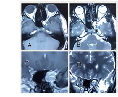
A 26-year-old healthy male was referred by an epi‐ leptologist with a 13-year history of seizures which oc‐ curred on a monthly basis despite taking several Anti-Epileptic Drugs (AEDs) (LTG, CBZ, LEV, CLB). His ictus consisted of olfactory and visual hallucinations, and de ja vu with loss of consciousness for 2-3 minutes, but without tonic and clonic phases. The diagnosis was con‐ firmed to be TLE. His drug history included lamotrigine 200 mg daily, imipramine 100 mg daily, alprazolam 0.5 mg at bedtime and trifluoperazine 4 mg daily. His fam‐ ily history was unremarkable for seizures or any other neurological disorders. Brain MRI revealed a left medial temporal cystic lesion, hypointense on T1- and hyperin‐ tense on T2- weighted images, respectively which had extended inferiorly to pterygopalatine fossa through
T
Highlights
● Mesial temporal Arachnoid cysts may rarely present with TLE.
● Transsphenoidal approach for fenestration of a mesial temporal arachnoid cyst is safely performed.
● Skull base reconstruction by the ENT surgeon following the fenestrations is mandatory to avoid CSF leakage.
● Transsphenoidal approach leaves the temporal cortex untouched and avoids the little, but considerable chance of iatrogenic foci for seizures.
Plain Language Summary
temporal fossa floor and also medially bulging into sphenoid sinus through its lateral wall (Figure 1).
Surgical procedure
The cystic lesion was approached endoscopically through transsphenoidal route. After visualizing via a bi-nostril exposure of the sphenoid sinus, the bulged area in the lateral wall of sphenoid sinus was seen. During removal of the eroded thin wall by curettes, the thinned cyst wall was ruptured followed by an abrupt flow of Ce‐ rebrospinal Fluid (CSF). After suctioning the fluid, a fat plug was inserted within the opening in cyst wall, and the closure was reinforced with fibrin glue sealants and middle turbinate vascularized flap [5]. Nasal pack was performed with tetracycline ointment 3% soaked mesh.
Pathology
The hematoxylin and eosin stained slides confirmed the diagnosis of arachnoid cyst (Figure 2).
Follow-up
During post-operative course, the nasal pack was re‐ moved the next day and the patient was discharged on the second post-operative day. The skull base closure was satisfactory and he did not develop CSF leakage. After a 9-month follow-up, the patient’s seizures were controlled with the previous medications, and post-operative MRI confirmed that the cyst had not recurred (Figure 3).
3. Discussion
Arachnoid cysts are CSF-filled cystic lesions which can present as an incidental neurologic finding, or headache, seizure and neurological deficits according to their size and location. Temporal fossa, which is the most com‐ mon site for arachnoid cysts, is the most common loca‐ tion that presents with seizures. According to hospital-based cohort studies, the odds for harboring arachnoid cysts in patients with focal epilepsy is five times of their Figure 1. Pre-operative MR images showing a cystic lesion at the anteromedial part of left temporal fossa, with extension to left sphenoid
sinus
Figure 2. Histopathology evaluation of the mass, Microscopic section shows a multilocular cystic lesion lined with flat cuboidal cells with scanty stroma in its wall.
A. Hematoxyllin and Eosin, X400; B. H&E, X40
Figure 3. Post-operative MR images showing resolution of temporal cystic lesion and reconstruction of the skull base with autologous fat tissue fragments
healthy counterparts, which is probably due to shared etiological ancestry rather than a causative association.
The source of epilepsy in patients with temporal arachnoid cysts is a matter of debate; whether it is due to intrinsic malformations and hippocampal dysgenesis or mass effect of the adjacent arachnoid cysts. Variable results have been reported regarding control of seizure after fenestration of temporal arachnoid cysts, with bet‐ ter results seen in those whose MRI does not show ab‐ normal signal intensities in mesial temporal structures.
Herein we presented a patient with temporal lobe epilepsy who was found to have an arachnoid cyst at the anteromedial part of his middle cranial fossa. This case is unique regarding two distinctive features; the first one: presentation with TLE. Seizures are reported to accompany temporal arachnoid cyst, probably due to developmental malformation and hypogenesis [6] or compression on adjacent brain tissue. However, his seizure symptoms of TLE (olfactory and visual hallucina‐ tions and de ja vu) were not previously reported in the literature.
The second distinguished feature is the surgical ap‐ proach; endoscopic transnasal transsphenoidal ap‐ proach can be considered in resection of variable pa‐ thologies within the sphenoid sinus [7]. We used this approach for fenestration of the cyst, which controlled TLE attacks during the 9-month follow-up period.
A few instances of temporal lobe arachnoid cysts with extension to sphenoid sinus have been reported in the neurosurgical and otolaryngology literature. In a recently published article titled: “Enlarging Temporal Arachnoid Cyst Extending Inside the Sphenoid Sinus” by Corona-Ruiz JM et al. [8], a 71-year-old lady presented with headache and was diagnosed to have a huge tem‐ poral arachnoid cyst. Her cyst had extension toward sphenoid sinus and erosion of temporal fossa floor, so, craniotomy was performed for resection of the arach‐ noid cyst and reconstruction of skull base.
Previous reports by Couvreur et al. [9] have shown the efficacy of endoscopic technology through fenestration of cysts via transcranial neuroendoscopy. They claimed seizure control in a third of patients with TLE. However, to our knowledge, endoscope has not been used in any reports from a transsphenoidal corridor for fenestra‐ tion of the temporal arachnoid cysts. We believe this approach should be considered in patients presenting with lesions in anteromedial part of middle fossa ex‐ tending to sphenoid sinus or eroding its lateral wall.
Theoretically, this approach avoids the cortical damage induced by craniotomy and transcranial neuroendosco‐ py, which have the potential of inducing seizure activ‐ ity. Despite the mentioned benefits of transsphenoidal endoscopic fenestration for such selected patients, this approach has some drawbacks; i.e., the risk of post-op‐ erative Cerebrospinal Fluid (CSF) leakage, which can be minimized by a teamwork between otolaryngologists and neurosurgeons for reconstruction of skull base.
A report by Bovenzi CD et al. [10] describing the en‐ doscopic transsphenoidal approach for fenestration of a symptomatic petrous apex arachnoid cyst, confirms that this surgical approach can be considered a safe corridor for fenestration of sellar, suprasellar, retrosel‐ lar and parasellar arachnoid cysts in selected individuals
[10].
4. Conclusion
Transsphenoidal endoscopic fenestration is a safe and feasible surgical approach for treatment of symptomatic arachnoid cysts in anteromedial part of middle fossa, when they erode the lateral wall of sphenoid sinus or have an intrasphenoidal extension. Despite the need for skull base reconstruction to prevent post-operative CSF leakage, this approach has the privilege of avoiding cortical damage accompanied by craniotomy and tran‐ scranial endoscopic procedures.
Ethical Considerations
Compliance with ethical guidelines
The Patient and his family gave informed consent about anonymous use of his surgical intervention, clinical data and MR images for purpose of publishing a research article. Writ‐ ten informed consent was signed by the patient and would be available to editorial office upon request.
Funding
No funding source was used in the preparation and pub‐ lication of this work.
Authors contributions
dipooya; Confirming the pathologic diagnosis: Amirreza De‐ hghanian. Writing – review & editing: Keyvan Eghbal.
Conflict of interest
The authors declared no conflict of interest.
References
[1] Bryson A, Gardner H, Wilson I, Rolfe T, Archer J. Temporal
lobe epilepsy following maintenance electroconvulsive ther-apy-Electrical kindling in the human brain? Epilepsia. 2016; 57(11):e216-e20. [DOI:10.1111/epi.13565] [PMID]
[2] Taghipour M, Ghaffarpasand F. Selective
amygdalohip-pocampectomy for mesial temporal sclerosis: Special consid-erations in geniuses. World Neurosurgery. 2018; 111:429-30. [DOI:10.1016/j.wneu.2017.11.135] [PMID]
[3] Armstrong DD. The neuropathology of temporal lobe
epi-lepsy. Journal of Neuropathology and Experimental Neurol-ogy. 1993; 52(5):433-43. [DOI:10.1097/00005072-199309000-00001] [PMID]
[4] Sztriha L, Gururaj AK, Bener A, Nork M. Temporal lobe
epilepsy in children: Etiology in a cohort with new‐onset
seizures. Epilepsia. 2002; 43(1):75-80. [DOI:10.1046/j.1528-1157.2002.24201.x] [PMID]
[5] Simal Julián JA, Miranda Lloret P, Cárdenas
Ruiz-Valde-peñas E, Barges Coll J, Beltrán Giner A, Botella Asunción C.
Middle turbinate vascularized flap for skull base reconstruc -tion after an expanded endonasal approach. Acta Neurochi-rurgica. 2011; 153(9):1827-32. [DOI:10.1007/s00701-011-1064-8] [PMID]
[6] Wester K. Arachnoid cysts-historical perspectives and
controversial aspects. In: Wester K, editor. Arachnoid Cysts Epidemiology, Biology, and Neuroimaging. Cambridge, MA: Academic Press; 2018. [DOI:10.1016/B978-0-12-809932-2.00001-6]
[7] Saffarian A, Derakhshan N, Taghipour M, Eghbal K,
Roshanfarzad M, Dehghanian A. Sphenoid aspergilloma with headache and acute vision loss. World Neurosurgery. 2018; 115:159-61. [DOI:10.1016/j.wneu.2018.04.054] [PMID]
[8] Corona-Ruiz JM, De Jesus O. Enlarging temporal arachnoid
cyst extending inside the sphenoid sinus. World Neurosur-gery. 2018; 115:1-4. [DOI:10.1016/j.wneu.2018.03.119] [PMID]
[9] Couvreur T, Hallaert G, Van Der Heggen T, Baert E,
De-waele F, Kalala Okito JP, et al. Endoscopic treatment of tem-poral arachnoid cysts in 34 patients. World Neurosurgery. 2015; 84(3):734-40. [DOI:10.1016/j.wneu.2015.04.053] [PMID]
[10] Bovenzi CD, Perez E, Ladner TR, Shrivastava R, Iloreta
AM. Petrous apex arachnoid cyst fenestration via an endo-scopic transsphenoidal approach: A case report and review of the literature. Journal of Neurological Surgery Part B: Skull