E D U C A T I O N A L R E V I E W
Open Access
Imaging indications and findings in
evaluation of lung transplant graft
dysfunction and rejection
Mnahi Bin Saeedan
1*, Sanjay Mukhopadhyay
2, C. Randall Lane
3and Rahul D. Renapurkar
1Abstract
Lung transplantation is a treatment option in end-stage lung disease. Complications can develop along a continuum in the immediate or longer post-transplant period, including surgical and technical complications, primary graft dysfunction, rejection, infections, post-transplant lymphoproliferative disorder, and recurrence of the primary disease. These complications have overlapping clinical and imaging features and often co-exist. Time of onset after transplant is helpful in narrowing the differential diagnosis. In the early post transplantation period, imaging findings are non-specific and need to be interpreted in the context of the clinical picture and other investigations. In contrast, imaging plays a key role in diagnosing and monitoring patients with chronic lung allograft dysfunction. The goal of this article is to review primary graft dysfunction, acute rejection, and chronic rejection with emphasis on the role of imaging, pathology findings, and differential diagnosis.
Keywords:Lung transplantation complications, Primary graft dysfunction, Acute rejection, Chronic lung allograft dysfunction, Imaging findings
Key points
Primary graft dysfunction is a syndrome of acute lung injury after lung transplant, which is
characterized by development of hypoxia and diffuse pulmonary opacities in the early post-operative period without another identifiable etiology.
Acute cellular rejection is the most common type of acute rejection. It is frequently asymptomatic, may occur at any time after transplant, and shows non-specific imaging findings which can vary considerably. Transbronchial biopsy remains the gold standard for diagnosis.
Antibody-mediated rejection is less common type of acute rejection, and the diagnosis is made by integrating clinical, serologic, radiographic, and pathologic findings.
Bronchiolitis obliterans syndrome is the most common form of chronic lung allograft dysfunction and characterized by fibrosis/scarring that
obliterates bronchioles. It typically develops after the 1st year following transplantation. Bronchiolitis obliterans syndromeis a clinical diagnosis defined by a persistent decline in FEV1 compared with baseline. Bronchiolitis obliterans manifests on CT with bronchial wall thickening, mosaic attenuation, and air trapping on expiratory images.
Restrictive allograft syndrome is a less common form of chronic lung allograft dysfunction. It carries a worse prognosis and is thought to be characterized pathologically by pleuroparenchymal fibroelastosis. Imaging reveals upper lobe fibrosis and signs of volume loss.
Introduction
Over the last few decades, lung transplantation has emerged as an accepted treatment option in end-stage lung disease. Diseases such as idiopathic pulmonary fibrosis, advanced emphysema, cystic fibrosis, and pul-monary arterial hypertension are being increasingly
© The Author(s). 2019Open AccessThis article is distributed under the terms of the Creative Commons Attribution 4.0 International License (http://creativecommons.org/licenses/by/4.0/), which permits unrestricted use, distribution, and reproduction in any medium, provided you give appropriate credit to the original author(s) and the source, provide a link to the Creative Commons license, and indicate if changes were made.
* Correspondence:binsaem@ccf.org
1Sections of Thoracic and Cardiovascular Imaging Laboratory, Imaging Institute, Cleveland Clinic, L-10, 9500 Euclid Avenue, Cleveland, OH 44195, USA
treated with lung transplantation. The number of lung transplants has been increasing over the last 20 years, with approximately 64,803 adult lung transplants per-formed worldwide so far. Improved survival following lung transplant has been linked to multiple factors, including improved surgical technique, drugs, and
improved patient selection [1]. Despite the improved
outcomes, several complications can develop during the early postoperative period as well as later in the time course, affecting quality of life and contributing to mor-tality. Important complications in the early postoperative course include surgical complications, infection, primary graft dysfunction, acute rejection, and in later course, chronic lung allograft dysfunction, infection, and post-transplant lymphoproliferative disorder [2,3]. Difficulties in diagnosis arise from the non-specific clinical presenta-tion of these entities, and the frequent coexistence of multiple complications in the same patient. Imaging plays an important role in evaluating graft dysfunction. Chest radiographs are routinely performed in the post-operative period and for periodic surveillance. Computed tomography (CT) is the imaging test of choice for evalu-ating lung graft complications and dysfunction. How-ever, because CT findings can be non-specific, especially in the early postoperative period, knowledge of the clinical context, including symptoms, time course of the presentation since transplant, and correlation with other investigations such as pulmonary function tests, echo-cardiography, and bronchoscopy helps in narrowing the
differential diagnosis (Table 1) [2–4]. Other imaging
tools such as magnetic resonance imaging (MRI) and positron-emission tomography (PET) are helpful in specific clinical scenarios.
The goal of this article is to review the pathophysi-ology and evaluation of lung transplant graft dysfunction along a time continuum with emphasis on the role of imaging.
CT protocol
The CT protocol used depends on the clinical question that needs to be addressed. For routine follow-up and evaluation of lung parenchyma, a routine chest CT is sufficient. Use of contrast is optional and preferred if there is a clinical concern of vascular complications. For acute graft dysfunction, CT angiography can be helpful if vascular complications such as pulmonary artery stenosis/occlusion or pulmonary venous stenosis are sus-pected. For evaluation of chronic lung allograft dysfunc-tion, CT protocol in patients in our institution includes high-resolution CT images from the lung apex to the diaphragm at end-inspiration with 1-mm slice thickness in 1-mm increments. End-inspiration imaging is usually followed by a free breathing imaging at different levels (mid trachea, carina, lung bases). Free breathing imaging
is helpful in assessing for air way malacia and air trapping. Axial (3 × 1.5 mm), sagittal (3 × 3 mm), and coronal (3 × 3 mm) images are routinely reconstructed. Continuous axial 1-mm images are available upon request to be uploaded to a 3-dimensional workstation for further evaluation including virtual bronchoscopy if needed but not routinely reconstructed.
Hyperacute rejection
Hyperacute rejection is a type of antibody-mediated re-jection. Hyperacute rejection after lung transplant is ex-ceedingly rare in the era of sensitive pre-transplant panel reactive antibody testing. Hyperacute rejection occurs in patients with pre-formed circulating antibodies to donor Table 1Summary of role of additional common tests in the workup of graft dysfunction
Workup Uses
Bronchoscopy BAL: to exclude infection. Infection is considered in the differential of PGD, acute rejection (ACR, AMR), and BO/BOS.
TBB:
ACR: histopathological findings are the gold standard diagnostic test.
AMR: histopathological findings and positive C4d stain are suggestive diagnostic features.
BO: pathological findings are characteristic; disease is patchy and biopsy may be negative; not required for diagnosis.
Recurrence of the primary lung disease such as sarcoidosis.
Malignancy such as post-transplant lymphoprolif-erative disorder.
Airway:
Dehiscence in early post-operative period.
Stenosis and malacia can cause abnormal spirometry; differential diagnosis of BOS.
Spirometry Sensitive in detecting graft dysfunction;
BOS: irreversible obstructive pattern; > > 20% decline in FEV1of baseline
RAS: irreversible restrictive pattern; > 20% decline in FEV1of baseline.
Echocardiography Cardiac dysfunction: cardiogenic edema or volume overload may cause diffuse lung opacities and should be considered in the differential of PGD in particular.
CTA Pulmonary embolism
Pulmonary venous thrombosis: in the differential diagnosis of PGD.
GERD Workup Aspiration: in the differential of PGD and acute rejection; may co-exist with either one; it is a potential risk factor of BO/BOS
BAL, bronchoalveolar lavage;PGD, primary graft dysfunction;ACR, acute cellular rejection;AMR, antibody-mediated rejection;BOS, bronchiolitis obliterans syndrome;TBB, transbronchial biopsy;FEV1, forced expiratory
human leukocyte antigen [HLA] that attack the graft. It develops during surgery or within the first 24 h after lung transplant. It can be treated with apheresis and augmented immunosuppression, but can be fatal despite treatment. Initial radiographs typically show diffuse opacities in the transplanted lung(s), typically of
pulmonary edema pattern [5–9].
Primary graft dysfunction
Primary graft dysfunction is a syndrome of acute lung injury in the early post-transplant period. It is a major cause of early morbidity and mortality, with an incidence
in the range of 30% [10]. Primary graft dysfunction is
thought to result from multifactorial injury to the trans-planted lung by the transplant process and other con-tributing factors. Transplant process-related factors include organ retrieval, preservation, implantation, and reperfusion. Acid aspiration, pneumonia, and micro-trauma from mechanical ventilation are thought to be
contributing factors. The term “primary graft
dysfunc-tion” has replaced other previously used terms such as
ischemia-reperfusion injury/edema, re-implantation
edema/response, and primary graft failure. The main pathologic manifestation of primary graft dysfunction is diffuse alveolar damage, characterized by hyaline
mem-branes in the acute stage (Fig. 1) and alveolar septal
thickening by fibroblasts [10, 11]. The pathologic
find-ings are identical to those seen in acute interstitial pneu-monia, except that they occur in the context of lung
transplantation [12]. Survivors of primary graft
dysfunc-tion have a higher incidence of development of chronic lung allograft dysfunction [10,13,14].
Primary graft dysfunction is characterized by the development of hypoxia and diffuse pulmonary radio-graphic opacities within the first 72 h after lung trans-plantation without another identifiable cause such a cardiogenic pulmonary edema, infection, or rejection. Assessment for this syndrome occurs at four time
points, starting at the time of reperfusion of the second lung, and then at 24, 48, and 72 h. The goal of the grad-ing system for primary graft dysfunction is to help iden-tify patients with potentially poor outcomes. The ratio of arterial fraction of oxygen (PaO2)/fraction of inspired oxygen (FiO2) determines the severity of primary graft dysfunction. Any PaO2/FiO2 ratio with a normal chest radiograph is considered grade 0. Grades 1, 2, and 3 are characterized by diffuse pulmonary opacities on chest radiograph with PaO2/FiO2 > 300, between 200 and 300, and < 200, respectively [10].
Radiographic and CT findings of primary graft dys-function are variable and non-specific. The most com-mon findings are perihilar and basilar heterogeneous opacities. The opacities typically appear within 3 days after transplantation, worsen for a few days, and usually start clearing between 5 and 10 days post-transplant. Persistent or worsening opacities should raise concern for acute rejection or infection. Typically, focal clustered
opacities raise the suspicion of an infectious process [2,
15–17]. Chest CT may show ground-glass opacities,
consolidation, and interstitial and interlobular septal thickening (Fig.1) [2,3,18].
Differential diagnostic considerations for graft dys-function during the first 72 h include pulmonary edema (due to volume overload or myocardial dysfunction), pneumonia, rejection, aspiration, pulmonary, and pulmon-ary vein stenosis or airway anastomotic complications. Pri-mary graft dysfunction may coexist with any of these processes. Therefore, the diagnostic workup of suspected primary graft dysfunction should include characterization of radiographic abnormalities, assessment of volume sta-tus, echocardiography, review of ventilator settings, review of recipient crossmatch, and screening for
donor-specific antibodies [4]. The primary role of imaging is to
exclude other process that may cause symptoms or radio-graphic abnormalities. For example, CT angiography can be used to diagnose pulmonary embolism or arterial or
Fig. 1Primary graft dysfunction: imaging and transbronchial biopsy findings. The patient was 2 days status-post left lung transplant and developed increasing hypoxemia. Axial CT images (a) shows smooth interlobular septal thickening with ground-glass opacities in the
venous anastomotic occlusion or stenosis (Fig.2) [2]. Im-aging also helps in selecting the most appropriate site for
bronchoalveolar lavage and transbronchial biopsy [19].
Flexible bronchoscopy with bronchoalveolar lavage is usually performed in cases of early graft dysfunction with infiltrates to obtain samples for microbiologic testing, to inspect the airway, and to evaluate for bronchial anasto-motic complications [4].
Acute allograft rejection
Acute rejection occurs in more than a third of lung trans-plant cases in the first year after transtrans-plant, and results in approximately 4% of deaths within the first 30 days after
transplantation [20–22]. Acute cellular rejection is the
most common type of acute rejection. It is caused by a T-lymphocyte-rich cellular response directed against major histocompatibility complexes (MHC) in the donor lung (also known as HLA in humans), or other donor antigens. Acute cellular rejection can occur at any time after trans-plant and is most commonly encountered within the first
year after lung transplantation [22]. Acute rejection may
present with symptoms of dyspnea, cough, or lower ex-tremity edema. A decline in forced expiratory volume in 1 s (FEV1) may be noted on pulmonary function testing.
Frequently, patients will be asymptomatic with stable pul-monary function testing, and the diagnosis is made with pathologic findings in transbronchial biopsies obtained during surveillance bronchoscopy. The pathologic findings
are well-characterized [22–24], and include patchy
lym-phohistiocytic inflammatory infiltrates centered on small blood vessels (arterioles/venules). Bronchioles may also be inflamed. The International Society for Heart and Lung Transplantation (ISHLT) has laid out standardized nomenclature for grading of acute cellular rejection based on the severity of the inflammatory process and the struc-tures involved. The vascular component, which is most common in practice, ranges from grade A0 (no rejection) to grade A4 (severe), most cases being A1 or A2. The airway component ranges from B0 (no rejection) to B2R (high grade). Acute vascular and airway rejection can be
seen independently (Fig. 3), or may co-exist in the same
patient [23].
Chest radiography is often ordered as a baseline test. It may show non-specific findings such as perihilar opacities and/or pleural effusion. It has low sensitivity and specifi-city for acute rejection, and is usually performed to look for diseases other than rejection. Chest CT is typically obtained to evaluate the severity and distribution of the
disease process. The CT findings of acute cellular rejection are non-specific. They include ground-glass opacities, con-solidation, and interstitial thickening with or without pleural effusions (Fig.4) [3,25,26]. Significant clinical and radiographic improvement after early administration of high dose of corticosteroids provides additional
support-ing evidence for acute rejection [27]. Given the
non-specific nature of imaging findings in acute cellular rejection, the main role of CT is to exclude other etio-logies and help identifying targets for bronchoscopic tissue
sampling [22, 25, 26]. Trans-bronchial biopsies are
typically obtained from the lower lobes if the lungs are normal on imaging or if the disease process is diffuse. When disease is patchy on imaging, biopsies are usually taken from the radiologically abnormal areas [19].
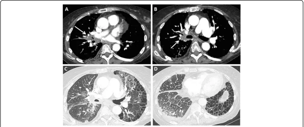
Antibody-mediated rejection is less common than acute cellular rejection. It is caused by antibodies against HLA antigens in the donor lung, or possibly to auto-antigens in the context of lung injury. Patients with antibody-mediated rejection usually present with respiratory symp-toms that typically manifest weeks or months following transplantation. The diagnosis is extremely challenging and needs careful correlation of the clinical, radiologic,
pathologic, and serologic findings (Fig.5). The pathologic
findings in this entity are poorly defined. Reported find-ings include acute lung injury and diffuse alveolar damage with or without capillaritis. Diagnostic criteria include detection of circulating donor specific antigen (DSA),
compatible histopathologic findings, and positive
immunohistochemical staining for complement compo-nent 4d (C4d). Cases with all three features are diagnosed as“definite.”The recommended designation is“probable”
if 2 features are present, and“possible” if any 1 feature
is present [5]. In practice, immunohistochemical
staining for C4d is associated with several problems, including low sensitivity and specificity, and high background staining. There is limited data in the literature regarding the imaging findings of acute antibody-mediated rejection.
The differential diagnosis of lung transplant patients with declining allograft function weeks to months after transplant includes acute rejection, infection, airway complications (air-way stenosis, tracheobronchomalacia, or granulation tissue), vascular anastomotic complications, chronic rejection, and cardiac dysfunction [4,22]. Evaluation includes a battery of laboratory testing for evidence of infection, imaging with chest radiography or CT, spirometry, and flexible bronchos-copy for bronchoalveolar lavage and transbronchial biopsy to confirm the diagnosis of acute rejection and to exclude processes such as infection, aspiration or organizing pneu-monia [4,22]. Even after a thorough evaluation, some cases can prove diagnostically challenging. The evaluation of these patients can be greatly complicated by presence of more
than one process in the same patient (Fig. 6).
Post-transplant lymphoproliferative disorder could rarely present early during the first weeks post lung transplant and should be considered in the differential diagnosis of persistent opacities during this period [28].
Chronic lung allograft dysfunction
Chronic lung allograft dysfunction (CLAD) following lung transplantation is a major cause of morbidity and
mortality [29]. It is an umbrella term that encompasses
bronchiolitis obliterans syndrome and restrictive allograft syndrome [29,30].
Bronchiolitis obliterans syndrome/obliterative bronchiolitis
Bronchiolitis obliterans syndrome/obliterative bronchio-litis is the most common form of chronic lung allograft dysfunction and reported in 48% of patients by 5 years
after transplant and 76% of patients after 10 years [1].
Obliterative bronchiolitis (also known as constrictive Fig. 4Patient with bilateral lung transplant and mild acute cellular rejection. Axial CT images (a–d) show asymmetric (right more than left) patchy and nodular peribronchial ground glass opacities (arrows) with basilar peribronchial consolidative opacities (arrowheads) and mild bibasilar septal thickening. Transbronchial biopsy of both lower lobes showed mild acute cellular rejection (grade A2 B0)
bronchiolitis) is a pathologic diagnosis characterized by fibrotic scarring that greatly narrows or completely
obliterates bronchiolar lumens (Fig. 7). Bronchiolitis
obliterans syndrome is the clinical manifestation of obliterative bronchiolitis. The hallmark of this syndrome is the presence of declining pulmonary function tests compared with baseline, in the absence of another
explanation such as acute rejection, infection, pulmonary edema, or airway stenosis. Defined in this way, the diag-nosis obviates the need for pathologic confirmation, which is limited by sampling issues and low sensitivity. Obliterative bronchiolitis/bronchiolitis obliterans syn-drome is the most common manifestation of chronic lung
allograft dysfunction [29]. Acute rejection, recurrent
Fig. 6Role of multimodality imaging in a 32-year-old male status post bilateral lung transplant presenting with shortness of breath. CT images in lung window setting (a,b) show multifocal bilateral consolidations, which are predominately seen in both lower lobes. Images from flexible bronchoscopy (c,d) show mucopurulent secretions at origin of right bronchus and in right middle lobe bronchi. Still image from esophagogram (e) shows small hiatal hernia (arrow); the patient had delayed esophageal emptying. Mycoplasma IgM titers were high and the patient was felt to have mycoplasma pneumonia with co-existing aspiration pneumonia. Trans-bronchial biopsy was negative for acute rejection
infections, primary graft dysfunction, HLA mismatch,
cytomegalovirus pneumonitis, aspiration, and
non-compliance with immunosuppressive therapy are thought to be potential risk factors for this disease [31]. Bronchio-litis obliterans syndrome often presents with non-specific symptoms; in advanced cases, patients may present with dyspnea at rest. The key feature is irreversible airflow limi-tation with a sustained decrease in FEV1 for > 3 weeks. The clinical staging of bronchiolitis obliterans syndrome is
based on the FEV1 compared with baseline (Table 2).
Pathologic examination shows obliterative bronchiolitis, as
described above [29, 32]. Bronchiolar involvement in this
disease is typically patchy, greatly limiting the yield of
transbronchial biopsy [33]. A major role of bronchoscopy
in this setting is to exclude other causes of decreased FEV1 such as acute rejection, infection, or bronchial stenosis [29].
Unlike assessment of acute rejection, imaging plays a crucial role in the diagnosis and stratification of patients with chronic lung allograft dysfunction. Chest radio-graphy is often the first investigation and is often normal in the early stage. Volume loss with fibrotic changes can be seen in advanced disease. CT scans may be normal during early stages of the process, or may show air trap-ping on expiratory imaging reflecting early obliterative
bronchiolitis [34]. Late stages show mosaic attenuation
caused by air trapping and may be associated with bron-chiectasis (Fig. 7) [35–38]. Spirometry is more sensitive in detecting the bronchiolitis obliterans syndrome than
chest CT [39]. Tree-in-bud and centrilobular nodules
can be seen and are thought to represent mucus impaction in distal bronchioles. Respiratory infection should be excluded based on clinical symptoms, bronchoalveolar lavage, and absence of concomitant suggestive imaging features such as consolidation before attributing these findings to the bronchiolitis obliterans syndrome [40].
Restrictive allograft syndrome
Restrictive allograft syndrome is the less common phenotype of irreversible chronic lung allograft dysfunc-tion; it has been associated with a worse prognosis compared with bronchiolitis obliterans syndrome. It
ultimately affects approximately 10% of lung transplant recipients. Patients present with restrictive lung function
tests [30]. Reported pathologic findings include
pleuro-parenchymal fibroelastosis with or without organizing
pneumonia [29,41]. Early imaging findings include
cen-tral and peripheral ground-glass opacities and organizing
pneumonia (Fig. 8) [42]. Imaging features that suggest
organizing pneumonia include peribronchial, perilobular, or subpleural consolidations with or without the classic
reverse halo sign [43]. Peripheral fibrotic changes with
upper lung predominance, volume loss, hilar retraction,
and traction bronchiectasis (Fig. 8) are the classic
advanced imaging findings [30,42, 44]. Some cases may
present with lower lobe or diffuse opacities and tend to have poor survival [30].
Survival estimates range from 6 to 18 months, com-pared with 3–5 years in patients with bronchiolitis oblit-erans syndrome. It is important to note that chronic lung allograft dysfunction is a heterogeneous umbrella term, and that these phenotypes can coexist, or evolve
from one to the other (Fig.9). Evolution from
obstruct-ive to restrictobstruct-ive chronic lung allograft dysfunction is
more common [30, 45]. The underlying severe
obstruc-tion poses a diagnostic challenge in cases that evolve from bronchiolitis obliterans syndrome to restrictive allograft syndrome. However, these cases carry a better prognosis compared with those without preceding
bronchiolitis obliterans syndrome [45].
Additional imaging techniques that may allow early detection and monitoring of chronic lung allograft dys-function include quantitative CT and low-dose CT volumetry. Quantitative CT is a density mapping with expiratory/inspiratory-ratio mean lung density to detect
pathologic air trapping in lung transplant patients [46].
Advanced quantitative analysis of CT in lung transplant patients can improve the accuracy of diagnosis of bronchiolitis obliterans syndrome and in predicting
functional parameters such as FEV [47]. Low-dose CT
volumetry shows decreased lung volume in restrictive allograft syndrome, whereas patients with bronchiolitis obliterans syndrome show no significant post-transplant
change [48]. Oxygen-enhanced T1-mapping MRI might
become a noninvasive tool for early detection and moni-toring of bronchiolitis obliterans syndrome. Oxygen is used as an inhaled contrast agent with T1 weighted images which may provide information regarding lung ventilation, perfusion, and diffusion capacity. The oxygen transfer function is a parameter using T1 relaxivity measured at room air and at 100% oxygen. Decreasing oxygen-induced relaxivity and increasing heterogeneity are suggestive features of bronchiolitis obliterans
syndrome [49]. This technique requires breath hold
and use of a closed facemask, which may not be well tolerated by some patients. Therefore, some simplified Table 2BOS classification system
FEV1 FEV25-75
BOS 0 > 90% of baseline > 75% of baseline
BOS 0-p 81–90% of baseline < or = 75% of baseline
BOS 1 66–80% of baseline
BOS 2 51–60% of baseline
BOS 3 < 50% of baseline
BOS, bronchiolitis obliterans syndrome;BOS 0-p, potential BOS;FEV1, forced
expiratory volume in 1 s;FEV25-75, forced expiratory flow at mid-expiratory
noncontrast-enhanced lung ventilation techniques have emerged including Fourier decomposition MRI
(FD-MRI) and phase resolved functional lung MRI
(PREFUL), which may have a potential applications in the future for early detection and severity assessment of
chronic lung allograft dysfunction [50, 51]. The uptake
of Fluorine-18-Fluorodeoxyglucose (FDG) on PET im-aging may be a promising diagnostic and prognostic tool in cases of chronic lung allograft dysfunction. FDG
uptake on fused PET/CT scan may distinguish restrict-ive allograft syndrome from bronchiolitis obliterans syndrome cases. Also, patients with restrictive allograft syndrome and areas of avid FDG uptake tend to have
worse outcome [52].
Conclusion
It is important to know the various types of lung
allo-graft dysfunction along a time continuum (Table 3).
Fig. 8Spectrum of imaging findings in restrictive allograft syndrome. CT images (aandb) show early to intermediate changes with upper lobe ground glass opacities and mild volume loss. Chest radiograph (c) in a patient with advanced restrictive allograft syndrome showing upper lobe predominant subpleural pleuroparenchymal fibrotic changes with volume loss. CT images (dande) in another patient with advanced restrictive allograft syndrome showing predominant subpleural fibrotic changes with associated pleural thickening
Fig. 9Imaging findings showing evolution of bronchiolitis obliterans to restrictive allograft syndrome. The patient initially presented with decreased FEV1 and mid-flows. Initial axial CT images (aandb) show mosaic attenuation, likely due to air-trapping. Transbronchial biopsy was negative for infection and acute rejection and the diagnosis of bronchiolitis obliterans syndrome was made. Follow-up CT images (candd) after 3 years show evolution of changes with upper lobe predominant subpleural pleuroparenchymal fibrotic changes typical of restrictive
Imaging findings in the acute setting (primary graft dys-function and acute rejection) are non-specific and need to be interpreted in conjunction with the clinical picture and other investigations. In contrast, imaging plays a key role in the diagnosis and follow-up of patients with chronic lung allograft dysfunction. Quantitative CT and T1-mapping MRI might evolve as non-invasive markers for chronic lung allograft dysfunction in the near future.
Acknowledgements
•Imaging Institute, Cleveland Clinic, Cleveland, Ohio.
•Pathology and Laboratory Medicine Institute, Cleveland Clinic, Cleveland Ohio.
•Respiratory Institute, Cleveland Clinic, Cleveland, Ohio
Authors’contributions
All authors contributed equally in writing and editing the manuscript. All authors read and approved the final manuscript.
Funding
No funding is needed or provided for writing this manuscript.
Availability of data and materials
Data sharing is not applicable to this manuscript as no datasets were generated or analyzed.
Ethics approval and consent to participate
Not applicable; this manuscript does not report on or involve the use of any animal or human data or tissue.
Consent for publication
Not applicable; this manuscript does not contain data from any individual person, please state.
Competing interests
The authors declare that they have no competing interests.
Author details
1Sections of Thoracic and Cardiovascular Imaging Laboratory, Imaging Institute, Cleveland Clinic, L-10, 9500 Euclid Avenue, Cleveland, OH 44195,
USA.2Department of Pathology, Cleveland Clinic, Cleveland, USA. 3
Department of Pulmonary and Critical Care Medicine, Cleveland Clinic, Cleveland, USA.
Received: 25 September 2019 Accepted: 29 November 2019
References
1. Chambers DC, Cherikh WS, Goldfarb SB et al (2018) The International Thoracic Organ Transplant Registry of the International Society for Heart and Lung Transplantation: thirty-fifth adult lung and heart-lung transplant report-2018; Focus theme: Multiorgan Transplantation. J Heart Lung Transplant 37(10):1169–1183
2. Krishnam MS, Suh RD, Tomasian A et al (2007) Postoperative complications of lung transplantation: radiologic findings along a time continuum. Radiographics 27(4):957–974
3. Habre C, Soccal PM, Triponez F et al (2018) Radiological findings of complications after lung transplantation. Insights Imaging 9(5):709–719 4. Ahmad S, Shlobin OA, Nathan SD (2011) Pulmonary complications of lung
transplantation. Chest 139(2):402–411
5. Levine DJ, Glanville AR, Aboyoun C et al (2016) Antibody-mediated rejection of the lung: a consensus report of the International Society for Heart and Lung Transplantation. J Heart Lung Transplant 35(4):397–406
6. Frost AE, Jammal CT, Cagle PT (1996) Hyperacute rejection following lung transplantation. Chest 110(2):559–562
7. Choi JK, Kearns J, Palevsky HI et al (1999) Hyperacute rejection of a pulmonary allograft. Immediate clinical and pathologic findings. Am J Respir Crit Care Med 160(3):1015–1018
8. Bittner HB, Dunitz J, Hertz M, Bolman MR 3rd, Park SJ (2001) Hyperacute rejection in single lung transplantation--case report of successful management by means of plasmapheresis and antithymocyte globulin treatment. Transplantation 71(5):649–651
9. Masson E, Stern M, Chabod J et al (2007) Hyperacute rejection after lung transplantation caused by undetected low-titer anti-HLA antibodies. J Heart Lung Transplant 26(6):642–645
10. Snell GI, Yusen RD, Weill D et al (2017) Report of the ISHLT Working Group on Primary Lung Graft Dysfunction, part I: Definition and grading-A 2016 Consensus Group statement of the International Society for Heart and Lung Transplantation. J Heart Lung Transplant 36(10):1097–1103
11. de Perrot M, Liu M, Waddell TK, Keshavjee S (2003) Ischemia-reperfusion-induced lung injury. Am J Respir Crit Care Med 167(4):490–511
Table 3Time onset, radiological findings, and diagnostic features of lung transplant primary graft dysfunction and different types of rejection
Complications Time onset Radiological findings Diagnostic criteria/features
Hyper-acute rejection < 24 h Diffuse pulmonary opacities. It is a type of antibody-mediated rejection with no gold standard diagnostic test.
Primary graft dysfunction 0–72 h Non-specific perihilar and basilar opacities and interstitial thickening.
Development of hypoxia and diffuse pulmonary radiographic opacities with no other identifiable cause.
Acute cellular rejection 1st week–1 year Non-specific ground-glass opacities, consolidation, and interstitial thickening with or without pleural effusions.
Diagnosed and graded based on pathologic findings on TBB specimens; lymphohistiocytic inflammatory infiltrates centered on small blood vessels; or bronchioles.
Acute antibody-mediated rejection
1st week–1 year No described specific imaging findings. Presence of DSA, characteristic lung histology, and positive C4d within the graft.
Bronchiolitis obliterans Syndrome
> 1 year Air trapping on expiratory images; bronchial wall thickening; centrilobular nodules; with or without bronchiectasis.
FEV1 decline≥20% from baseline; irreversible obstructive PFT pattern. Pathology findings are characteristic but not required for diagnosis.
Restrictive allograft syndrome
> 1 year Early: central and peripheral ground-glass opacities.
Late: peripheral and upper lung predominant reticulation, architecture distortion, and traction bronchiectasis with hilar retraction.
FEV1 decline≥20% from baseline; total lung capacity decline≥10% from baseline; irreversible restrictive PFT pattern.
12. Mukhopadhyay S, Parambil JG (2012) Acute interstitial pneumonia (AIP): relationship to Hamman-Rich syndrome, diffuse alveolar damage (DAD), and acute respiratory distress syndrome (ARDS). Semin Respir Crit Care Med 33(5):476–485
13. Fiser SM, Tribble CG, Long SM et al (2002) Ischemia-reperfusion injury after lung transplantation increases risk of late bronchiolitis obliterans syndrome. Ann Thorac Surg 73(4):1041–1047
14. Bharat A, Kuo E, Steward N et al (2008) Immunological link between primary graft dysfunction and chronic lung allograft rejection. Ann Thorac Surg 86(1):189–195
15. Anderson DC, Glazer HS, Semenkovich JW et al (1995) Lung transplant edema: chest radiography after lung transplantation--the first 10 days. Radiology 195(1):275–281
16. Kundu S, Herman SJ, Winton TL (1998) Reperfusion edema after lung transplantation: radiographic manifestations. Radiology 206(1):75–80 17. Marom EM, Choi YW, Palmer SM, DeLong DM, Stuart MD, McAdams HP
(2001) Reperfusion edema after lung transplantation: effect of daclizumab. Radiology 221(2):508–514
18. Belmaati EO, Steffensen I, Jensen C et al (2012) Radiological patterns of primary graft dysfunction after lung transplantation evaluated by 64-multi-slice computed tomography: a descriptive study. Interact Cardiovasc Thorac Surg 14(6):785–791
19. Hopkins PM, Aboyoun CL, Chhajed PN et al (2002) Prospective analysis of 1,235 transbronchial lung biopsies in lung transplant recipients. J Heart Lung Transplant 21(10):1062–1067
20. Yusen RD, Edwards LB, Kucheryavaya AY et al (2015) The Registry of the International Society for Heart and Lung Transplantation: thirty-second official adult lung and heart-lung transplantation report--2015; Focus Theme: Early Graft Failure. J Heart Lung Transplant 34(10):1264–1277 21. Martinu T, Pavlisko EN, Chen DF, Palmer SM (2011) Acute allograft rejection:
cellular and humoral processes. Clin Chest Med 32(2):295–310 22. Martinu T, Chen DF, Palmer SM (2009) Acute rejection and humoral
sensitization in lung transplant recipients. Proc Am Thorac Soc 6(1):54–65 23. Stewart S, Fishbein MC, Snell GI et al (2007) Revision of the 1996 working formulation for the standardization of nomenclature in the diagnosis of lung rejection. J Heart Lung Transplant 26(12):1229–1242
24. De Vito DA, Hoffman LA, Iacono AT, Zullo TG, McCurry KR, Dauber JH (2004) Are symptom reports useful for differentiating between acute rejection and pulmonary infection after lung transplantation? Heart Lung 33(6):372–380 25. Gotway MB, Dawn SK, Sellami D et al (2001) Acute rejection following lung
transplantation: limitations in accuracy of thin-section CT for diagnosis. Radiology 221(1):207–212
26. Park CH, Paik HC, Haam SJ et al (2014) HRCT features of acute rejection in patients with bilateral lung transplantation: the usefulness of lesion distribution. Transplant Proc 46(5):1511–1516
27. Fuehner T, Simon A, Dierich M et al (2009) Indicators for steroid response in biopsy proven acute graft rejection after lung transplantation. Respir Med 103(8):1114–1121
28. Lewis AJ, Jagadeesh D, Mukhopadhyay S, Budev M, Mehta AC (2018) Corrigendum to: Post-transplant lymphoproliferative disorder presenting on post-transplant day 35 as a pulmonary parenchymal infiltrate-a case report. Oxf Med Case Reports. 2018(12):omy100
29. Verleden GM, Raghu G, Meyer KC, Glanville AR, Corris P (2014) A new classification system for chronic lung allograft dysfunction. J Heart Lung Transplant 33(2):127–133.https://doi.org/10.1016/j.healun.2013.10.022Epub 2013 Oct 24
30. Sato M, Waddell TK, Wagnetz U et al (2011) Restrictive allograft syndrome (RAS): a novel form of chronic lung allograft dysfunction. J Heart Lung Transplant 30(7):735–742
31. Husain AN, Siddiqui MT, Holmes EW et al (1999) Analysis of risk factors for the development of bronchiolitis obliterans syndrome. Am J Respir Crit Care Med 159(3):829–833
32. Estenne M, Maurer JR, Boehler A et al (2002) Bronchiolitis obliterans syndrome 2001: an update of the diagnostic criteria. J Heart Lung Transplant 21(3):297–310 33. Kramer MR, Stoehr C, Whang JL et al (1993) The diagnosis of obliterative
bronchiolitis after heart-lung and lung transplantation: low yield of transbronchial lung biopsy. J Heart Lung Transplant 12(4):675–681 34. Bankier AA, Van Muylem A, Knoop C, Estenne M, Gevenois PA (2001)
Bronchiolitis obliterans syndrome in heart-lung transplant recipients: diagnosis with expiratory CT. Radiology 218(2):533–539
35. Morrish WF, Herman SJ, Weisbrod GL, Chamberlain DW (1991) Bronchiolitis obliterans after lung transplantation: findings at chest radiography and high-resolution CT. The Toronto Lung Transplant Group. Radiology 179(2):487–490 36. Worthy SA, Park CS, Kim JS, Muller NL (1997) Bronchiolitis obliterans after
lung transplantation: high-resolution CT findings in 15 patients. AJR Am J Roentgenol 169(3):673–677
37. Lee ES, Gotway MB, Reddy GP, Golden JA, Keith FM, Webb WR (2000) Early bronchiolitis obliterans following lung transplantation: accuracy of expiratory thin-section CT for diagnosis. Radiology 216(2):472–477 38. Choi YW, Rossi SE, Palmer SM, DeLong D, Erasmus JJ, McAdams HP (2003)
Bronchiolitis obliterans syndrome in lung transplant recipients: correlation of computed tomography findings with bronchiolitis obliterans syndrome stage. J Thorac Imaging 18(2):72–79
39. Konen E, Gutierrez C, Chaparro C et al (2004) Bronchiolitis obliterans syndrome in lung transplant recipients: can thin-section CT findings predict disease before its clinical appearance? Radiology 231(2):467–473 40. Gunn ML, Godwin JD, Kanne JP, Flowers ME, Chien JW (2008)
High-resolution CT findings of bronchiolitis obliterans syndrome after hematopoietic stem cell transplantation. J Thorac Imaging 23(4):244–250 41. Ofek E, Sato M, Saito T et al (2013) Restrictive allograft syndrome post lung
transplantation is characterized by pleuroparenchymal fibroelastosis. Mod Pathol 26(3):350–356
42. Verleden SE, de Jong PA, Ruttens D et al (2014) Functional and computed tomographic evolution and survival of restrictive allograft syndrome after lung transplantation. J Heart Lung Transplant 33(3):270–277
43. Arakawa H, Kurihara Y, Niimi H, Nakajima Y, Johkoh T, Nakamura H (2001) Bronchiolitis obliterans with organizing pneumonia versus chronic eosinophilic pneumonia: high-resolution CT findings in 81 patients. AJR Am J Roentgenol 176(4):1053–1058
44. Konen E, Weisbrod GL, Pakhale S, Chung T, Paul NS, Hutcheon MA (2003) Fibrosis of the upper lobes: a newly identified late-onset complication after lung transplantation? AJR Am J Roentgenol 181(6):1539–1543
45. Verleden SE, Vos R, Vanaudenaerde BM, Verleden GM (2017) Chronic lung allograft dysfunction phenotypes and treatment. J Thorac Dis 9(8):2650–2659
46. Solyanik O, Hollmann P, Dettmer S et al (2015) Quantification of pathologic air trapping in lung transplant patients using CT density mapping: comparison with other CT air trapping measures. PLoS One 10(10) 47. Mortani Barbosa EJ Jr, Shou H, Simpsom S, Gee J, Tustison N, Lee JC (2018)
Quantitative computed tomography metrics from the transplanted lung can predict forced expiratory volume in the first second after lung
transplantation. J Thorac Imaging 33(2):112–123
48. Saito T, Horie M, Sato M et al (2016) Low-dose computed tomography volumetry for subtyping chronic lung allograft dysfunction. J Heart Lung Transplant 35(1):59–66
49. Renne J, Lauermann P, Hinrichs JB et al (2015) Chronic lung allograft dysfunction: oxygen-enhanced T1-mapping MR imaging of the lung. Radiology 276(1):266–273
50. Voskrebenzev A, Greer M, Gutberlet M et al (2018) Detection of chronic lung allograft dysfunction using ventilation-weighted Fourier decomposition MRI. Am J Transplant 18(8):2050–2060
51. Moher Alsady T, Voskrebenzev A, Greer M et al (2019) MRI-derived regional flow-volume loop parameters detect early-stage chronic lung allograft dysfunction. J Magn Reson Imaging 27(10):26799
52. Verleden SE, Gheysens O, Goffin KE et al (2019) Role of 18F-FDG PET/CT in restrictive allograft syndrome after lung transplantation. Transplantation 103(4):823–831
Publisher’s Note