Volume 9 | Issue 2 | June 2017
IJBC 2017; 9(2): 37-43
Brain MRI Findings in Children with Acute Lymphoblastic Leukemia
Karmella Kamali1, Reza Taghavinasab2, Sezaneh Haghpanah3, Mohammadreza Bordbar4*,
Parsa Kamalipour5
1Assistant professor of Radiology, Radiology Research Center, Shiraz University of Medical Sciences, Shiraz, Iran 2Resident of Radiology, Shiraz University of Medical Sciences, Shiraz, Iran
3Associate professor of Community Medicine, Hematology Research Center, Shiraz University of Medical Sciences, Shiraz, Iran 4Associate professor of Pediatric Hematology-Oncology, Hematology Research Center, Shiraz University of Medical Sciences, Shiraz, Iran 5Medical Student, Shiraz University of Medical Sciences, Shiraz, Iran
A R T I C L E I N F O
Original Article
Article History: Received: 01.12.2016 Accepted: 03.02.2017
Keywords: Childhood leukemia Magnetic resonance imaging Complications
Brain abnormalities
CNS findings
*Corresponding author: Mohammadreza Bordbar, MD Address: Hematology Research Center, Shiraz University of Medical Sciences, Shiraz, Iran
Tel/Fax: +98 71 36281528 Email: bordbarm@sums.ac.ir
ABSTRACT
Background: Patients with leukemia are facing more complications in order to achieve longer survival. We aimed to evaluate the frequency of central nervous system abnormalities (CNS) on MRI of children with acute lymphoblastic leukemia (ALL).
Methods: Sixty-six children with diagnosis of ALL aged 2-18 years were recruited. Non-contrast sequences of brain MRI in addition to diffusion weighted imaging of brain were obtained with 1.5 T (Siemens medical system) scanners in their maintenance phase of treatment. The age of onset, type of leukemia, protocol of treatment, and elapsed time from diagnosis were recorded. Chi-square test was used to compare the groups and t-test was used to evaluate the effect of not normally distributed variables.
Results: 19 (28.8%) had abnormal CNS findings identified on MRI images including: nonspecific white matter high signal intensity in flair images with normal DWI, white matter ischemia proved on DWI, generalized brain atrophy, isolated mild enlargement of lateral ventricle and extracerebral complications including sinus thrombosis and sinusitis. Brain abnormalities were correlated with leukemia type, chemotherapy protocol and radiotherapy (P=0.006, 0.036, and 0.01, respectively).
Conclusion: The wide spectrum of CNS abnormalities that were observed in children with ALL showed correlation with treatment methods and type of leukemia in this study. Combination of radiation therapy and chemotherapy increased CNS complications. Among extracerebral complications, dural sinus thrombosis proved by MRV was seen more frequently in T-cell leukemia patients treated with multiple high doses of the chemotherapy agent “L-asparaginase”. Since some neurological complications of leukemia are treatable, early diagnosis sounds essential.
Introduction
Leukemia is the most common cancer in children and has increased in frequency in the twentieth century.1
The most common signs and symptoms include fever, anemia, thrombocytopenia, hepatosplenomegaly, and lymphadenopathy.2 Acute lymphocytic leukemia
(ALL) comprises about two-third of cases, while acute myeloblastic leukemia (AML), chronic myelogenous leukemia (CML), and juvenile myelomonocytic leukemia (JMML) are less prevalent subtypes in children. Despite the current improvements in diagnosis and treatment, induction failure and relapse has been reported in up to
Iranian Journal of Blood & Cancer
Journal Home Page: www.ijbc.ir
Please cite this article as: Kamali K, Taghavinasab R, Haghpanah S, Bordbar MR, Kamalipour P. Brain MRI Findings in Children with Acute Lymphoblastic Leukemia. IJBC 2017; 9(2): 37-43.
10% of the cases.3 The improved cure rate and survival of
the patients has caused the complications to become more noticeable.4 Chemotherapy, radiotherapy and stem cell
transplantation in specific cases are the main therapeutic options for childhood leukemia,5 which in turn have their
own complications and adverse effects that might have some impact on patients’ survival.6,7 Among the most
important complications are central nervous system (CNS)-related, as they mainly affect child’s cognition and neuropsychological function.8 Damage to normal brain
tissue is hypothesized to be mainly due to treatments such as irradiation.9 In addition, neurotoxicity and
leukoencephalopathy is proposed to be induced by some chemotherapy drugs especially systemic and intrathecal
methotrexate (MTX) injections.10-13 Studies have
confirmed beneficial role for imaging modalities such as magnetic resonance imaging (MRI) and computed tomography (CT) in diagnosing CNS abnormalities.14,15
Evaluating these complications thoroughly, in terms of incidence, risk factors, diagnosis, and treatment reveals the priority of each treatment modality for physicians and researchers.
We aimed to evaluate the frequency of CNS abnormalities on MRI of patients with ALL which may help diagnosis and management of their neurological side effects in a timely manner.
Materials and Methods
In this cross-sectional study, 66 children of 2-18 years with diagnosis of ALL based on flowcytometry analysis of bone marrow aspirate were recruited. They were visited regularly in an outpatient pediatric oncology clinic affiliated to Shiraz University of Medical Sciences in Shiraz, south of Iran. The patients were at different time courses of their chemotherapy treatments and had been treated for at least 6 months. The treatment strategy was determined based on the type of leukemia and the risk category. Some patients had received cranial irradiation if they had CNS involvement or prophylactically in cases of T-cell ALL. Those with underlying neurological and metabolic diseases were excluded from the study.
Non-contrast sequences of the brain MRI, obtained with 1.5 T (Siemens medical system) scanners, in addition to diffusion weighted imaging of brain were obtained in
the maintenance phase of treatment and the results were recorded by a single radiologist.
The frequency of abnormalities on the patients’ brain MRI were determined according to the age at diagnosis, type of leukemia, treatment protocol and elapsed time from treatment. Moreover, it was assessed whether radiotherapy had an additional effect on brain MRI including intra and extracerebral findings.
Data were analyzed by SPSS software, version 21. Descriptive data were presented as mean, standard deviation, frequency and percentage. Qualitative and quantitative variables were compared by Chi-square test and Student t-test, respectively between two groups of patients. Level of significance less than 0.05 was considered statistically significant.
Results
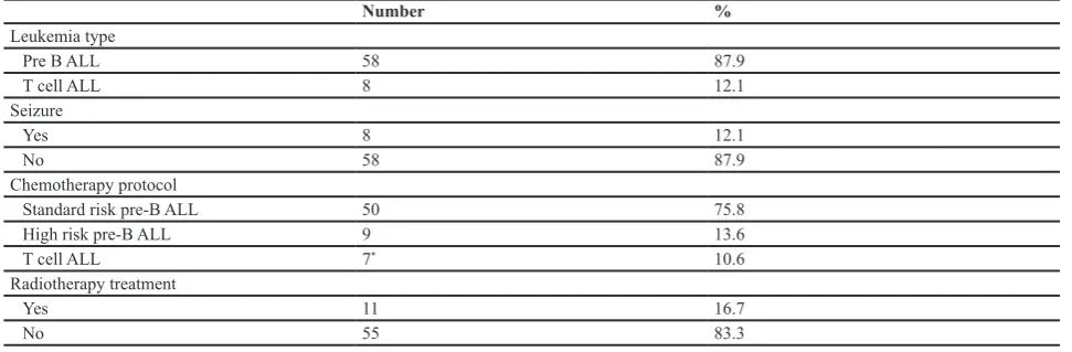
The study group included 66 patients (42 boys, M/F ratio 1.75), with the mean age of 5.74±3.65 years, (range 15-192 months). The mean time elapsed from treatment until MRI assessment was 27.85±14.62 months. The clinical characteristics as well as the treatment protocols of the studied population are demonstrated in table 1.
As identified on MRI (Figure 1), sinusitis with or without retention cyst was the most common finding. Air-fluid level and mucosal thickening more than 2 mm was considered as criteria for diagnosis of sinusitis on MRI (30.2%, n=20).16 Other extracerebral findings included
mastoiditis (n=6) and otitis media (n=1). Nineteen patients (28.8%) had CNS complications including: nonspecific white matter high signal intensity in flair images with no associated DWI abnormalities (n=7), brain atrophy (n=6), white matter acute ischemia approved with DWI (n=5), sinus thrombosis on MRV (n=4), and mild enlargement of lateral ventricles without any dilatation of extra-axial fluid (n=3). Some patients showed more than one abnormal MRI finding. There was also one case with incidentally found a parietal arachnoid cyst most probable having no relation to the patient’s treatment.
The patients were treated according to ALL-BFM protocols for pre-B and T-cell leukemia.17,18 They were all
in their maintenance phase of treatment which included monthly injections of vincristine, oral mercaptopurine, weekly methotrexate and boosts of prednisolone for 5
Table 1: Clinical characteristics of the study population
Number %
Leukemia type
Pre B ALL 58 87.9
T cell ALL 8 12.1
Seizure
Yes 8 12.1
No 58 87.9
Chemotherapy protocol
Standard risk pre-B ALL 50 75.8
High risk pre-B ALL 9 13.6
T cell ALL 7* 10.6
Radiotherapy treatment
Yes 11 16.7
No 55 83.3
Volume 9 | Issue 2 | June 2017
days after each vincristine injection. They also received intrathecal injection of methotrexate every two months. Eleven cases were also given craniospinal irradiation; including 8 cases with T-cell ALL prophylactically, and 3 patients with pre-B cell ALL who had CNS involvement.
The patients were classified into two groups based on their MRI finding. Those with normal brain imaging and extracerebral findings were classified as group 1, while those with CNS abnormalities were classified as group 2.
Table 2 demonstrates the association between MRI findings and different variables including age at diagnosis, age at the time of imaging, gender, type of leukemia, treatment protocols, time elapsed since
diagnosis/treatment, and radiation therapy between the two treatment groups.
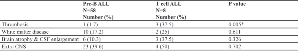
There was a significant correlation between leukemia type, chemotherapy protocol and brain abnormalities on MRI (P=0.006 and 0.036, respectively), indicating more abnormalities in T-cell leukemia. Radiotherapy was also associated with more brain abnormalities (P=0.01). Among extracerebral findings, thrombosis of dural sinus was seen more frequently in T-cell ALL patients (P=0.009), while other MRI findings were not correlated with the type of leukemia (table 3). Moreover, there was no significant correlation between gender, age, treatment duration and seizure (seen in 8 cases) with brain abnormalities on MRI (table 2).
Figure 1: Frequency of MRI findings in the study population
Table 2: Comparison of clinical and demographic characteristics between the two groups of leukemia patients with and without
CNS MRI findings
Variables Without MRI findings
N=47 With MRI findingsN=19 P value
Gender
Male 29 13 0.779
Female 18 6
Leukemia type
Pre B cell ALL 45 13 0.006*
T cell ALL 2 6
Seizure
Yes 4 4 0.099
No 43 15
Chemotherapy protocols
Standard risk pre-B ALL 38 12 0.036*
High risk pre-B ALL 7 2
T cell ALL 2 5
Radiotherapy
Yes 4 7 0.01*
No 43 12
Age (month) (mean±SD) 95.1±45.9 112.3± 49.8 0.183
Age at diagnosis (month) (mean±SD) 63.1±41.8 83.2±47.6 0.094
Treatment duration (month) (mean±SD) 28.1±13 27.4±18.7 0.866
¶ One patient with T-cell ALL was treated with high risk pre-B –cell ALL protocol
Discussion
The male to female ratio in our study was 1.75, which was similar to another Iranian study, assessing 368 cases that reported a male/female ratio of 1.5.2 Among 66 cases
who were assessed by MRI, 40 patients (60.6%) had MRI abnormalities, but 19 cases showed MRI findings which might be attributed to the treatment. Chen and colleagues have also reported 12 cases of brain abnormalities among 19 cases on MR and CT scans.19
Ulu and colleagues have retrospectively evaluated CNS imaging findings of 15 patients with acute leukemia. They found that most of abnormalities (17/22) were attributed to the disease itself, and a minority (4/22) were treatment related. The most common complications in decreasing order of frequency were meningeal leukemia, orbital chloroma, posterior reversible encephalopathy syndrome (PRES), retinal and intracranial hemorrhage.20 While
we did not observe any documented case of leukemic meningitis, 34.3% of our patients had extracerebral abnormalities, with sinusitis being the most prevalent finding.
Some authors have attributed the cerebrovascular complications such as sinus thrombosis to the chemotherapy agent L-asparaginase.19,21-23 Others have
also ascribed white matter brain changes to MTX, which was not associated with neuropsychological deficit.24
There was not any case of periventricular complication probably due to MTX among our patients.
Our results did not show any association between the type of leukemia and brain abnormalities, except dural sinus thrombosis which was seen more frequently in T-cell ALL patients who had received multiple doses of high-dose L-asparaginase in their treatment regimen. Cerebral venous thrombosis is a unique adverse effect of “L-asparaginase” which has been reported in up to 3% of cases.21,25 It has been ascribed to the depletion
of anticoagulant proteins mainly antithrombin III. The dose, frequency and preparation of the drug have been proposed to play some role in its coagulant activity.21,25,26
Nevertheless, as our patients had not been treated with high-dose MTX, and they were all receiving low-dose oral weekly MTX, no conclusion could be reached on the association of MTX and brain abnormalities.
It was noted previously that leukemic infiltration of the meninges and orbits were the most common CNS findings in Ulu’s patients, although their patients had a wide age range (1-22 years) and the sample size was too small (15 patients). Meanwhile, Chen and colleagues have reported the most common early CNS complications to be cerebrovascular and infections. They also concluded
in a case report that leukemic infiltration of paranasal sinuses should be considered in cases with sinusitis.27 Thus,
infection should be highly considered in patients with leukemia undergoing treatment in a timely manner which may prevent functional brain sequels in these children.
Chan et al. have reported cerebral hemorrhage as the most common finding in ALL cases, 15% of which were detected by T2 MRI and was mainly evident only on gradient echo imaging.28 Chu and colleagues have
also used MR spectroscopy and have concluded that it demonstrates metabolic changes in brain after
high-dose MTX, which was not obvious by normal MRI.29
Regarding different imaging methods used in different centers, which may not completely reveal the associated pathology, periodic neurocognitive tests are recommended for such children to prevent cognition deficits.15 We found
no case of intracerebral hemorrhage in our study.
Moreover, combination therapy (radiation plus chemotherapy), performed for patients with T-cell ALL or patients with CNS leukemia gave rise significantly to more brain abnormalities on MRI. Chan et. al used radiation for 58% of their cases and established the significant relationship between hemorrhage and radiation.28 Although they had used different imaging
methods which might cause them to reach to a different conclusion in terms of complications, we have also found a significant association between radiation and brain abnormalities. Vazquez et. al have also demonstrated that radiation leads to white matter disease, microangiopathy, vascular malformations, telangiectasia and secondary neoplasms.30 Thus, it is suggested to pay more attention
to patients undergoing radiation in order to diagnose and treat the complications earlier. Furthermore, some experts recommend to replace radiation with safer alternatives such as high-dose MTX to avoid its late side effects particularly in growing children.31,32
In the current study, 8 out of 66 studied patients reported to have experienced seizure at least once, but only half of them occurred during their present illness. Seizure was observed in two patients with T cell leukemia with sagittal sinus thrombosis. Another patient who had seizure was a boy with pre-B cell leukemia who developed decreased level of consciousness, headache, visual symptoms and convulsion in the induction phase of chemotherapy, which was attributed to PRES confirmed by MRI. He was found to have hypersignal images in deep white matter of left parietal and occipital lobes about 18 months after the incident. The last one was a case of pre-B cell ALL who presented with fever, convulsion and hemiparesis in the induction phase of treatment. MRI showed a large brain Table 3:MRI findings in the study population with regards to their diagnosis
Pre-B ALL N=58 Number (%)
T cell ALL N=8 Number (%)
P value
Thrombosis 1 (1.7) 3 (37.5) 0.005*
White matter disease 10 (17.2) 2 (25) 0.611
Brain atrophy & CSF enlargement 6 (10.3) 3 (37.5) 0.326
Extra CNS 23 (39.6) 4 (50) 0.702
Extra CNS: sinusitis, mastoiditis, otitis media, retention cyst
Volume 9 | Issue 2 | June 2017
abscess at that time which was successfully treated with drainage and broad-spectrum antibiotics. His MRI at the time of our study which was a few years later showed severe cortical atrophy.
This study investigated the detailed information of children with leukemia, and aimed to give physicians and researchers a broader view on CNS complications or abnormalities detected by MRI. In the current study sinusitis was the most common extracerebral pathology. This finding is clinically important due to devastating ophthalmological and neurological complications in
the immunocompromised children.33 Moreover, MRI
abnormalities especially dural sinus thrombosis were observed in T-cell leukemia which emphasizes the necessity of more frequent CNS monitoring in this group of leukemia patients. Additionally, patients receiving adjuvant radiotherapy had higher brain abnormalities. Therefore, families should be informed about the long-term sequels of treatments and radiotherapy. The long-term sequels in particular, might adversely affect children’s quality of life including academic performance. Taking into account the high frequency of adverse effects associated with anti-leukemic treatments, some investigators have recommended to consider some refinements in treatment modalities.34,35
To our knowledge, this is the first local study investigating CNS findings detected by MRI in pediatric patients with ALL. As a result, in parallel to concerns regarding the intensity of the treatment and quality of life of the patients, safety of the treatments and the long-term consequences are noteworthy as well.
Our study had also some flaws and limitations in some aspects. We did not follow the patients to study the outcome of each of these complications in long-term. Moreover, we did not assess the psychosocial and cognitive consequences of CNS abnormalities. In addition, the patients were not studied by the imaging modalities before starting any kind of chemotherapy at the beginning of thir disease and hence, the abnormalities detected on MRI could not be attributable exactly to the treatment. Larger cohort studies with longer periods of follow up and doing imaging studies at baseline are needed to disclose the outcome of these lesions and their impact on the quality of life and cognitive function and neurologic status of the survivors of leukemia.
The choice of treatment in each patient was also based on the physician’s experience; as there are different local treatment strategies, which make the comparison between study groups difficult. Multicenter clinical trials with similar treatment protocols are advised to overcome these shortcomings.
In addition, our study included pediatric ALL patients, and patients with AML were not investigated. As patients with AML receive different chemotherapy drugs from ALL patients, and their treatment usually lack radiotherapy and intrathecal or high-dose systemic MTX, investigating CNS related side effects in this subgroup of patients may reveal diverse MRI abnormalities.
In conclusion, the wide spectrum of CNS abnormalities occurring during or after treatment of leukemia could be
related to the treatment protocol or leukemia subtype. Additional radiotherapy also increases cerebral and exrtacerebral complications. Dural sinus thrombosis was associated with multiple doses of “L-asparaginase”.
As long as most of the neurologic complications can be debilitating while treatable, early diagnosis by imaging methods and cognitive tests is essential.
Acknowledgment
We would like to appreciate our patients and their families who let us do this investigation. We would also thank MS. S. Parand for improving the language of the manuscript. This manuscript was relevant to the thesis of Reza Taghavinasab and was supported by the Vice-Chancelleor of Research and Technology of the Shiraz University of Medical Sciences with grant number 46964.
Conflict of Interest: None declared.
References
1. Belson M, Kingsley B, Holmes A. Risk factors for acute leukemia in children: a review. Environ Health Perspect. 2007;115(1):138-45. PubMed PMID:17366834. PubMed Central PMCID: PMC1817663.
2. Karimi M, Mehrabani D, Yarmohammadi H, Jahromi FS. The prevalence of signs and symptoms of childhood leukemia and lymphoma in Fars Province, Southern Iran. Cancer Detect Prev. 2008;32(2):178-83. doi: 10.1016/j.cdp.2008.06.001. PubMed PMID: 18632219.
3. Oudot C, Auclerc MF, Levy V, Porcher R, Piguet C, Perel Y, et al. Prognostic factors for leukemic induction failure in children with acute lymphoblastic leukemia and outcome after salvage therapy: the FRALLE 93 study. J Clin Oncol. 2008; 26(9):1496-503. doi: 10.1200/JCO.2007.12.2820. PubMed PMID: 18349402.
4. Brenner H, Kaatsch P, Burkhardt-Hammer T, Harms DO, Schrappe M, Michaelis J. Long-term survival of children with leukemia achieved by the end of the second millennium. Cancer. 2001;92(7):1977-83. 5. Hoelzer D, Gokbuget N. Recent approaches in
acute lymphoblastic leukemia in adults. Crit Rev Oncol Hematol. 2000;36(1):49-58. PubMed PMID: 10996522.
6. Creutzig U, Zimmermann M, Reinhardt D, Dworzak M, Stary J, Lehrnbecher T. Early deaths and treatment-related mortality in children undergoing therapy for acute myeloid leukemia: analysis of the multicenter clinical trials AML-BFM 93 and AML-BFM 98. J Clin Oncol. 2004;22(21):4384-93.
7. Lange BJ, Gerbing RB, Feusner J, Skolnik J, Sacks N, Smith FO, et al. Mortality in overweight and underweight children with acute myeloid leukemia. JAMA. 2005;293(2):203-11. doi: 10.1001/ jama.293.2.203. PubMed PMID: 15644547.
8. Langer T, Martus P, Ottensmeier H, Hertzberg H, Beck JD, Meier W. CNS late-effects after ALL therapy in childhood. Part III: neuropsychological
performance in long-term survivors of childhood ALL: impairments of concentration, attention, and memory. Med Pediatr Oncol. 2002;38(5):320-8. doi: 10.1002/mpo.10055.
9. Schroeder H, Garwicz S, Kristinsson J, Siimes MA, Wesenberg F, Gustafsson G. Outcome after first relapse in children with acute lymphoblastic leukemia: a population-based study of 315 patients from the Nordic Society of Pediatric Hematology and Oncology (NOPHO). Med Pediatr Oncol. 1995;25(5):372-8. doi: 10.1002/mpo.2950250503. 10. Fisher MJ, Khademian ZP, Simon EM, Zimmerman
RA, Bilaniuk LT. Diffusion-weighted MR imaging of early methotrexate-related neurotoxicity in children. AJNR Am J Neuroradiol. 2005;26(7):1686-9. PubMed PMID: 16091514.
11. Gupta A, Swaroop C, Rastogi R, Garg R, Bakhshi S. Simultaneous occurrence of posterior reversible leukoencephalopathy syndrome in two cases of childhood acute lymphoblastic leukemia induction chemotherapy. Pediatr Hematol Oncol. 2008;25(4):351-8. doi: 10.1080/08880010802016052. 12. Shuper A, Stark B, Kornreich L, Cohen IJ, Aviner
S, Steinmetz A, et al. Methotrexate treatment protocols and the central nervous system: significant cure with significant neurotoxicity. J Child Neurol. 2000;15(9):573-80. doi:10.1177/088307380001500902. PubMed PMID: 11019787.
13. Ziereisen F, Dan B, Azzi N, Ferster A, Damry N, Christophe C. Reversible acute methotrexate leukoencephalopathy: atypical brain MR imaging features. Pediatr Radiol. 2006;36(3):205-12. doi: 10.1007/s00247-005-0015-z. PubMed PMID: 16369780.
14. Ginsberg LE, Leeds NE. Neuroradiology of leukemia. AJR Am J Roentgenol. 1995;165(3):525-34. doi: 10.2214/ajr.165.3.7645463. PubMed PMID: 7645463. 15. Iuvone L, Mariotti P, Colosimo C, Guzzetta F, Ruggiero A, Riccardi R. Long-term cognitive outcome, brain computed tomography scan, and magnetic resonance imaging in children cured for acute lymphoblastic leukemia. Cancer. 2002;95(12):2562-70.doi: 10.1002/ cncr.10999. PubMed PMID: 12467071.
16. Kristo A, Uhari M, Luotonen J, Koivunen P, Ilkko E, Tapiainen T, et al. Paranasal sinus findings in children during respiratory infection evaluated with magnetic resonance imaging. Pediatrics. 2003;111(5 Pt 1):e586-9. PubMed PMID: 12728114.
17. Goldberg JM, Silverman LB, Levy DE, Dalton
VK, Gelber RD, Lehmann L, et al. Childhood T-cell acute lymphoblastic leukemia: the Dana-Farber Cancer Institute acute lymphoblastic leukemia consortium experience. J Clin Oncol. 2003;21(19):3616-22. doi: 10.1200/JCO.2003.10.116. PubMed PMID: 14512392.
18. Seibel NL, Steinherz PG, Sather HN, Nachman JB, Delaat C, Ettinger LJ, et al. Early postinduction intensification therapy improves survival for children and adolescents with high-risk acute lymphoblastic leukemia: a report from the Children’s Oncology
Group. Blood. 2008;111(5):2548-55. doi: 10.1182/ blood-2007-02-070342. PubMed PMID: 18039957. PubMed Central PMCID: PMC2254538.
19. Chen CY, Zimmerman RA, Faro S, Bilaniuk LT, Chou TY, Molloy PT. Childhood leukemia: central nervous system abnormalities during and after treatment. AJNR Am J Neuroradiol. 1996;17(2):295-310. PubMed PMID: 8938302.
20. Ulu EM, Tore HG, Bayrak A, Gungor D, Coskun M. MRI of central nervous system abnormalities in childhood leukemia. Diagn Interv Radiol. 2009;15(2):86-92. PubMed PMID: 19517377. 21. Couturier MA, Huguet F, Chevallier P, Suarez
F, Thomas X, Escoffre-Barbe M, et al. Cerebral venous thrombosis in adult patients with acute lymphoblastic leukemia or lymphoblastic lymphoma during induction chemotherapy with l-asparaginase: The GRAALL experience. Am J Hematol. 2015; 90(11):986-91. doi: 10.1002/ajh.24130. PubMed PMID: 26214580.
22. Diaz Diaz J, Nunez Enamorado N, Martinez de Aragon A, Barrios Lopez M, Camacho Salas A, Simon de la Heras R. [Cerebral sinovenous thrombosis in children due to L-asparaginase]. An Pediatr (Barc). 2015;82(2):113-4.
23. Eden D, Hipkins R, Bradbury CA. Cerebral Thrombotic Complications Related to l-Asparaginase Treatment for Acute Lymphoblastic Leukemia: Retrospective Review of 10 Cases. Clin Appl Thromb Hemost. 2016; 22(6):589-93. doi: 10.1177/1076029615572464. PubMed PMID: 25693917.
24. Paakko E, Harila-Saari A, Vanionpaa L, Himanen S, Pyhtinen J, Lanning M. White matter changes on MRI during treatment in children with acute lymphoblastic leukemia: correlation with neuropsychological findings. Med Pediatr Oncol. 2000;35(5):456-61. PubMed PMID: 11070477.
25. Ranta S, Tuckuviene R, Makipernaa A, , Albertsen BK, Frisk T, Tedgård U, et al. Cerebral sinus venous thromboses in children with acute lymphoblastic leukaemia - a multicentre study from the Nordic Society of Paediatric Haematology and Oncology. Br J Haematol. 2015;168(4):547-52. doi: 10.1111/ bjh.13162. PubMed PMID: 25288392.
26. Alsaid Y, Gulab S, Bayoumi M, Baeesa S. Cerebral Sinus Venous Thrombosis due to Asparaginase Therapy. Case Rep Hematol. 2013;2013:841057. doi: 10.1155/2013/841057.
27. Chang BH, Chen YL, Lee TJ, Lee LA, Liao SK. Paranasal sinus involvement in acute lymphoblastic leukemia. Chang Gung Med J. 2004;27(12):924-9. PubMed PMID: 15754783.
28. Chan MS, Roebuck DJ, Yuen MP, Li CK, Chan YL. MR imaging of the brain in patients cured of acute lymphoblastic leukemia--the value of gradient echo imaging. AJNR Am J Neuroradiol. 2006;27(3):548-52. PubMed PMID: 16551991.
29. Chu WC, Chik KW, Chan YL, Yeung DK, Roebuck DJ, Howard RG, et al. White matter and cerebral
Volume 9 | Issue 2 | June 2017
metabolite changes in children undergoing treatment for acute lymphoblastic leukemia: longitudinal study with MR imaging and 1H MR spectroscopy. Radiology. 2003; 229(3):659-69.doi: 10.1148/ radiol.2293021550. PubMed PMID: 14576448. 30. Vazquez E, Lucaya J, Castellote A, Piqueras J, Sainz
P, Olivé T, et al. Neuroimaging in pediatric leukemia and lymphoma: differential diagnosis. Radiographics. 2002;22(6):1411-28. doi: 10.1148/rg.226025029. PubMed PMID: 12432112.
31. Nathan PC, Whitcomb T, Wolters PL, Steinberg SM, Balis FM, Brouwers P, et al. Very high-dose methotrexate (33.6 g/m(2)) as central nervous system preventive therapy for childhood acute lymphoblastic leukemia: results of National Cancer Institute/ Children’s Cancer Group trials CCG-191P, CCG-134P and CCG-144P. Leuk Lymphoma. 2006;47(12):2488-504. doi: 10.1080/10428190600942769. PubMed PMID: 17169794.
32. Spiegler BJ, Kennedy K, Maze R, Greenberg ML, Weitzman S, Hitzler JK, et al. Comparison of long-term neurocognitive outcomes in young
children with acute lymphoblastic leukemia treated with cranial radiation or dose or very high-dose intravenous methotrexate. J Clin Oncol. 2006; 24(24):3858-64. doi: 10.1200/JCO.2006.05.9055. PubMed PMID:16921038.
33. Bhargava D, Sankhla D, Ganesan A, Chand P. Endoscopic sinus surgery for orbital subperiosteal abscess secondary to sinusitis. Rhinology. 2001;39(3):151-5. PubMed PMID: 11721506.
34. Hill FG, Richards S, Gibson B, , Hann I, Lilleyman J, Kinsey S, et al. Successful treatment without cranial radiotherapy of children receiving intensified chemotherapy for acute lymphoblastic leukaemia: results of the risk-stratified randomized central nervous system treatment trial MRC UKALL XI (ISRC TN 16757172). Br J Haematol. 2004;124(1):33-46. PubMed PMID:14675406.
35. Pui CH, Pei D, Sandlund JT, Campana D, Ribeiro RC, Razzouk BI, et al. Risk of adverse events after completion of therapy for childhood acute lymphoblastic leukemia. J Clin Oncol. 2005;23(31):7936-41. doi: 10.1200/jco.2004.01.0033.