Original article www.RBMB.net
Quantitation of Vascular Endothelial Growth
Factor and Interleukin-6 in Different
Stages of Breast Cancer
Kabbathi Raghunathachar Sahana
1, Prashant Akila*
1, Vishwanath Prashant
1,
Bellekere Sharath Chandra
2, Maduvanahalli Nataraj Suma
1Abstract
Background: Determination of the impact of angiogenesis on tumor development and progression is essential. This study aimed to determine the serum levels of Vascular endothelial growth factor (VEGF) and Interleukin 6 (IL-6) in breast carcinoma, and to correlate them with tumor size, lymph node involvement, and cancer stage.
Methods: Under aseptic precautions 5 ml of venous blood was collected from 37 breast cancer patients and 20 healthy females after obtaining due consent and ethical committee clearance. Serum levels of VEGF and IL-6 were determined by enzyme-linked immunosorbent assay (ELISA).
Results: Serum IL-6 and VEGF levels were both significantly greater in patients than controls (P = 0.001, P = 0.001, respectively). The serum IL-6 and VEGF levels also significantly correlated with TNM staging (P = 0.001, P = 0.001). Serum IL-6 and VEGF positively correlated with each other (r2 = 0.668, P = 0.01). Serum IL-6 and VEGF levels did not correlate with tumor size (P = 0.45, P = 0.17) or lymph node metastasis (P = 0.95, P = 0.68).
Conclusions: Serum IL-6 and VEGF were greater in breast cancer patients than controls. The levels increased with advanced tumor, nodes, metastasis (TNM) staging, thus correlating with the patients’ prognoses. Serum IL-6 and VEGF levels can be used as diagnostic tools and prognostic factors in breast cancer.
Keywords: Breast cancer, ELISA,IL-6, TNM staging system, VEGF
Introduction
Breast cancer is malignant proliferation of epithelial cells lining the ducts or lobules of the breast. Epithelial malignancies of the breast are the most common cause of cancer and cancer-related mortality among women worldwide (1). A woman who lives to the age of 90 has a one in eight chance of developing breast cancer(2).
Many cancers arise from sites of chronic inflammation. Inflammatory cells that exist in the tumor microenvironment play an important role in cancer progression (3). It is essential for tumors to acquire new blood vessels when they grow beyond the size of 2–3 mm. Angiogenesis plays an essential rolein
tumor growth, invasion, and metastasis. This process is mainly regulated by increased activity of angiogenic factors such as vascular endothelial growth factor (VEGF), interleukin-6 (IL-6), and other inflammatory markers (4, 5).
Vascular endothelial growth factor is a homodimeric glycoprotein with a molecular weight of approximately 45 kDa. It is a selective cytokine, acting exclusively on vascular endothelial cells. Its level has been correlated to the clinical course of the disease in several tumor types (6, 7). Activation of the VEGF-receptor pathway triggers a network of signaling processes that promote endothelial cell growth, migration, and survival from pre-existing
Rep. Biochem. Mol. Biol, Vol. 6, No. 1, Oct 2017 34
vasculature. Interleukin-6 is a proinflammatory cytokine produced by macrophages and T, B, endothelial, and tumor cells. It stimulates tumor cell proliferation by upregulating anti-apoptotic proteins and inducing pro-angiogenic cytokines (8, 9). Overproduction of IL-6 is commonly seen in a variety of cancer cells, and elevated serum IL-6 levels correlate with poor outcome in cancer patients (10). Interleukin-6 enhances the production of VEGF. Determination of the impact of angiogenesis on tumor development and progression is essential.
The present study was undertaken to measure serum VEGF and IL-6 levels in different stages of breast cancer, which may help in better understanding the pathogenesis of breast cancer and correlate the values with clinicopathological features.
Materials and Methods
Patient selectionThirty-seven women aged 35-60 with confirmed cases of breast cancer and 20 age-matched healthy females as controls were included in the study. Patients with breast cancer who had received chemotherapy or radiation or undergone mastectomies were excluded from the study.
Sample collection
Using aseptic precautions 5 ml of venous blood was collected from the patients and controls after obtaining due consent and clearance from the Institutional Ethical Committee before the commencement of the study. All the blood samples were stored for 30 min at room temperature in the vacutainer, centrifuged at 3,000 x g for 5 min, and the supernatants were aliquoted and stored at -80°C until further analyses. The serum samples were thawed shortly before determination of VEGF and IL-6 levels, which were determined by enzyme-linked immunosorbent assay (ELISA).
VEGF and IL-6 immunoassays
Serum VEGF and IL-6 levels of all samples were measured using an ELISA kit from PeproTech, USA. Both assays employ the quantitative sandwich enzyme immunoassay technique. The color intensity in each well was measured at 450 nm for VEGF and IL-6 using an automated iMark ELISA reader from Biorad laboratories. The detectable concentrations
ranged from 16-1000 pg/ml for VEGF and 24-1500 pg/ml for IL-6 as quoted by the manufacturer.
Statistical analysis
The data was entered into Microsoft Excel and analyzed using SPSS version 22. Descriptive statistical measures included percentage, mean, and standard deviation. Inferential statistical tests included the chi square test, unpaired t-test, one way analysis of variance (ANOVA), and Pearson’s co-relation. Associations and differences were interpreted as statistically significant at P < 0.05.
Results
The 37 patients and 20 controls were selected from JSS Medical College Hospital, Mysore, between January 2014 and August 2015. Patient characteristics are shown in Table 1. The subjects’ ages ranged from 34 to 60 years. The patients’ mean age was 51.84 ± 8.01 years and the controls’ mean age was 49.21 ± 8.87 years. Two patients’ (5.4%) tumors were in stage IA, fourteen (37.8%) were stage IIA, four (10.8%) were in stage IIB, nine (24.3%) were in stage IIIA, and eight (21.6%) were in stage IIIC.
Serum VEGF level in breast cancer patients
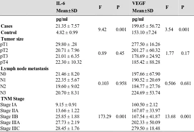
Mean serum VEGF levels were significantly greater in patients than controls (P = 0.001) (Table 2). The mean levels increased proportionally from the control group to higher TNM stages (P = 0.001) (Table 2). Serum VEGF concentrations did not differ significantly with tumor size (F = 1.77, P = 0.17) or lymph node metastasis (F = 0.506, P = 0.681) (Table 2). Comparison of mean serum VEGF concentrations between groups revealed statistically significant differences between controls and stage IIIA, controls and stage IIIC, stage IIA and stage IIIC, and stage IIB and stage IIIC. No other differences were statistically significant (Table 3). The mean VEGF concentrations in estrogen-positive (199.73 pg/ml) and estrogen-negative (199.59 pg/ml) tumors were not statistically different, nor were those of progesterone-positive (193.39 pg/ml) and progesterone-negative (203.92 pg/ml), or Human epidermal growth factor receptor (Her2)/neu-positive (193.94 pg/ml) and Her2/neu-negative (206.37 pg/ml) tumors (Data not shown in the tables).
Table 1. Basic characteristics of study subjects
Number %
Patients 37
Controls 20
Median Age (Range) 54 (34-60)
Laterality
Left 15 40.5
Right 22 59.5
Tumor size
pT1 2 5.4
pT2 25 67.6
pT3 8 21.6
pT4 2 5.4
Lymph node metastasis
N0 21 56.8
N1 7 18.9
N2 3 8.1
N3 6 16.2
Distant metastasis
Metastasis (-) 37 100 Metastasis (+) 0 0
TNM Stage
Stage IA 2 5.4 Stage IIA 14 37.8 Stage IIB 4 10.8 Stage IIIA 9 24.3 Stage IIIC 8 21.6
Table 2. Relationship between IL-6 and VEGF with clinicopathological parameters
IL-6
Mean±SD F P
VEGF
Mean±SD F P
pg/ml pg/ml
Cases 21.35 ± 7.57
9.42 0.001 199.65 ± 56.72 3.54 0.001
Control 4.82 ± 0.99 153.10 ±7.24
Tumor size
pT1 29.80 ± .28
0.89 0.45
277.50 ± 16.26
1.77 0.17 pT2 20.71 ± 7.96 201.27 ± 60.32
pT3 21.01 ± 6.35 178.69 ± 24.92 pT4 22.30 ± 10.32 185.42 ± 88.28
Lymph node metastasis
N0 21.46 ± 8.20
0.103 0.958
197.66 ± 67.90
0.506 0.681 N1 22.35 ± 5.67 190.52 ± 20.69
N2 19.60 ± 9.02 184.77 ± 27.76 N3 20.70 ± 8.31 224.69 ± 53.74
TNM Stage
Stage IA 9.15 ± 0.91
173.29 0.001
160.50 ± 2.12
13.68 0.001 Stage IIA 13.66 ± 1.22 167.07 ± 33.97
Stage IIB 25.85 ± 1.88 167.54 ± 41.87 Stage IIIA 27.73 ± 2.19 202.33 ± 50.09 Stage IIIC 28.45 ± 1.76 279.50 ± 18.48 F – Fischer’s value which is the combined figure of variances between and within groups. P – P value is the corresponding probability value of F.
Rep. Biochem. Mol. Biol, Vol. 6, No. 1, Oct 2017 36
Table 3. Stages comparisons of serum VEGF
Subject No. Comparison Mean difference SE P value
1 Control vs Stage I 7.39 5.25 0.176 2 Control vs Stage IIA 13.46 7.95 0.101 3 Control vs Stage IIB 13.89 9.40 0.154 4 Control vs Stage IIIA 49.22 11.50 0.000 5 Control vs Stage IIIC 126.39 4.87 0.000 6 Stage I vs Stage IIA -6.07 24.6 0.809 7 Stage I vs Stage IIB -6.50 31.20 0.845 8 Stage I vs Stage IIIA -41.83 36.92 0.287 9 Stage I vs Stage IIIC -119.0 13.68 0.000 10 Stage IIA vs Stage IIB -0.429 20.07 0.983 11 Stage IIA vs Stage IIIA -35.76 17.43 0.053 12 Stage IIA vs Stage IIIC -112.9 13.02 0.000 13 Stage IIB vs Stage IIIC -112.5 16.86 0.000 14 Stage IIB vs Stage IIIA -35.33 28.79 0.245 15 Stage IIIA vs Stage IIIC -77.16 18.80 0.001
Serum IL-6 level in breast cancer patients
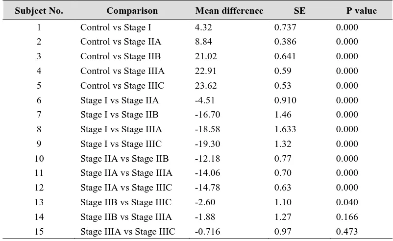
Mean serum IL-6 levels were also significantly greater in patients than controls (Table 2). The IL-6 concentrations increased from the control group to higher stages of breast carcinoma. The lowest level in patients was seen in stage IA, and highest in stage IIIC (p = 0.001) (Table 2). Serum IL-6 concentrations did not differ significantly with tumor size (F = 0.89, P = 0.45) or lymph node metastasis (F = 0.103, P = 0.958) (Table 2). Analysis of variance revealed statistically significant differences between the groups based
on stage (Table 4). The mean IL-6 concentrations in the control and stage I groups were significantly less than those of all the other stages. IL-6 levels were not significantly different between the stage IIIA and stage IIB or stage IIIC and stage IIIA groups. The mean IL-6 concentrations in estrogen-positive (21.38 pg/ml) and estrogen-negative (21.33 pg/ml) tumors were not significantly different, nor were those of progesterone-positive (22.20 pg/ml) and progesterone negative (20.78 pg/ml), or Her2/neu-positive (21.89 pg/ml) and Her2/neu-negative (20.72 pg/ml) tumors (Data not shown in the tables).
Table 4. Stages comparisons of serum IL-6
Subject No. Comparison Mean difference SE P value
1 Control vs Stage I 4.32 0.737 0.000 2 Control vs Stage IIA 8.84 0.386 0.000 3 Control vs Stage IIB 21.02 0.641 0.000 4 Control vs Stage IIIA 22.91 0.59 0.000 5 Control vs Stage IIIC 23.62 0.53 0.000 6 Stage I vs Stage IIA -4.51 0.910 0.000 7 Stage I vs Stage IIB -16.70 1.46 0.000 8 Stage I vs Stage IIIA -18.58 1.633 0.000 9 Stage I vs Stage IIIC -19.30 1.32 0.000 10 Stage IIA vs Stage IIB -12.18 0.77 0.000 11 Stage IIA vs Stage IIIA -14.06 0.70 0.000 12 Stage IIA vs Stage IIIC -14.78 0.63 0.000 13 Stage IIB vs Stage IIIC -2.60 1.10 0.040 14 Stage IIB vs Stage IIIA -1.88 1.27 0.166 15 Stage IIIA vs Stage IIIC -0.716 0.97 0.473
Association between IL-6 and VEGF
The correlation between serum IL-6 and
VEGF was strongly positive (r2 = 0.668, p < 0.01) (Fig. 1).
Fig.1. Correlation between IL-6 and VEGF in breast cancer.
Discussion
Our study was designed to measure VEGF and IL-6 in patients with breast cancer to evaluate their potential prognostic importance. Many studies have reported elevated serum VEGF concentrations in cancer patients and claimed that the measurement of circulating VEGF is a surrogate marker of angiogenesis and/or metastasis (4-6). To determine the utility of VEGF measurement in the diagnosis and prognosis of breast cancer, we measured its concentration in healthy controls and women with different stages of breast cancer.
Vascular endothelial growth factor is a potent angiogenic cytokine in normal and tumor tissues, stimulating endothelial cell proliferation in vitro and inducing angiogenesis in vivo. (6) The highest concentrations of serum VEGF were found in metastatic breast cancer, particularly among
patients who did not receive cancer therapy for metastatic disease. A significant correlation between VEGF concentration and microvessel density has been reported (11). A study of 29 invasive breast carcinomas revealed that VEGF expression in the peritumoral endothelial cells correlated with angiogenesis, lymphangiogenesis, and higher pathologic stage (12).In a study of 50 invasive ductal carcinomas, VEGF mRNA and protein expression correlated with tumor size, lymph node metastasis, and TNM staging. The microvessel density counts correlated with VEGF expression and axillary lymph node metastasis (13). Cox analysis revealed that intratumoral VEGF was an independent prognostic factor for node-negative breast cancer. High VEGF and low soluble VEGFR-1 (an intrinsic negative counterpart of VEGFA) levels were significantly associated with poor prognoses (14).
Rep. Biochem. Mol. Biol, Vol. 6, No. 1, Oct 2017 38
Interleukin-6 is a 30 kDa glycoprotein characterized by functional pleiotropy (15).Interleukin 6 is produced by a variety of cell types, including endothelial and normal haematopoietic cells. High IL-6 serum levels correlate with decreased survival rates in patients with hematological malignancies, renal cell carcinoma, and prostate cancer(16-18). High IL-6 levels have been found to indicate poor prognoses in patients with breast cancer (19).
The TNM classification system is used to stage the disease, which has a strong influence on patient prognoses. Interleukin-6 is one of many cytokines secreted by breast tumors. Interleukin-6 binds to the IL-6 receptor, activates the Janus kinase (JAK), and subsequently phosphorylates the signal transducers and activators of transcription (STAT). The phosphorylated STAT protein translocates into the nucleus and activates target genes, including VEGF and Rho, which increase the aggressiveness of the tumor. This involvement of IL-6 at a cellular level with the processes of cancer control is reflected by the results of serum studies of cancer patients, where IL-6 may reflect prognosis and tumor load. Elevated IL-6 levels have been associated with advanced stages and metastasis-related morbidity (20).
Interleukin-6 is associated with angiogenesis by virtue of its ability to induce VEGF mRNA expression, which induces angiogenesis. Additionally, IL-6 activates the Rho protein, which is associated with cell-cell adhesion and invasion in malignancy. Together these factors increase tumor aggressiveness. Interleukin-6 may promote cell migration by activating the mitogen-dependent protein kinase pathway, and increase chemotherapeutic resistance by inhibiting the activation of proteases involved in apoptosis.
Because IL-6 induces VEGF transcription and augments both platelet production and platelet storage of VEGF, it was formerly hypothesised that it could be regarded as an indirect angiogenic factor acting mainly on
VEGF metabolism. On the other hand, the IL-6 serum level is an independent prognosis factor, which agrees with results obtained by Zhang et. al.(19) on a smaller series. This suggests that, apart from the regulatory effects of IL-6 on VEGF production, other mechanisms are responsible for the deleterious effects of high serum IL-6 levels on tumor growth, thus explaining their strong correlation with prognosis in breast cancer patients.
In our study, serum IL-6 and VEGF levels were higher in the breast cancer cases than in the controls. Higher concentrations of IL-6 and VEGF were found in patients with advanced cancer stages than in those with lower stages. No correlations were found between IL-6 or VEGF concentrations and primary tumor size or lymph node involvement.
In this study, we showed that serum IL-6 and VEGF are greater in patients with breast cancer than controls. We compared the IL-6 and VEGF serum levels with the TNM staging system of breast cancer and correlated them with the patients’ prognoses. Our results suggest that IL-6 and VEGF play important roles in metastatic breast carcinoma progression in vivo, have a significant impact on patient prognosis, and can be used to identify higher-risk breast cancers. Thus serum IL-6 and VEGF can be used as diagnostic tools and prognostic factors in breast cancer. We propose further studies with these markers for early detection of breast cancer recurrence. The small sample size is one limitation of our study; however, this may be the basis for large scale multicentric studies.
Acknowledgments
We would also like to acknowledge Department of Science and Technology, Government of India for funding the CEMR lab under their Funds for Improvement of S&T Infrastructure (DST-FIST) programme in the year 2012.
We also acknowledge the financial assistance rendered by JSS University for the post graduate student towards her dissertation.
References
1. Jemal A, Siegel R, Ward E, Hao Y, Xu J, Thun MJ. Cancer statistics, 2009. CA Cancer J Clin. 2009;59:225–249.
2. Lester SC. The Breast. Kumar V, Abbas AK, Fausto N, Aster JC, Robbins and Cotran Pathologic Basis of Disease. 8th ed. Philadelphia: Elsevier;2010:1065-95. 3. Grivennikov SI, Greten FR, Karin M. Immunity, inflammation, and cancer. Cell. 2010; 140:883– 899. 4. Senger DR, Brown LF, Claffey KP, Dvorak HF. Vascular permeability factor, tumor angiogenesis and stroma generation. Invasion Metastasis 1994;14 385-394.
5. Angelo LS, Kurzrock R. Vascular endothelial
growth factor and
its relationship to inflammatory mediators. Clin Cancer Res 2007;13:2825-30.
6. Byrne GJ, McDowell G, Agarawal R, Sinha G, Kumar S, Bundred NJ. Serum Vascular Endothelial Growth Factor in Breast Cancer. Anti cancer research. 2007:27;3481-3488.
7. Ilhan N1, Ilhan N, Ilhan Y, Akbulut H, Kucuksu M. C-reactive protein procalcitonin, interleukin-6, vascular endothelial growth factor
and oxidative metabolites in
diagnosisof infection and staging in patients with gastric cancer. World J Gastroenterol. 2004;10: 1115-20.
8. Schneider MR, Hoeflich A, Fischer JR, Wolf E, Sordat B, Lahm H. Interleukin-6 stimulates clonogenic growth of primary and metastatic human colon carcinoma cells. Cancer Lett 2000;151:31– 8.
9. Wei LH, Kuo ML, Chen CA, Cheng WF, Cheng SP, Hsieh FJ, Hsieh CY. Interleukin-6 in cervical cancer: the relationship with vascular endothelial growth factor. GynecolOncol2001;82:49 –56.
10.Sansone P, Storci G, Tavolari S, Guarnieri T, Giovannini C, Taffurelli M, et al. IL-6 triggers malignant features in mammospheres from human ductal breast carcinoma and normal mammary gland. J. Clin. Invest. 2007;117:3988–4002.
11. Obermair A, Kucera E, Mayerhofer K, Speiser P, Seifert M, Czerwenka K, and et al. Vascular endothelial growth factor (VEGF) in humanbreast cancer: correlation with disease-free survival. Int. J. Cancer 1997;74:455–458.
12.Ghosh S, Sullivan CA, Zerkowski MP, Molinaro AM, Rimm DL, Camp RL, Chung GG. High levels of vascular endothelial growth factor and its receptors (VEGFR-1, VEGFR-2, neuropilin-1) are associated with worse outcome in breast cancer. Human Pathol2008;39:1835–1843.
13.Xie XD, Qu SX, Liu ZZ, Zhang F, Zheng ZD. Study on relationship between angiogenesis and micrometastases of peripheral blood in breast cancer. J. Cancer Res Clin2009;135:413–419.
14.Toi M, Bando H, Ogawa T, Muta M, Hornig C, Weich HA. Significance of vascular endothelial growth factor (VEGF)/soluble VEGF receptor-1 relationship in breast cancer. Int J Cancer 2002;98:14–18.
15.Kishimoto T. Interleukin-6: discovery of a pleiotropic cytokine. Arthritis Res Ther 2006;8Suppl 2:S2.
16.Kishimoto T, Akira S, Narazaki M, Taga T. Interleukin-6 family of cytokines and gp130. Blood. 1995:86;1243–1254.
17.Blay JY, Negrier S, Combaret V, Attali S, Goillot E, Merrouche Y, et.al. Serum level of interleukin 6 as a prognosis factor in metastatic renal cell carcinoma. Cancer Res 1992:52;3317–3322.
18.Nakashima J, Tachibana M, Horiguchi Y, Oya M, Ohigashi T, Asakura H, et.al. Serum interleukin 6 as a prognostic factor in patients with prostate cancer. Clin Cancer Res 2000:6;2702–2706.
19.Zhang GJ, Adachi I. Serum interleukin-6 levels correlate to tumor progression and prognosis in metastatic breast carcinoma. Anticancer Res 1999:19;1427–32.
20. Ravishankaran p, Karunanithi R. Clinical significance of preoperative serum interleukin-6 and C-reactive protein level in breast cancer patients. World Journal of Surgical Oncology 2011:9;18.