P R I M A R Y R E S E A R C H
Open Access
Hypoxic regulation of cytoglobin and
neuroglobin expression in human normal and
tumor tissues
Marwan Emara, A Robert Turner, Joan Allalunis-Turner
*Abstract
Background:Cytoglobin (Cygb) and neuroglobin (Ngb) are recently identified globin molecules that are expressed in vertebrate tissues. Upregulation of Cygb and Ngb under hypoxic and/or ischemic conditionsin vitroandin vivo increases cell survival, suggesting possible protective roles through prevention of oxidative damage. We have previously shown that Ngb is expressed in human glioblastoma multiforme (GBM) cell lines, and that expression of its transcript and protein can be significantly increased after exposure to physiologically relevant levels of hypoxia. In this study, we extended this work to determine whether Cygb is also expressed in GBM cells, and whether its expression is enhanced under hypoxic conditions. We also compared Cygb and Ngb expression in human primary tumor specimens, including brain tumors, as well as in human normal tissues. Immunoreactivity of carbonic anhydrase IX (CA IX), a hypoxia-inducible metalloenzyme that catalyzes the hydration of CO2to bicarbonate, was used as an endogenous marker of hypoxia.
Results:Cygb transcript and protein were expressed in human GBM cells, and this expression was significantly increased in most cells following 48 h incubation under hypoxia. We also showed that Cygb and Ngb are expressed in both normal tissues and human primary cancers, including GBM. Among normal tissues, Cygb and Ngb expression was restricted to distinct cell types and was especially prominent in ductal cells. Additionally, certain normal organs (e.g.stomach fundus, small bowel) showed distinct regional co-localization of Ngb, Cygb and CA IX. In most tumors, Ngb immunoreactivity was significantly greater than that of Cygb. In keeping with previousin vitroresults, tumor regions that were positively stained for CA IX were also positive for Ngb and Cygb, suggesting that hypoxic upregulation of Ngb and Cygb also occursin vivo.
Conclusions:Our finding of hypoxic up-regulation of Cygb/Ngb in GBM cell lines and human tumor tissues suggests that these globin molecules may be part of the repertoire of defense mechanisms that allow cancer cells to survive in hypoxic microenvironments.
Background
A third member of the vertebrate globin family, neuro-globin (Ngb), was discovered in 2000 and so-named because it is primarily expressed in neuronal tissue, including retina [1]. Shortly thereafter, a fourth verte-brate globin–cytoglobin (Cygb), was described indepen-dently by three groups [2-4]. Cygb is expressed ubiquitously in human tissue [2], however, low cellular levels of Cygb and Ngb (μM range) may have impeded their earlier discovery [5]. The amino acid sequences of
Cygb and Ngb show little similarity to that of hemog-lobin (Hb) or myoghemog-lobin (Mb) (< 30% and <25% identity for Cygb and Ngb, respectively). However, amino acids that confer Hb and Mb function are conserved together with all features of the globin fold [2,4,6]. Unlike Hb and Mb, the physiological roles of Ngb and Cygb are incompletely understood and several functions are con-ceivable. Ngb and Cygb may function as a Mb-type molecule to store O2 thus facilitating O2 diffusion to mitochondria [1,6,7]. However, the lower O2 affinity of Ngb (P50 of 7.5 torr under physiological conditions of pH and temperature) [8] compared to that of Mb (P50 of 2-3 torr) [7] does not support an O2storage function
* Correspondence: joan.turner@ualberta.ca
Department of Oncology, University of Alberta, Cross Cancer Institute, 11560 University of Alberta, Edmonton, Alberta, T6G 1Z2, Canada
for Ngb in neuronal tissue, including retina, as only a small fraction of Ngb (~ 12%) will be O2 saturated under normal conditions [5,8-10].
Similar to Mb, but in contrast to Ngb, the O2binding of Cygb is pH-independent [8] with higher O2 affinity values (P50 of 0.7-1.8 torr) [4,8,11], thus suggesting a possible physiological role to supply O2. However, due to its low concentrationin vivo, Cygb function may be restricted to O2-requiring cellular reactions unrelated to mitochondrial respiration [8].
In brain, Ngb is upregulated under hypoxic/ischemic conditions [12] and may function to scavenge reactive oxygen (ROS) and nitrogen species (RNS) that are a major cause of cellular damage [12,13]. It has been shown that in the Fe2+-NO form, Ngb reacts more rapidly with peroxynitrite than does Hb [14]. Addition-ally, in contrast to Hb and Mb, the reaction of met(Fe3+) Ngb with peroxynitrite or hydrogen peroxide does not appear to generate the cytotoxic ferryl (Fe4+) species, and this may contribute to cellular survival [5,14]. However, evidence for Ngb’s neuroprotective functionin vivo is inconsistent [10,12,13,15-17]. Similarly, Cygb was found to be upregulated following oxidative stress and hypoxic/ ischemic conditions in vitro and in vivo [17-21], and overexpression of Cygb in hepatic stellate cells, human SH-SY5Y neuroblastoma cells and MIN6 cells is protec-tive [17,22,23]. The function of Cygb has not yet been intensively investigated. In fibroblasts and related cells expressing Cygb, its expression has been linked to col-lagen production and organ fibrosis [24-27]. Recently, it has been proposed that CYGB may function as a tumor suppressor gene as hypermethylation of its promoter was detected in primary human non-small cell lung cancers (48%) [28] and oral cancers (65%) [29], and in lung (8 of 10) and breast (4 of 4) cancer cell lines [30].
Glioblastoma multiforme (GBM) is the most common brain tumor among adults comprising 25% of all malig-nant nervous system tumors. Its resistance to multimod-ality therapy confers a poor prognosis and the 2-year survival rate remains only 10-25% [31]. Necrosis, and by inference hypoxia, is a diagnostic feature of human GBM tumors [32]. Hypoxic microenvironments fre-quently occur in human tumors, and tumor cells that are hypoxic are resistant to ionizing radiation and cer-tain chemotherapeutic agents, are genetically unstable and metastasize frequently. Further, hypoxic microenvir-onments also select for tumor cells with reduced apop-totic potential. The presence of tumor hypoxia is an indicator of poor prognosis for both local-regional con-trol and progression-free survival, and there is also evi-dence that hypoxiaper seselects for a more aggressive tumor phenotype (reviewed in [33] and references therein). Tumor cells that survive in hypoxic microen-vironments must first sense changes in [O2] and then
activate defense and adaptation mechanisms [34]. Pre-vious work suggests that increased Ngb expression may be part of the repertoire of hypoxia defense mechanisms in normal brain. Although some studies have reported that Ngb is expressed exclusively in neurons but not in glia [35,36], others have reported Ngb expression in astrocytes cultured from newborn mouse brain [37], and we have shown that Ngb is expressed and upregulated by hypoxia in human GBM cell lines [38]. Here, we examined whether Cygb is also expressed in human GBM cell lines and tested whether physiologically rele-vant levels of hypoxia can modulate its expression as was previously demonstrated for Ngb. In addition to these in vitro analyses, we used human tissue microar-rays (TMA) to assess whether Ngb/Cygb is more broadly expressed among human primary tumors, including brain cancers, and their adjacent normal tissues. CA IX immunoreactivity was used as an endo-genous marker for tissue hypoxia.
Results
Cygb transcript and protein are expressed in GBM cells
in vitro
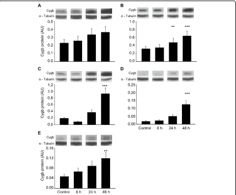
Cygb mRNA was detected in all aerobic GBM cell lines (controls), and when cultured under hypoxic conditions (0.6% O2 × 48 h), all cell lines showed significantly increased expression of Cygb mRNA (M006x, M010b and U87T, p < 0.05; M006xLo and M059J, p < 0.001) (Figure 1). Two cell lines also showed significant increases in Cygb transcript at earlier time points (M006xLo at 6 and 24 h, p < 0.001; M059J at 24 h, p < 0.01). Consistent with qRT-PCR results, Cygb pro-tein was detected in all aerobic GBM cell lines with hypoxia-tolerant cells (M006x, M006xLo) showing the highest basal levels of Cygb. After 48 h of hypoxia, Cygb protein was significantly increased in four of five cell lines (M006xLo, M010B, and M059J, p < 0.001; U87 T, p < 0.01), with the greatest relative increases seen in hypoxia-sensitive cells M010b and M059J (Figure 2). Cygb protein was also significantly increased in M006xLo cells after 24 h of hypoxia (p < 0.01). M006x cells showed a modest increase in Cygb protein after hypoxia, but this was not significant (Figure 2A). There was no correlation between the magnitude of Cygb pro-tein increase after hypoxia and the respective basal levels of each cell line.
Cygb and Ngb are expressed in human normal tissues and cancers
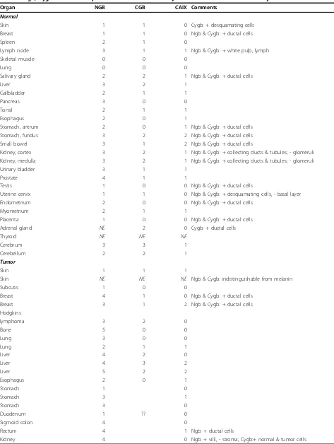
normal tissues, Ngb expression was greatest in bone, liver, sigmoid colon, rectum and kidney, with low or negligible levels in skin, muscle and lung (Table 1). Tis-sues with highest Cygb expression were stomach fundus, kidney tubules and cerebellum. However, compared to Ngb, Cygb levels were reduced overall and were low or absent in approximately half of the tissues. There was no significant correlation between levels of Cygb and Ngb protein in these normal tissues (p < 0.07). However, two features of Cygb and Ngb expression in normal tis-sues were of particular interest. First, in several normal tissues, expression of Cygb and Ngb was not uniform throughout the tissue. Rather, distinct cell types or tis-sue structures frequently showed strong positive staining while the remainder of the tissue showed weak or absent staining. Among the structures strongly stained for Cygb were ductal cells of the breast and kidney, secretory cells of the salivary gland, white pulp/lymph of the spleen, and tips of desquamating cells found in
several types of normal tissue. Ngb staining was also prominent among ductal cells of breast, endometrium, testis, prostate and salivary gland. Both Ngb and Cygb were generally absent from stroma. Second, most nor-mal tissues showed low or absent CA IX immunoreac-tivity. However, stomach, small bowel, and gallbladder showed nearly identical patterns of Ngb, Cygb and CA IX staining (Figure 3).
Tumor sections showed three distinct staining patterns for Cygb/Ngb: (1) a uniform expression throughout the non-stromal tissue; (2) distinct regions of positively stain-ing cells among otherwise negative or weakly stainstain-ing tis-sue; or (3) densely staining focal areas frequently including intensely positive foci. Among individual tumor sections included in the‘tumor/matched normal tissue’TMA, Ngb and Cygb expression was generally increased compared to levels observed in corresponding normal tissues. As well, Cygb and Ngb immunoreactivity showed similar patterns of distribution. Most tumor
Figure 1Cygb mRNA expression in human GBM cells. Cygb mRNA expression was assessed by qRT-PCR after exposure to hypoxia (0.6% O2)
sections (24/29) showed low or absent CA IX immunor-eactivity. However, the breast, liver, bladder and thyroid tumors that contained regions strongly positive for CA IX also showed in matching sections similar staining patterns for Cygb and Ngb (Figure 4).
Cygb and Ngb are expressed in human brain tumors
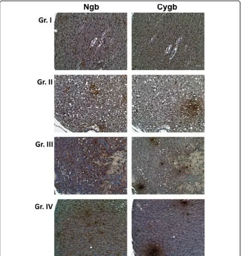
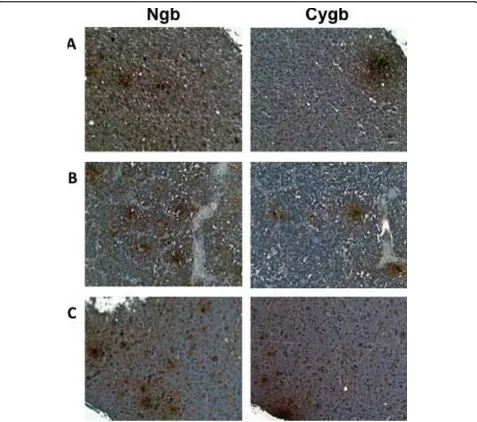
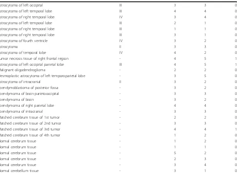
Both Cygb and Ngb were detected in all human brain tumors, including grades I-IV astrocytomas (Figure 5), and ependymoblastomas, gangliogliomas and oligoden-drogliomas (Figure 6). Among low grade astrocytomas (Grades I and II), Ngb staining intensity was signifi-cantly greater than that of Cygb (p < 0.05). Grade III astrocytomas and GBMs also showed relatively greater Ngb staining but these differences were not significant
(Tables 2 and 3). In tumors that showed distinct pat-terns of Ngb and Cygb expression, a comparison of tissue sections showed that these proteins were expressed in the same regions of the tumor. Large, posi-tively staining Ngb and Cygb foci were commonly observed in GBM tumors. Approximately 60% of the GBM sections were also positive for CA IX staining which was confined to regions of the tumor also positive for Cygb and Ngb.
Discussion
Cygb mRNA and protein were detected in five human GBM cell lines cultured under aerobic conditions. How-ever, basal Cygb protein levels varied ~19-fold. The rank-order of Cygb protein expression in these cell lines
Figure 2Cygb protein expression in human GBM cells. Cygb expression was assessed by Western blot analyses after exposure to hypoxia (0.6% O2) for 0, 6, 24 and 48 h (n = 4). The integrated intensities of Cygb anda- tubulin (control) bands were determined and expressed in
Table 1 Ngb, Cygb and CA IX expression in tissue microarrays of human solid tumors and adjacent normal tissues
Organ NGB CGB CAIX Comments
Normal
Skin 1 1 0 Cygb: + desquamating cells
Breast 1 1 0 Ngb & Cygb: + ductal cells
Spleen 2 1 0
Lymph node 3 1 1 Ngb & Cygb: + white pulp, lymph
Skeletal muscle 0 0 0
Lung 0 0 0
Salivary gland 2 2 1 Ngb & Cygb: + ductal cells
Liver 3 2 1
Gallbladder 2 1 1
Pancreas 3 0 0
Tonsil 2 1 1
Esophagus 2 0 1
Stomach, antrum 2 0 1 Ngb & Cygb: + ductal cells
Stomach, fundus 3 2 2 Ngb & Cygb: + ductal cells
Small bowel 3 1 2 Ngb & Cygb: + ductal cells
Kidney, cortex 3 2 1 Ngb & Cygb: + collecting ducts & tubules; - glomeruli Kidney, medulla 3 2 1 Ngb & Cygb: + collecting ducts & tubules; - glomeruli
Urinary bladder 3 1 1
Prostate 4 1 1
Testis 1 0 0 Ngb & Cygb: + ductal cells
Uterine cervix 1 1 0 Ngb & Cygb: + desquamating cells, - basal layer
Endometrium 2 0 0 Ngb & Cygb: + ductal cells
Myometrium 2 1 1
Placenta 1 0 0 Ngb & Cygb: + ductal cells
Adrenal gland NE 2 0 Cygb: + ductal cells
Thyroid NE NE NE
Cerebrum 3 3 1
Cerebellum 2 2 1
Tumor
Skin 1 1 1
Skin NE NE NE Ngb & Cygb: indistinguishable from melanin
Subcutis 1 0 0
Breast 4 1 0 Ngb & Cygb: + ductal cells
Breast 3 1 2 Ngb & Cygb: + ductal cells
Hodgkins
lymphoma 3 2 0
Bone 5 0 0
Lung 3 0 0
Lung 2 1 1
Liver 4 2 0
Liver 4 3 2
Liver 5 2 2
Esophagus 2 0 1
Stomach 1 0
Stomach 3 1
Stomach 3 0
Duodenum 1 ?? 0
Sigmoid colon 4 0
Rectum 4 1 Ngb: + ductal cells
Table 1 Ngb, Cygb and CA IX expression in tissue microarrays of human solid tumors and adjacent normal tissues (Continued)
Urinary bladder 1 3 Ngb: + infiltrating cells
Prostate 3 1 Ngb: + ductal cells
Testis 2 0 Ngb: + ductal cells
Uterine cervix 2 1
Endometrium 1 1 Ngb: some + ductal cells
Ovary 2 0
Ovary 3 0
Ovary 1 1
Thyroid 2 3
Tissue microarrays constructed from cores obtained from various human solid tumors and normal adjacent tissues were purchased from IMGENEX Corporation (San Diego, CA). Immunopositive staining for Ngb, Cygb and CA IX was assessed by two observers who noted the overall tissue staining and the presence of positively stained focal regions. Each section was assigned an overall score, where 0 = absence of positively stained cells; 1 = a few positive cells/regions ( < 10%); 2 = weak positive staining, occasional positive foci; 3 = intermediate staining, occasional positive foci ( < 50%); 4 = strongly positive in most of the section, several positive foci; and 5 = strongly positive throughout with many intensely positive foci. Distinct features of the immunostaining patterns in individual tissue cores are noted in the comments column, where“+”= positive staining,“-”= negative staining andNE= not evaluable.
is the reverse order of that of Ngb [38], with hypoxia-tolerant cells having highest Cygb, but lowest Ngb, levels. We are not aware of any other comparisons of Cygb/Ngb concentrations in human tumor cell lines. While our results in GBM cell lines hints at a reciprocal relationship between Cgyb/Ngb, at least in GBM cell lines, this would have to be confirmed in a larger study. Co-expression of Ngb and Cygb has been reported among various structures of the anterior segment of human and canine eyes, including the cornea, iris, irido-corneal angle and the cilliary body [40], and in human retinal neurons and pigmented epithelium [41]. How-ever, the relative expression of each protein within spe-cific cellular structures was not quantified. In the murine retina, divergent expression of Cygb and Ngb
has been reported, with Ngb levels being significantly higher [42].
Both Cygb transcript and protein were significantly increased in four of five GBM cell lines cultured under conditions that simulate in vivo O2 concentrations found in the hypoxic regions of human tumors [43,44]. Others have also shown that the hypoxia up-regulates Cygb transcript and protein in cell lines of neuronal ori-gin– HN33 [18] and (SH-SY5) [45]. Hypoxia-tolerant M006x cells that express high basal level of Cygb pro-tein showed a small, but non-significant, increase in Cygb after exposure to reduced [O2]. Nonetheless, M006x cells are hypoxia-responsive as we have shown that expression of another hypoxia-inducible gene,
VEGF, can be up-regulated at equivalent levels of
hypoxia [46]. It is interesting to note that while Cygb levels varied widely among GBM cells under aerobic condition (19-fold), when exposed to hypoxia, the differ-ences among the cell lines were notably reduced. This would suggest that mechanisms exist to allow cells to
titrate intracellular concentrations of Cygb to optimally meet the demands of given levels of oxidative stress.
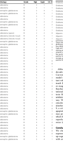
In addition to GBM cell lines, Cygb expression was also observed in both low and high grade human astro-cytomas, including GBM. To our knowledge, this is the
first report of Cygb expression in human primary brain cancers although high Cygb levels have been reported in brain metastases of two patients with alveolar soft part sarcomas [47]. Both Cygb transcript and protein have been reported in normal neurons, but not glia [41]. To date, there is little information concerning the co-expression of these two globin molecules in malignant human tissues. In this study, Ngb protein levels in Grades I and II astrocytomas were significantly greater than Cygb levels (p < 0.05). There was also a trend toward increased Cygb expression as tumor grade increased from I - IV, however, this would have to be confirmed in a larger study. A similar pattern of greater
Ngb/Cygb positivity was also observed among non-brain cancers specimens examined, with Ngb expression sig-nificantly higher than that of Cygb (p <0.001). Regions of necrosis are a pathognomonic feature of GBM tumors, and in many sections, enhanced Cygb/Ngb staining was observed adjacent to necrotic tissue. The microenvironment of these perinecrotic regions is likely to be severely hypoxic and thus, hypoxia-inducible pro-teins would be expected to be upregulated. In order to test this, we used CA IX staining as an endogenous marker of tumor hypoxia. CA IXis a target gene of the hypoxia-inducible transcription factor, HIF-1a, and some, but not all, studies have shown a correlation
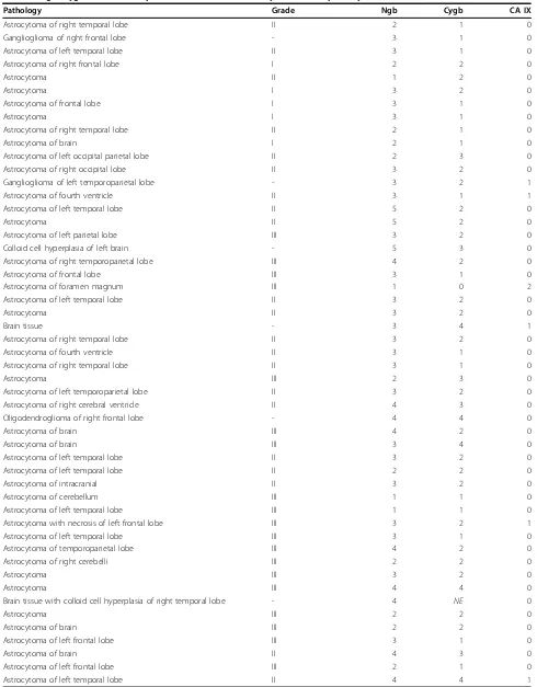
Table 2 Ngb, Cygb and CA IX expression in tissue microarrays of human primary brain tumors
Pathology Grade Ngb Cygb CA IX
Astrocytoma of right temporal lobe II 2 1 0
Ganglioglioma of right frontal lobe - 3 1 0
Astrocytoma of left temporal lobe II 3 1 0
Astrocytoma of right frontal lobe I 2 2 0
Astrocytoma II 1 2 0
Astrocytoma I 3 2 0
Astrocytoma of frontal lobe I 3 1 0
Astrocytoma I 3 1 0
Astrocytoma of right temporal lobe II 2 1 0
Astrocytoma of brain I 2 1 0
Astrocytoma of left occipital parietal lobe II 2 3 0
Astrocytoma of right occipital lobe II 3 2 0
Ganglioglioma of left temporoparietal lobe - 3 2 1
Astrocytoma of fourth ventricle II 3 1 1
Astrocytoma of left temporal lobe II 5 2 0
Astrocytoma II 5 2 0
Astrocytoma of left parietal lobe III 3 2 0
Colloid cell hyperplasia of left brain - 5 3 0
Astrocytoma of right temporoparietal lobe III 4 2 0
Astrocytoma of frontal lobe III 3 1 0
Astrocytoma of foramen magnum III 1 0 2
Astrocytoma of left temporal lobe II 3 2 0
Astrocytoma II 3 2 0
Brain tissue - 3 4 1
Astrocytoma of right temporal lobe II 3 2 0
Astrocytoma of fourth ventricle II 3 1 0
Astrocytoma of right temporal lobe II 3 1 0
Astrocytoma III 2 3 0
Astrocytoma of left temporoparietal lobe II 3 2 0
Astrocytoma of right cerebral ventricle II 4 3 0
Oligodendroglioma of right frontal lobe - 4 4 0
Astrocytoma of brain III 4 2 0
Astrocytoma of brain III 3 4 0
Astrocytoma of left temporal lobe II 3 2 0
Astrocytoma of left temporal lobe II 2 2 0
Astrocytoma of intracranial II 3 2 0
Astrocytoma of cerebellum III 1 1 0
Astrocytoma of left temporal lobe III 1 1 0
Astrocytoma with necrosis of left frontal lobe III 3 2 1
Astrocytoma of left temporal lobe III 3 1 0
Astrocytoma of temporoparietal lobe III 4 2 0
Astrocytoma of right cerebelli III 2 2 0
Astrocytoma III 3 2 0
Astrocytoma III 4 4 0
Brain tissue with colloid cell hyperplasia of right temporal lobe - 4 NE 0
Astrocytoma III 2 2 0
Astrocytoma of brain III 2 2 0
Astrocytoma of left frontal lobe III 3 1 0
Astrocytoma of brain II 4 3 0
Astrocytoma of left frontal lobe III 2 1 0
between CA IX expression and tumor hypoxia (reviewed in [48]). In these TMAs, tumor regions that were posi-tive for CA IX also showed Cygb/Ngb expression. How-ever, not all regions that were Ngb/Cygb positive also showed CA IX immunoreactivity. A possible explanation is the fact that in tumors, CA IX expression is generally associated with perinecrotic regions that are likely to be severely hypoxic [48]. In contrast, the up-regulation of both Ngb and Cygb expression has been observed at relatively higher O2tensions (~1% O2).
In addition to the brain tumor TMAs, we also studied Ngb and Cygb expression in several normal tissues and their malignant counterparts. Although Cygb has been described as a ubiquitous cellular globin, it was reduced or absent from many normal tissues, but prominent in ductal cells in gallbladder, stomach, small bowel and kidney. Shigematsu et al. [49] previously reported a similar distribution for Cgyb in normal tissues. However,
the most striking feature observed in our study was the distinct regional co-localization of Ngb, Cygb and CA IX in several normal tissues, including stomach, small bowel and salivary gland. CA IX expression in human alimentary tract has been previously described [50], but to our knowledge, this is the first demonstration of co-expression of Ngb/Cygb/CA IX in human tissue.
To corroborate these protein expression patterns, we queried NCBI Gene Expression Omnibus (GEO) Pro-files–a public repository of microarray gene expression data [51]. This search indicated that there is robust expression of Ngb and Cygb mRNA in a variety of human primary non-malignant and tumor samples [See Additional files 1 and 2]. However, there is con-siderable heterogeneity in the expression Ngb and Cygb mRNA, both within a given histological type of non-malignant or tumor tissue, and among different types of tissue.
Table 2 Ngb, Cygb and CA IX expression in tissue microarrays of human primary brain tumors(Continued)
Astrocytoma of left occipital III 3 3 0
Astrocytoma of left temporal lobe III 4 4 0
Astrocytoma of right temporal lobe IV 3 4 0
Astrocytoma of left temporal lobe III 2 1 0
Astrocytoma of right temporal lobe III 1 1 0
Astrocytoma of right temporal lobe III 3 1 0
Astrocytoma of fourth ventricle IV 3 2 0
Astrocytoma II 3 3 0
Astrocytoma of temporal lobe IV 4 2 0
Tumor necrosis tissue of right frontal region - 4 5 1
Astrocytoma of left occipital parietal lobe III 4 5 1
Malignant oligodendroglioma - 2 2 0
Desmoplastic astrocytoma of left temporoparietal lobe - 3 5 0
Astrocytoma of intracranial II 3 2 0
Ependymoblastoma of posterior fossa - 3 2 0
Ependymoma of brain parietooccipital - 3 3 0
Ependymoma of brain - 3 2 0
Ependymoma of right parietal lobe - 4 4 0
Ependymoma of intracranial - 3 3 0
Matched cerebrum tissue of 1st tumor - 2 2 0
Matched cerebrum tissue of 2nd tumor - 3 3 0
Matched cerebrum tissue of 3rd tumor - 4 4 1
Matched cerebrum tissue of 4th tumor - 1 2 0
Normal cerebrum tissue - 1 2 0
Normal cerebrum tissue - 1 1 0
Normal cerebrum tissue - 5 2 0
Normal cerebrum tissue - 2 3 0
Normal cerebrum tissue - 3 4 0
Normal cerebellum tissue - 3 1 0
Although Ngb and Cygb were discovered almost a decade ago, their functions continue to be explored. Currently, evidence from both in vitro and in vivo model systems indicates that these globins play impor-tant roles in protecting cells from death during ischemic insult (hypoxia-reperfusion), oxygen/glucose deprivation, or exogenous oxidative stress [17,22,45,52-55]. Recently, Raychaudhuriet al. [56] used experimental and compu-tational analyses to show that Ngb overexpression pro-tects SH-SY5Y neuroblastoma cells from cell death by inhibiting the initiation of the intrinsic apoptotic path-way. They hypothesize that the ability of Ngb to bind to cytochromecprovides a mechanism whereby Ngb could interfere with cytochromec-induced activation caspase-9, a critical step required for initiation of the intrinsic apoptotic cascade. Accordingly, Ngb or Cygb expression may provide the same pro-survival effect in tumors in which both chronic and intermittent hypoxia (ischemia/ reperfusion) as well as glucose deprivation commonly occur (reviewed in [33].
Conclusions
We showed two novel globins–Cygb and Ngb, are expressed in human primary tumors and cell lines. Their up-regulation by hypoxiain vitro, and their association with an endogenous hypoxia marker in tumor tissue
Table 3 Assessment of Cygb, Ngb and CA IX expression in tissue microarrays of human glioblastoma multiforme tumors
Pathology Grade Ngb Cygb CA IX
Glioblastoma IV 3 1 1
Glioblastoma IV 3 1 1
Pleomorphic glioblastoma IV 5 4 1
Pleomorphic glioblastoma IV 5 4 1
Glioblastoma IV 3 1 2
Glioblastoma IV 2 1 1
Pleomorphic glioblastoma IV 5 2 2
Pleomorphic glioblastoma IV 5 2 2
Glioblastoma IV 2 2 1
Glioblastoma IV 2 2 1
Glioblastoma (sparse) IV 2 2 0
Glioblastoma (necrotic tissue) IV 4 5 1 Glioblastoma (necrotic tissue) IV 4 5 1
Pleomorphic glioblastoma IV 4 3 0
Pleomorphic glioblastoma IV 4 3 0
Glioblastoma III 2 2 0
Glioblastoma III 2 2 0
Glioblastoma IV 2 3 1
Glioblastoma IV 3 2 1
Glioblastoma IV 3 2 1
Glioblastoma III 5 4 0
Glioblastoma III 5 4 0
Glioblastoma IV 5 2 1
Glioblastoma IV 5 2 2
Glioblastoma IV 4 2 0
Glioblastoma IV 5 2 1
Glioblastoma IV 5 2 3
Glioblastoma IV 3 2 0
Glioblastoma IV 3 3 1
Glioblastoma IV 4 3 0
Glioblastoma IV 4 2 2
Glioblastoma IV 4 2 0
Glioblastoma IV 3 2 0
Glioblastoma III 5 2 0
Glioblastoma III 5 3 1
Glioblastoma IV 4 2 3
Glioblastoma IV 4 2 0
Glioblastoma IV 5 2 0
Pleomorphic glioblastoma IV 4 2 1
Pleomorphic glioblastoma IV 4 3 2
Glioblastoma III 4 3 2
Glioblastoma III 4 3 2
Glioblastoma III 4 2 0
Glioblastoma III 4 3 0
Glioblastoma IV 4 4 0
Glioblastoma IV 5 3 0
Glioblastoma IV 4 3 0
Pleomorphic glioblastoma IV 5 4 1
Pleomorphic glioblastoma IV 5 4 1
Table 3 Assessment of Cygb, Ngb and CA IX expression in tissue microarrays of human glioblastoma multiforme tumors(Continued)
Glioblastoma III 3 2 0
Glioblastoma III 3 2 0
Glioblastoma IV 4 3 3
Glioblastoma IV 4 3 3
Glioblastoma IV 4 4 0
Glioblastoma IV 3 2 0
Glioblastoma IV 4 3 0
Glioblastoma IV 3 4 0
Glioblastoma IV 3 4 0
Glioblastoma IV 2 3 0
Glioblastoma IV 3 2 0
Adjacent normal brain - 4 3 0
Adjacent normal brain - 3 2 1
Adjacent normal brain - 3 4 0
sections suggests that these globins may contribute to the pro-survival mechanisms utilized by these cancers to sur-vive under conditions of oxidative stress.
Methods
Cell lines and in vitro culture conditions
The origin and characterization of the GBM cell lines have been published previously: the M059J (ATCC num-ber CRL2366) and M010b cell lines are hypoxia-sensitive; the M006x and M006xLo cell lines are hypoxia-tolerant [46,57,58]. The U87T cell line was established following serialin vivoselection in a glioma mouse model [59] and was kindly provided by Dr. Donna Senger (University of Calgary). Its relative sensitivity to hypoxia has not been determined. All cells were maintained as monolayer cul-tures in DMEM/F12 media supplemented with 10% fetal calf serum and 1 mM L-glutamine in a humidified atmosphere of 5% CO2in air at 37°C. All tissue culture supplies were purchased from GIBCO.
Generation of hypoxia in vitro
A de-gassing manifold was used to generate hypoxia
in vitro [60]. Exponential phase cells (~2 × 10⁵) were seeded onto 60-mm glass plates and then incubated under standard laboratory culture conditions (5% CO2 in air) for 4 days. The medium was then replenished and the plates were transferred to aluminum chambers from which the air was evacuated and then replaced with 5% CO2/balance N2 until an O2 tension of 0.6% was achieved. The sealed, air-tight aluminum chambers were then incubated at 37°C for 6-48 h. At the end of each incubation interval, the aluminum chambers were unsealed, the tissue culture plates removed, RNA and protein isolated from the cells as described in Section 2.4 and 2.5.
Quantitative real-time reverse transcription-PCR
Total RNA from cultured cell lines was isolated using the RNeasy mini kit (QIAGEN). Reverse transcription (RT) was carried out with 1 μg total RNA. per 20 ml reaction volume using QuantiTect reverse transcription kit (QIAGEN). RT experiments were performed with GeneAmp PCR system 9700 (Applied Biosystems). Quantitative real-time PCR (qRT-PCR) analysis was per-formed with a 7900 HT Fast Real-Time PCR System (Applied Biosystems) using TaqMan fast universal PCR master mix and a validated TaqMan Gene Expression assay (Applied Biosystems) for the human CYGB gene (assay ID: Hs00370478_ml). Human 18 S rRNA gene (part no.: 4333760T, Applied Biosystems) was used as endogenous control. Amplification data were analyzed with SDS RQ Manager 1.2. Relative quantities of Cygb transcripts were normalized against relative quantities of
the 18 S rRNA transcripts, and fold-expression changes calculated using the expression 2-ΔΔCT.
Western blotting
Whole-cell lysates were prepared using complete Lysis-M buffer (Roche Diagnostics) and protein content deter-mined using a protein assay kit (Pierce). Equal amounts of protein (50μg) were resolved using 13% SDS -PAGE under reducing condition and electro-transferred to polyvinylidene difluoride membranes (Bio-Rad Labora-tories, CA). Membranes were blocked with 5% skim milk then incubated with Cygb antibody (1:60 rabbit anti-human polyclonal antibody [sc-66855] Santa Cruz Biotechnology, Inc, CA) ora-tubulin antibody (1:5000, mouse anti-human a-tubulin monoclonal antibody, SIGMA) as loading control. Membranes were incubated with polyclonal goat anti-rabbit IgG (H+L) horseradish peroxidase (HRP)-conjugated (1:25000, Jackson Immu-noResearch Laboratories, Inc. PA) or polyclonal goat anti-mouse IgG HRP-conjugated (1:6000, DakoCytoma-tion Denmark A/S, Demark). Bound proteins were detected using chemiluminescence reagents (SuperSignal West Pico Chemiluminescent Substrate, Thermo Scien-tific, IL) and visualized by exposing to X-ray film (Fuji Photo Film, Japan) that was developed using a Kodak X-OMAT 2000A processor (Eastman Kodak Company, Japan). X-ray films were scanned using an Artixscan 1800f scanner (MICROTEK, TAIWAN) and bands were analyzed using Quantity One 1-D analysis software (Bio-Rad Laboratories). The integrated areas of bands were determined and expressed in arbitrary units (AU).
Tissue micorarrays and immunostaining
specificity of the primary antibodies was demonstrated by using Cygb or Ngb peptides to block positive immu-nostaining [Additional file 3]. Tissue endogenous peroxi-dase was quenched by incubation with 3% H2O2 for 15 min. Tissue sections were incubated with HRP-con-jugated goat anti-mouse antibodies (DakoCytomation) and positive staining visualized by the chromogenic reaction of HRP with DAB (3,3’diaminobenzidine tetra-hydrochloride) (DakoCytomation). Relative Cygb and Ngb expression in each tissue core was assessed by two observers who noted the overall tissue staining and the presence of positively stained focal regions. Each section was assigned an overall score, where 0 = absence of positively stained cells; 1 = a few positive cells/regions (< 10%); 2 = weak positive staining, occasional positive foci; 3 = intermediate staining, occasional positive foci (< 50%); 4 = strongly positive in most of the section, several positive foci; and 5 = strongly positive through-out with many intensely positive foci.
Statistics
Data from four replicate experiments were expressed as mean ± standard error. Statistical analyses were per-formed using SigmaPlot 11 software (Systat Software, Inc). Differences between groups were compared using one-way ANOVA or ANOVA on ranks (Kruskal-Wallis) based on the normality and equal variance tests. To determine exactly which groups are different and the size of the difference, multiple comparisons versus con-trol group were carried out using Bonferroni t-test and Dunnett’s or Dunn’s test for one-way ANOVA and ANOVA on ranks (Kruskal- Wallis), respectively, as post-hoc tests.
Additional material
Additional file 1: Table S1. Results of GEO profiles query of Cygb expression in human non-malignant and tumor tissues. This table provides the range of the intra-sample percentile rank values for Cygb expression in a variety of human primary tissue samples. These values provide an indication of the relative expression of Cygb compared to that of all other genes expressed in that sample.
Additional file 2: Table S2. Results of GEO profiles query of Ngb expression in human non-malignant and tumor tissues. This table provides the range of the intra-sample percentile rank values for Ngb expression in a variety of human primary tissue samples. These values provide an indication of the relative expression of Ngb compared to that of all other genes expressed in that sample.
Additional file 3: Figure S1. Antibody specificity demonstrated by competitive immunostaining. This figure shows that incubation of primary Ngb and Cygb antibodies with the relevant recombinant proteins effectively blocked positive immunostaining.
Acknowledgements
We thank Bonnie Andrais for assistance with tissue culture and Gerry Barron for help with the preparation of the photomicrographs. This study was
supported by an award from the Canadian Cancer Society Research Institute with funds provided by the Canadian Cancer Society.
Authors’contributions
EM and JAT conceived of the study. EM performed thein vitroanalyses and TMA immunostaining, did the statistical analyses and wrote the first draft of the manuscript. ART and JAT analyzed the TMAs. All authors contributed to the final draft of the manuscript.
Competing interests
The authors declare that they have no competing interests.
Received: 17 March 2010 Accepted: 9 September 2010 Published: 9 September 2010
References
1. Burmester T, Weich B, Reinhardt S, Hankeln T:A vertebrate globin expressed in the brain.Nature2000,407(6803):520-523.
2. Burmester T, Ebner B, Weich B, Hankeln T:Cytoglobin: a novel globin type ubiquitously expressed in vertebrate tissues.Mol Biol Evol2002, 19(4):416-421.
3. Kawada N, Kristensen DB, Asahina K, Nakatani K, Minamiyama Y, Seki S, Yoshizato K:Characterization of a stellate cell activation-associated protein (STAP) with peroxidase activity found in rat hepatic stellate cells. J Biol Chem2001,276(27):25318-25323.
4. Trent JT, Hargrove MS:A ubiquitously expressed human hexacoordinate hemoglobin.J Biol Chem2002,277(22):19538-19545.
5. Fago A, Hundahl C, Malte H, Weber RE:Functional properties of neuroglobin and cytoglobin. Insights into the ancestral physiological roles of globins.IUBMB Life2004,56(11-12):689-696.
6. Pesce A, Bolognesi M, Bocedi A, Ascenzi P, Dewilde S, Moens L, Hankeln T, Burmester T:Neuroglobin and cytoglobin. Fresh blood for the vertebrate globin family.EMBO Rep2002,3(12):1146-1151.
7. Wittenberg JB, Wittenberg BA:Myoglobin function reassessed.J Exp Biol 2003,206(Pt 12):2011-2020.
8. Fago A, Hundahl C, Dewilde S, Gilany K, Moens L, Weber RE:Allosteric regulation and temperature dependence of oxygen binding in human neuroglobin and cytoglobin. Molecular mechanisms and physiological significance.J Biol Chem2004,279(43):44417-44426.
9. Wangsa-Wirawan ND, Linsenmeier RA:Retinal oxygen: fundamental and clinical aspects.Arch Ophthalmol2003,121(4):547-557.
10. Hundahl C, Stoltenberg M, Fago A, Weber RE, Dewilde S, Fordel E, Danscher G:Effects of short-term hypoxia on neuroglobin levels and localization in mouse brain tissues.Neuropathol Appl Neurobiol2005, 31(6):610-617.
11. Hamdane D, Kiger L, Dewilde S, Green BN, Pesce A, Uzan J, Burmester T, Hankeln T, Bolognesi M, Moens L,et al:The redox state of the cell regulates the ligand binding affinity of human neuroglobin and cytoglobin.J Biol Chem2003,278(51):51713-51721.
12. Sun Y, Jin K, Mao XO, Zhu Y, Greenberg DA:Neuroglobin is up-regulated by and protects neurons from hypoxic-ischemic injury.Proc Natl Acad Sci USA2001,98(26):15306-15311.
13. Sun Y, Jin K, Peel A, Mao XO, Xie L, Greenberg DA:Neuroglobin protects the brain from experimental stroke in vivo.Proc Natl Acad Sci USA2003, 100(6):3497-3500.
14. Herold S, Fago A, Weber RE, Dewilde S, Moens L:Reactivity studies of the Fe(III) and Fe(II)NO forms of human neuroglobin reveal a potential role against oxidative stress.J Biol Chem2004,279(22):22841-22847. 15. Hundahl C, Kelsen J, Kjaer K, Ronn LC, Weber RE, Geuens E, Hay-Schmidt A,
Nyengaard JR:Does neuroglobin protect neurons from ischemic insult? A quantitative investigation of neuroglobin expression following transient MCAo in spontaneously hypertensive rats.Brain Res2006,1085(1):19-27. 16. Fraser J, de Mello LV, Ward D, Rees HH, Williams DR, Fang Y, Brass A,
Gracey AY, Cossins AR:Hypoxia-inducible myoglobin expression in nonmuscle tissues.Proc Natl Acad Sci USA2006,103(8):2977-2981. 17. Fordel E, Thijs L, Martinet W, Lenjou M, Laufs T, Van Bockstaele D, Moens L,
Dewilde S:Neuroglobin and cytoglobin overexpression protects human SH-SY5Y neuroblastoma cells against oxidative stress-induced cell death. Neurosci Lett2006,410(2):146-151.
vitro and in vivo study by quantitative real-time PCR.Biochem Biophys Res Commun2004,319(2):342-348.
19. Burmester T, Haberkamp M, Mitz S, Roesner A, Schmidt M, Ebner B, Gerlach F, Fuchs C, Hankeln T:Neuroglobin and cytoglobin: genes, proteins and evolution.IUBMB Life2004,56(11-12):703-707. 20. Guo X, Philipsen S, Tan-Un KC:Study of the hypoxia-dependent
regulation of human CYGB gene.Biochem Biophys Res Commun2007, 364(1):145-150.
21. Li D, Chen XQ, Li WJ, Yang YH, Wang JZ, Yu AC:Cytoglobin up-regulated by hydrogen peroxide plays a protective role in oxidative stress. Neurochem Res2007,32(8):1375-1380.
22. Stagner JI, Parthasarathy SN, Wyler K, Parthasarathy RN:Protection from ischemic cell death by the induction of cytoglobin.Transplant Proc2005, 37(8):3452-3453.
23. Xu R, Harrison PM, Chen M, Li L, Tsui TY, Fung PC, Cheung PT, Wang G, Li H, Diao Y,et al:Cytoglobin overexpression protects against damage-induced fibrosis.Mol Ther2006,13(6):1093-1100.
24. Schmidt M, Gerlach F, Avivi A, Laufs T, Wystub S, Simpson JC, Nevo E, Saaler-Reinhardt S, Reuss S, Hankeln T,et al:Cytoglobin is a respiratory protein in connective tissue and neurons, which is up-regulated by hypoxia.J Biol Chem2004,279(9):8063-8069.
25. Hankeln T, Ebner B, Fuchs C, Gerlach F, Haberkamp M, Laufs TL, Roesner A, Schmidt M, Weich B, Wystub S,et al:Neuroglobin and cytoglobin in search of their role in the vertebrate globin family.J Inorg Biochem2005, 99(1):110-119.
26. Nakatani K, Okuyama H, Shimahara Y, Saeki S, Kim DH, Nakajima Y, Seki S, Kawada N, Yoshizato K:Cytoglobin/STAP, its unique localization in splanchnic fibroblast-like cells and function in organ fibrogenesis.Lab Invest2004,84(1):91-101.
27. Hankeln T, Burmester T:Neuroglobin and Cytoglobin.InThe Smallest, Biomolecules, Diatomics and their Interactions with Heme Proteins.Edited by: Ghosh A. Elsevier BV; 2008:203-218.
28. Xinarianos G, McRonald FE, Risk JM, Bowers NL, Nikolaidis G, Field JK, Liloglou T:Frequent genetic and epigenetic abnormalities contribute to the deregulation of cytoglobin in non-small cell lung cancer.Hum Mol Genet2006,15(13):2038-2044.
29. Shaw RJ, Hall GL, Woolgar JA, Lowe D, Rogers SN, Field JK, Liloglou T, Risk JM:Quantitative methylation analysis of resection margins and lymph nodes in oral squamous cell carcinoma.Br J Oral Maxillofac Surg 2007,45(8):617-622.
30. Shivapurkar N, Stastny V, Okumura N, Girard L, Xie Y, Prinsen C, Thunnissen FB, Czerniak B, Frenkel E,et al:Cytoglobin, the newest member of the globin family, functions as a tumor suppressor gene. Cancer Res2008,68(18):7448-7456.
31. Stupp R, Mason WP, van den Bent MJ, Weller M, Fisher B, Taphoorn MJ, Belanger K, Brandes AA, Marosi C, Bogdahn U,et al:Radiotherapy plus concomitant and adjuvant temozolomide for glioblastoma.N Engl J Med 2005,352(10):987-996.
32. Wilden J, Moore I:Histological Factors in the Prognosis of Malignant Glioma. Brain Oncology: Biology, Diagnosis and Therapy.Martinus Nijhoff, Dordrecht 1987.
33. Bertout JA, Patel SA, Simon MC:The impact of O2 availability on human cancer.Nat Rev Cancer2008,8(12):967-975.
34. Hochachka PW, Buck LT, Doll CJ, Land SC:Unifying theory of hypoxia tolerance: molecular/metabolic defense and rescue mechanisms for surviving oxygen lack.Proc Natl Acad Sci USA1996,93(18):9493-9498. 35. Laufs TL, Wystub S, Reuss S, Burmester T, Saaler-Reinhardt S, Hankeln T: Neuron-specific expression of neuroglobin in mammals.Neurosci Lett 2004,362(2):83-86.
36. Ostojic J, Sakaguchi DS, de Lathouder Y, Hargrove MS, Trent JT, Kwon YH, Kardon RH, Kuehn MH, Betts DM, Grozdanic S:Neuroglobin and cytoglobin: oxygen-binding proteins in retinal neurons.Invest Ophthalmol Vis Sci2006,47(3):1016-1023.
37. Chen XQ, Qin LY, Zhang CG, Yang LT, Gao Z, Liu S, Lau LT, Fung YW, Greenberg DA, Yu AC:Presence of neuroglobin in cultured astrocytes. Glia2005,50(2):182-186.
38. Emara M, Salloum N, Allalunis-Turner J:Expression and hypoxic up-regulation of neuroglobin in human glioblastoma cells.Mol Oncol2009, 3(1):45-53.
39. Hankeln T, Wystub S, Laufs T, Schmidt M, Gerlach F, Saaler-Reinhardt S, Reuss S, Burmester T:The cellular and subcellular localization of
neuroglobin and cytoglobin–a clue to their function?IUBMB Life2004, 56(11-12):671-679.
40. Ostojic J, Grozdanic S, Syed NA, Hargrove MS, Trent JT, Kuehn MH, Kardon RH, Kwon YH, Sakaguchi DS:Neuroglobin and cytoglobin distribution in the anterior eye segment: a comparative
immunohistochemical study.J Histochem Cytochem2008,56(9):863-872. 41. Ostojic J, Grozdanic SD, Syed NA, Hargrove MS, Trent JT, Kuehn MH,
Kwon YH, Kardon RH, Sakaguchi DS:Patterns of distribution of oxygen-binding globins, neuroglobin and cytoglobin in human retina.Arch Ophthalmol2008,126(11):1530-1536.
42. Schmidt M, Laufs T, Reuss S, Hankeln T, Burmester T:Divergent distribution of cytoglobin and neuroglobin in the murine eye.Neurosci Lett2005, 374(3):207-211.
43. Olive PL, Trotter T, Banath JP, Jackson SM, Le Riche J:Heterogeneity in human tumour hypoxic fraction using the comet assay.Br J Cancer Suppl 1996,27:S191-195.
44. Vaupel P, Schlenger K, Knoop C, Hockel M:Oxygenation of human tumors: evaluation of tissue oxygen distribution in breast cancers by
computerized O2 tension measurements.Cancer Res1991, 51(12):3316-3322.
45. Fordel E, Thijs L, Martinet W, Schrijvers D, Moens L, Dewilde S:Anoxia or oxygen and glucose deprivation in SH-SY5Y cells: a step closer to the unraveling of neuroglobin and cytoglobin functions.Gene2007, 398(1-2):114-122.
46. Allalunis-Turner MJ, Franko AJ, Parliament MB:Modulation of oxygen consumption rate and vascular endothelial growth factor mRNA expression in human malignant glioma cells by hypoxia.Br J Cancer 1999,80(1-2):104-109.
47. Genin O, Rechavi G, Nagler A, Ben-Itzhak O, Nazemi KJ, Pines M: Myofibroblasts in pulmonary and brain metastases of alveolar soft-part sarcoma: a novel target for treatment?Neoplasia2008,10(9):940-948. 48. Vordermark D, Brown JM:Endogenous markers of tumor hypoxia
predictors of clinical radiation resistance?Strahlenther Onkol2003, 179(12):801-811.
49. Shigematsu A, Adachi Y, Matsubara J, Mukaide H, Koike-Kiriyama N, Minamino K, Shi M, Yanai S, Imamura M, Taketani S,et al:Analyses of expression of cytoglobin by immunohistochemical studies in human tissues.Hemoglobin2008,32(3):287-296.
50. Kivela AJ, Kivela J, Saarnio J, Parkkila S:Carbonic anhydrases in normal gastrointestinal tract and gastrointestinal tumours.World J Gastroenterol 2005,11(2):155-163.
51. Barrett T, Troup DB, Wilhite SE, Ledoux P, Rudnev D, Evangelista C, Kim IF, Soboleva A, Tomashevsky M, Marshall KA,et al:NCBI GEO: archive for high-throughput functional genomic data.Nucleic Acids Res2009, ,37 Database:D885-890.
52. Burmester T, Hankeln T:What is the function of neuroglobin?J Exp Biol 2009,212(Pt 10):1423-1428.
53. Duong TT, Antao S, Ellis NA, Myers SJ, Witting PK:Supplementation with a synthetic polyphenol limits oxidative stress and enhances neuronal cell viability in response to hypoxia-re-oxygenation injury.Brain Res2008, 1219:8-18.
54. Fago A, Mathews AJ, Brittain T:A role for neuroglobin: resetting the trigger level for apoptosis in neuronal and retinal cells.IUBMB Life2008, 60(6):398-401.
55. Li RC, Morris MW, Lee SK, Pouranfar F, Wang Y, Gozal D:Neuroglobin protects PC12 cells against oxidative stress.Brain Res2008,1190:159-166. 56. Raychaudhuri S, Skommer J, Henty K, Birch N, Brittain T:Neuroglobin
protects nerve cells from apoptosis by inhibiting the intrinsic pathway of cell death.Apoptosis2010,15(4):401-411.
57. Allalunis-Turner MJ, Barron GM, Day RS, Fulton DS, Urtasun RC: Radiosensitivity testing of human primary brain tumor specimens.Int J Radiat Oncol Biol Phys1992,23(2):339-343.
58. Parliament MB, Allalunis-Turner MJ, Franko AJ, Olive PL, Mandyam R, Santos C, Wolokoff B:Vascular endothelial growth factor expression is independent of hypoxia in human malignant glioma spheroids and tumours.Br J Cancer2000,82(3):635-641.
60. Koch CJ, Howell RL, Biaglow JE:Ascorbate anion potentiates cytotoxicity of nitro-aromatic compounds under hypoxic and anoxic conditions.Br J Cancer1979,39(3):321-329.
doi:10.1186/1475-2867-10-33
Cite this article as:Emaraet al.:Hypoxic regulation of cytoglobin and neuroglobin expression in human normal and tumor tissues.Cancer Cell International201010:33.
Submit your next manuscript to BioMed Central and take full advantage of:
• Convenient online submission
• Thorough peer review
• No space constraints or color figure charges
• Immediate publication on acceptance
• Inclusion in PubMed, CAS, Scopus and Google Scholar • Research which is freely available for redistribution