http://www.arjournals.org/index.php/ijpm/index
Original Research Article
Ethanolic Extracts Of A. Paniculata (burm. f.) Nees And Its Active Compound,
Andrographolide, Decrease The Expression Of Glucose Transporters (glut 4)
In High Fuctose-Fat Fed Rats
Agung Endro Nugroho1, Mohamad Andrie1, Rina Susilowati2, Arief Nurrochmad1, Endang Lukitaningsih3, Suwijiyo
Pramono4
*Corresponding author:
Agung Endro Nugroho
1
Department of Pharmacology and Clinical Pharmacy, Faculty of Pharmacy, Universitas Gadjah Mada, Yogyakarta, Indonesia, 55281.
2
Department of Histology and Cell Biology, Faculty of Medicine, Universitas Gadjah Mada, Yogyakarta, Indonesia, 55281. 3
Department of Pharmaceutical Chemistry, Faculty of Pharmacy, Universitas Gadjah Mada, Yogyakarta, Indonesia, 55281. 4
Department of Pharmaceutical Biology, Faculty of Pharmacy, Universitas Gadjah Mada, Yogyakarta, Indonesia, 55281.
A B S T R A C T
Andrographolide is a major compound (> 4%) of Andrographis paniculata (Burm. f.) Nees. The compound is most active than the other compounds contained in the plant. Previously, the compound was reported showing hypoglycemic activity in type 1 diabetes mellitus (DM) rats. The compound also improved the uptake of glucose in isolated soleus muscle of type 1 DM rats by increasing the GLUT4 expression. The disruption of GLUT4 translocation is a major factor of insulin resistance. In the study, we investigated the effect of andrografolid and ethanolic extract of A. paniculata (Burm. f.) Nees on the GLUT4 expression in high fuctose-fat fed rats, a model of insulin resistant rats. Insulin resistance in rats was induced by high fructose-fat containing 36% fructose, 15% lard and 5% egg yolks in 0.36 g/200g BW for 55 days. In the study, andrographolide and ethanolic extract of A. paniculata (Burm. f.) showed potent hypoglycaemic effects in insulin resistant rats. In addition, these treatments and metformin could restore the depleted GLUT-4 expression in insulin resistant rats. In conclusion, andrographolide and ethanolic extract of A. paniculata (Burm. f.) decreased the blood glucose levels by increasing the GLUT-4 expression in insulin resistant rats.
Keywords : Andrographis paniculata (Burm. f.) Nees, andrographolide, insulin resistance, GLUT-4.
Introduction
Diabetes mellitus (DM) is a chronic disordered metabolism related to a deficiency of insulin secretion or/and a decrease in tissues sensitivity such as skeletal muscle, adipose tissue to the presence of insulin (insulin resistance). DM is characterized by hyperglycemia, a high blood glucose level. There are two type of DM according to dependence on exogenous insulin : type 1 DM, also named as insulin-dependent diabetes mellitus (IDDM), and type 2 DM, also named as non-insulin-dependent DM (NIDDM) (1,
2). Type 1 DM is characterized by an absolute insulin deficiency due to destruction of pancreatic beta cells. The main cause of this destruction is the autoimmune processes. Pathogenesis of the autoimmune DM involves macrophages, beta cell autoantigens, dendritic cells, T lymphocytes, and B lymphocytes (3). Type 2 DM is related to insulin resistance and/or impaired insulin secreation. Type 2 DM is common in patients over the age of 40. Type 2 DM with insulin resistance is associated with obesity and decreased physical activity, and long term consumption of high calorie. Uncontrolled type 2 DM would develop into ISSN: 0975-0185
487
type 1 DM. Therefore, type 2 DM patients should consider the diet, lifestyle and oral hypoglycaemic drugs (2, 4).
It has been reported that more than 400 traditional plants have been used in the treatment of diabetes mellitus. However, only a few plants that were scientifically evaluated for their efficacy (5). Some of them are spices often used in everyday life such as fenugreek seeds, garlic, onion, turmeric and coriander (6). Most research on medicinal plants were associated with hypoglycemic effects. To date, several medicinal plants have been used both as a traditional medicine or supplements in diabetic patients.
Indonesia is the second largest country in the world after Brazil in terms of biodiversity including medicinal plants. One of the plants potentially developed as an antidiabetic agent is is Andrographis paniculata (Burm. f.) Nees. This plant originates from India, and growing extends southward to Southeast Asia including Indonesia. In Indonesia, these plants are widespread in many areas. Traditionally, this plant is used for several purposes, primarily preventing diabetes mellitus (DM) (7). Previously, ethanolic extracts of A. paniculata (Burm. f.) Nees was reported to decrease the blood glucose levels in type 1 DM rats potently (8). In addition, the water soluble extract of A. paniculata (Burm. f.) Nees showed an antioxidant activity by increasing the activities of superoxide dismutase (SOD) and catalase in type 1 DM rats (9).
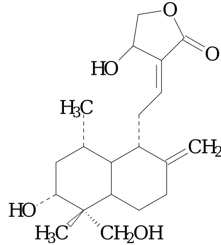
Andrographolide (fig. 1) is a major compound (> 4%) of A. paniculata (Burm. f.) Nees, and most active than the other compounds (10, 11, 12). Previously, andrographolide obviously decreased the blood glucose level in both normal and type 1 DM rats. The compound also increased the glucose utilization through increase of mRNA and protein levels of GLUT 4 in type 1 DM rats (13). In addition, andrographolide could stimulate the insulin release, and inhibited the absorption of glucose through inhibition of the enzyme alpha-glucosidase and alpha-amylase (14).
Figure 1. Chemical structure of andrographolide.
In the study, we investigated the effect of andrographolide and ethanolic extract of Andrographis paniculata (Burm. f.) Nees in expression of glucose transporters (GLUT 4) in high fuctose-fat fed rats, a model of type 2 DM associated with insulin resistance. The result of these studies may provide useful information for further discovering pharmacologically traditional plants isolated-active compounds for treatment of type 2 DM with insulin resistance.
Materials And Methods
Materials.
Andrographolide, glibenclamide and metformin were purchased from Sigma Chemical Co. (St.Louis, MO, USA). Sodium carboxymethyl cellulose, fructose, glucose were obtained from E. Merck, Darmstadt, Germany. Glucose level were measuerd using colorimetric method (GOD/PAP) with glucose oxidase and 4-aminoantipyrine (DiaSys, Diagnostic Systems GmbH, Holzheim, Germany). Antibodies for the determination of GLUT-4 expression were primary anti-GLUT4 antibody (Santa Cruz Biotechnologies, California, USA) and secondary chicken anti-goat IgG antibody (Invitrogen Carlsbad, CA, USA).
CH
2
HO
H
3
C
CH
2
OH
O
O
HO
488
Animals
Wistar rats weighing 100-150 g were used in the study. They were housed at a constant temperature (22 ± 2°C) with a constant relative humidity (55 ± 10%) on the an automatically controlled 12:12 h light-dark cycle (light on at 7:00 a.m.). They were fed with a standard laboratory food and water as libitum. The animal handling protocols of this study were in accordance with the guidelines of the animal care of the Department of Pharmacology and Clinical Pharmacy, Faculty of Pharmacy, Universitas Gadjah Mada, Indonesia.
Preparation of ethanolic extract of Andrographis paniculata (Burm. f.) Nees
Andrographis paniculata (Burm. f.) Nees was collected from area around Yogyakarta, Indonesia. The plant was identified by a botanist at Department of Pharmaceutical Biology, Universitas Gadjah Mada, Indonesia and the voucher specimen was stored in herbarium of the department.
In brief, dried ground powder of A. paniculata (Burm. f.) Nees was extracted with 90 % ethanol for 24 hours, and filtered. The extract was fractionated with n-hexane at a ratio of 1:20 (extract:n-n-hexane). The insoluble fraction of n-hexane was then fractionated using ethyl acetate at a ratio of 1:10 (insoluble fraction of n-hexane: ethyl acetate). The insoluble fraction was concentrated by rotary vacuum evaporator to obtain viscous extract. The insoluble fraction was then washed with hot water, and diluted with ethanol 90 % to yield an ethanolic extract.
Experimental design.
The rats were placed in animal laboratory for one week for acclimatization. The animals were administered with high fuctose-fat for the total period of 55 days to induce insulin resistance. This diet contained 36 % fructose, 15% lard and 5% egg yolks in 0.36 g/200 g BW. Control rats were fed with standard chow diet (normal rats). The rats were treated with the drug or its vehicle (negative control) at day 50. The high fuctose-fat fed rats were divided into several groups consisting of four-six rats.
Group I : the rats received oral saline 10 ml/kg BW (control group) from the day 50 onwards for the next 5 days.
Group II and III : the rats orally received puried extract of Andrographis paniculata (Burm. f.) Nees dose 434.6 and 1308.8 mg/kg BW, respectively. It was administered twice daily from the day 50 onwards for the next 5 days.
Group IV and V : the rats orally received andrographolide dose 1.5 and 4.5 mg/kg BW orally, respectively. It was administered twice daily from the day 50 onwards for the next 5 days. Group VI : the rats orally received metformin dose 45 mg/kg BW orally, respectively. It was administered twice daily from the day 50 onwards for the next 5 days.
All the rats were fasted for eight hours prior to drug administration. Blood samples were collected from retro-orbital plexus at the day of 0 (basal value), 50 and 55 for determination of preprandial blood glucose levels. Two hours after being fed, the blood was collected for determination of postprandial blood glucose levels. At the end of the experiment (day 55), the rats were anaesthetized by diethyl eter, then sacrificed by decapitation. Soleus muscles were quickly removed, separated from connective tissue, and frozen in a liquid nitrogen or a -80˚C refrigerator.
Insulin resistance test
Previously, long-term high fructose feeding in rats could cause an insulin resistance (15, 16). In the study, insulin resistance induced by high fuctose-fat fed was confirmed using the loss of glibenclamide-induced hypoglycemic action according the previous study (17). In the study, the rats (normal and insulin resistance) were fasted for 8-10 hours. Afterward, they were orally administered by a single dose of 5 mg/kg glibencamide. After 60 min, the blood was collected for determination of blood glucose levels.
Immunohistochemical study
489
the tissues were cleared using clearing agents (xylol) because the paraffin and ethanol are immiscible. The agent functions to mix the paraffin and ethanol. The tissues were then embedded in paraffine wax prior to subsequent sectioning. Tissue sections 2-3 µm in thickness were mounted on glass slides.
Activity of endogenous peroxidase was blocked using 3% H2O2 in methanol for 15 min, and then washed with aquadest. To prevent non-specific binding, the sections were incubated with 20% horse serum for at least 10 min. The sections were then incubated with primary antibody against GLUT-4 at a 1:250 dilution for one hour at room temperature. Subsequently, the sections was incubated with peroxidase-conjugated secondary antibody at a 1:500 dilution for one hour at room temperature. Expression of GLUT-4 would be visualized after incubation with substrate for 15 min. The sections were counterstained with hematoxylin, and then mounted. The expression of GLUT-4 was
semiquantified by two parameters : area and intensity of staining. The intensity of staining was categorized by assessing the following categories: 0 (0–4%), 1 (5– 24%), 2 (25 – 49%), 3 (50 – 74%), or 4 (75 – 100%) according to previous study (Katja et al., 1999). The staining area of GLUT-4 was calculated using macbiophotonics image J. Immunoreactive score was calculated by multiplication of area and intensity of staining.
Statistical analysis
All data were presented as mean ± the standard error of the mean (SEM). One-way analysis of variance (ANOVA) followed by the least significant difference (LSD) test was used for statistical analysis to compare more than two groups. While the unpaired t test was used to compare the mean of two groups. P-values of less than 0.05 were considered significant.
Figure 2. The hypoglycemic activity of glibenclamid in normal rats and in insulin resistant rats (high-fructose fat rats). In addition, metformin was used as positive control in hypoglycemic activity test in insulin resistant rats. Data represent mean±SEM, and are five-six independent experiments. *Significant difference (P<0.05) compared to the normal rats (rats fed normal chow).
0 10 20 30 40 50 60 70 80
Normal rats high fuctose-fat fed rats high fuctose-fat fed rats +
Metformin
Hypog
ly
ca
emic
ac
tivity
(%)
*
*
490
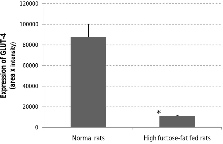
Figure 3. Effect of high fructose-fat feeding on the GLUT-4 expression in soleus muscles. administered once a day in rats for 55 days. Data represent mean±SEM, and are four-five independent experiments. *Significant difference (P<0.05) compared to the control value (rats fed normal chow).
Result
Induction of insulin resistance
In the study, glibenclamide, an sulfonylurea antidiabetic drug, was used to confirm an insulin resistance condition in the rats. Glibenclamide (5 mg/kg BW) was orally administered to fasted normal and high fuctose-fat fed rats. The blood samples were collected from retro-orbital plexus at 60 min after drug administration. In fig. 2, glibenclamide obviously decreased the blood glucose levels with hypogycemic activity of 47.86±2.75%. In contrast, hypoglycemic activity of glibenclamide in high fuctose-fat fed rats was only 16.08±0.84%. It indicates that hypoglycaemic activity of glibenclamide was reduced when administered in high fuctose-fat fed rats. However, metformin, an extrapancreatic antidiabetic drug, markedly decreased the blood glucose levels in high fuctose-fat fed rats with hypogycemic activity of 65.29±3.64%. In the immunohistochemical study as showed in fig 4, administration of high fuctose-fat for the total period of 55 days depleted the expression of GLUT-4 in soleus muscle sections. In the study, the diet contained 36 % fructose, 15% lard and 5% egg
yolks in 0.36 g/200 g BW. In fig.3, high fuctose-fat diet markedly decreased the expression of GLUT-4 by 87% in comparison to normal rats (P<0.05).
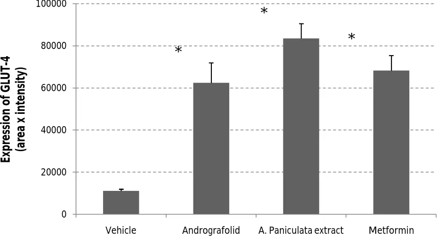
Effect of andrographolide on GLUT-4 expression Andrographolide (1.5 and 4.5 mg/kg BW) could decrease both preprandial and postprandial glucose levels significantly by 37.46±4.99 and 42.34±6.29 (P<0.05). Ethanolic extract of Andrographis paniculata (Burm. f.) Nees dose 434.6 and 1308.8 mg/kg BW also succeeded to decrease both preprandial and postprandial glucose levels significantly by 41.5±7.43 and 48.98±9.02 (P<0.05). In the study, we investigated the effect of andrographolide on GLUT-4 expression that its activation is altered in insulin resistance. Fig. 5 shows that andrograholide (4.5 mg/kg BW), ethanolic extract of Andrographis paniculata (Burm. f.) Nees (1308.8 mg/kg BW) and metformin (45 mg/kg BW) could increase the GLUT-4 expression in soleus muscles in comparison to control group (vehicle) (P<0.05). The expression of GLUT-4 was semiquantified based on area and intensity of staining. The area of GLUT-4 expression was calculated using macbiophotonics image J., and the intensity of staining 0
20000 40000 60000 80000 100000 120000
Normal rats High fuctose-fat fed rats
Expr
ession of GL
UT
-4
(a
rea
x
int
e
ns
ity)
491
was scored according to Katja et al. (18). Immunoreactive score representing GLUT-4 expression was calculated by multiplication of area and intensity of staining. In the study, andrograholide (4.5 mg/kg BW), ethanolic extract of Andrographis paniculata (Burm. f.) Nees (1308.8 mg/kg BW) and metformin (45 mg/kg BW) succeeded to restore the
depleted GLUT-4 expression in insulin resistance rats by 67.06±12.47%, 94.50±9.29% and 74.64±9.41%, respectively. Based on the results, treatment of ethanolic extract of Andrographis paniculata (Burm. f.) Nees at the dose of 1308.8 mg/kg BW for five days almost normalized the GLUT-4 expression in insulin resistance rats.
Figure 4. Immunohistochemical observation of GLUT4 expression in skeletal muscle both in normal rats
(a) and in insulin resistant rats (high-fructose fat rats) (b). The expression of GLUT4 was stained brown pointed by black arrow.
(a)
492
Figure 5. Effect of ethanolic extract of Andrographis paniculata (Burm. f.) Nees (1303.8 mg/kg BW), andrographolide (4.5 mg/kg BW) or metformin (45 mg/kg BW) twice daily on GLUT-4 expression in high fructose-fat fed rats (insulin resistant rats). The drug was administered on the day 50 for 5 days. Data represent mean±SEM, and are four-five independent experiments. *Significant difference (P<0.05) compared to the negative control value (vehicle).
Disscussion
Insulin resistance is actually present for many years before the onset of diabetes mellitus. The condition is stimulated by some factors such as family history, lifestyle (smoking, caffeine, alcohol, stress), obesity, other diseases (hypertention, arteriosclerosis, metabolic syndrome), and medication (glucocorticoids) (19, 20, 21, 22). Among them, obesity is a major factor in the development of insulin resistance (23). Normally, insulin interacts with insulin receptor on muscle or fat cells, and then stimulates the translocation of GLUT-4 into the cell membranes (24). GLUT-4 functions to transport the glucose from extracelluler side to intracelluler side (25, 26). Obesity can alter the activation of GLUT-4 translocation by insulin, and then the pancreas was stimulated to produce more insulin (hyperinsulinemia). Consequently, the muscle and fat cells become insensitive to the insulin (insulin resistance) (27). As long as the pancreas produce
more insulin, the muscle and fats continue to be insensitive to the insulin. Briefly, insulin resistance is related to the condition in which insulin become less effective to decrease the blood glucose levels.
Long-term administration of high fructose in rats can be used to induce an insulin resistance (15, 16). Reportly, fructose feeding consisting of 66% fructose, 22% casein and 12% lard for 8 weeks could stimulate insulin resistance in rats characterized by increased levels of insulin, glucose and triglyceride significantly (28). Long-term consumption of fructose contributes factors to development of obesity and metabolic abnormalities related to insulin resistance syndrome (29). In the study, diet containing 36 % fructose, 15% lard and 5% egg yolks in 0.36 g/200 g BW was used to stimulate insulin resistance.
0 20000 40000 60000 80000 100000
Vehicle Andrografolid A. Paniculata extract Metformin
Expr
ession of GL
U
T-4
(a
rea
x i
nt
ensit
y)
*
*
493
(a)
494
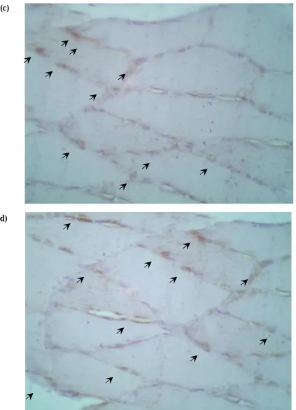
Figure 6. Immunohistochemical observation of GLUT4 expression in skeletal muscle of insulin resistant rats (high-fructose fat rats) after 5-days pretreatment with vehicle (a), ethanolic extract of Andrographis paniculata (Burm. f.) Nees (1303.8 mg/kg BW) (b), andrographolide (4.5 mg/kg BW) (c) or metformin (45 mg/kg BW) twice daily (d). The expression of GLUT4 was stained brown pointed by black arrow.
(c)
495
To confirm the condition of insulin resistance in rats, two parameters were used in the study. These are : 1). the loss of glibenclamide-induced hypoglycemic action (17), and 2). The disruption of GLUT-4 translocation into the cell membranes. In the study, glibenclamide showed a potent hypogycemic activity in normal rats, but the activity was decreased three times in insulin resistant rats. The drug acts dominantly by stimulating insulin release from the beta cells in the pancreas (pancreatic action) (30). Therefore, the use of the drug in insulin resistance showed mild hypogycemic activity. In the other hand, metformin succeeded to decrease the blood glucose levels in high fuctose-fat fed rats. Metformin acts to stimulate insulin-induced components of glucose uptake such as GLUT-4 into skeletal muscle and adipocytes (extrapancreatic action) (31). In imunohistochemical study, administration of high fuctose-fat for the total period of 55 days could deplete the expression of GLUT-4 in soleus muscle sections. The diet decreased the expression of GLUT-4 by 87% in comparison to normal rats. These facts indicate that administration of high fuctose-fat for the total period of 55 days could induce insulin resistance in rats.
Andrographolide is a major compound and abundance found in Andrographis paniculata (Burm. f.) Nees. The compound has an important role in the biological activities of the plant. A. paniculata is medicinal plant growing widely in many areas in South East Asia countries including Indonesia (32). The plant are traditionally used primarily as an antidiabetes, antiinflammatory, hepatoprotective, antispasmodic and antioxidant agents (Niranjan et al., 2010). A. paniculata contains major constituents such as diterpenoids, flavonoids and polyphenol (33). The other diterpenoids are deoxyandrographolide, 19-O-acetylanhydro-andrographolide, neoandrogra-pholide, 14-deoxy-didehydroandrographolide and homoandrographolide (34).
Andrographolide was reported showing hypoglycemic activity in streptozotocin-diabetics rats, a model of type 1 diabetes mellitus (DM) (13). The compound improved the uptake of glucose in isolated soleus muscle of type 1 DM rats by increasing the GLUT4
expression (13, 35). In the study, andrographolide and ethanolic extract of A. paniculata showed potent hypoglycaemic effects in insulin resistant rats. In addition, these treatments and metformin could restore the depleted GLUT-4 expression in insulin resistant rats. The effect of ethanolic extract of A. paniculata was higher than this of andrographolide and metformin. The ethanolic extract of almost normalized the GLUT-4 expression in insulin resistance rats. The other diterpenoids contained in the ethanolic extract might contribute in its hypoglycemic activity.
Conclusion
In conclusion, andrographolide and ethanolic extract of Andrographis paniculata (Burm. f.) decreased the blood glucose levels by increasing the GLUT-4 expression in insulin resistant rats. The result of these studies may provide useful information for further discovering pharmacologically traditional plants isolated-active compounds for treatment of type 2 diabetes mellitus.
Author’s Contribution
AEN was responsible to make a research concept and design of the study, data collection, acquisition of data,
analysis of data, statistical of data, drafted and corresponding author the manuscript. AN was responsible to analysis of data and statistical of data. MA and SP contributed to providing ethanolic extracts of A. paniculata (Burm. f.) Nees. AEN, RS and MA helped to observe the expression of GLUT-4 in soleus muscles imunohistochemically.
Acknowledgements
496
References
1. Greenspan FS, Strewler GJ. Basic and Clinical Endocrinology. 5th Ed. Prentice-Hall International Inc. Sydney. 1997;605-609. 2. Rang HP, Dale MM, Ritte JM. Pharmacology.
4th Ed. Churchill Livingstone. Melbourne. 2003;385-340.
3. Tisch R, McDevitt H. Insulin-Dependent Diabetes Mellitus. Cell, 1996;85:291-297. 4. Bastaki S. Diabetes Mellitus and Its
Treatment. Int J Diabetes & Metabolism. 2005;13:111-134
5. Bailey CJ, Day C. Traditional plant medicines as treatments for diabetes. Diabetes Care, 1989;12(8):553-564.
6. 6.Srinivasan K. Plant foods in the management of diabetes mellitus: spices as beneficial antidiabetic food adjuncts. Int J Food Sci Nutr. 2005;56(6):399-414.
7. Niranjan A, Tewari S, Lehri A. Biological activities of Kalmegh (Andrograpis paniculata Nees. Indian J Nat Proc Resour, 2010;1(2):125-135.
8. Zhang XF, Tan BK, Anti-diabetic property of ethanolic extract of Andrographis paniculata in streptozotocin-diabetic rats. Acta Pharmacol Sin, 2000;1(12):1157-1164.
9. Dandu AM, Inamdar NM. Evaluation of beneficial effects of antioxidant properties of aqueous leaf extract of Andrographis paniculata in STZ-induced diabetes. Pak J Pharm Sci, 2009;22(1):49-52.
10. Cheung HY, Cheung CS, Kong CK. Determination of bioactive diterpenoids from Andrographis paniculata by micellar electrokinetic chromatography. J Chromatogr A 2001;930(1-2):171-176.
11. Pholphana N, Rangkadilok N, Thongnest S, Ruchirawat S, Ruchirawat M, Satayavivad J. Determination and variation of three active diterpenoids in Andrographis paniculata (Burm.f.) Nees. Phytochem Anal, 2004;15(6):365-371.
12. Burgos RA, Caballero EE, Sánchez NS, Schroeder RA, Wikman GK, Hancke JL. Testicular toxicity assessment of Andrographis
paniculata dried extract in rats. J Ethnopharmacol, 1997;58(3):219-224.
13. Yu BC, Hung CR, Chen WC, Cheng JT. Antihyperglycemic effect of andrographolide in streptozotocin-induced diabetic rats. Planta Med, 2003;69(12):1075-1079.
14. Subramanian R, Asmawi MZ, Sadikun A. In vitro alpha-glucosidase and alpha-amylase enzyme inhibitory effects of Andrographis paniculata extract and andrographolide. Acta Biochim Pol, 2008;55(2):391-398.
15. Suga A, Hirano T, Kageyama H, Osaka T, Namba Y, Tsuji M, Miura M, Adachi M, Inoue. Effects of fructose and glucose on plasma leptin, insulin, and insulin resistance in lean and VMH-lesioned obese rats. Am J Physiol Endocrinol Metab, 2000;278:E677-E683. 16. Pooranaperundevi M, Sumiyabanu MS,
Viswanathan P, Sundarapandiyan R, Anuradha CV. Insulin resistance induced by a high- fructose diet potentiates thioacetamide hepatotoxicity. Singapore Med, 2010;J51(5):389-398.
17. Chi TC, Liu IM, Cheng JT. Less of insulin desensitization in sympathetic nerve terminals from wistar rats with insulin resistance. J Auton Nerv Syst, 2000;80(1-2):80-84.
18. Katja CZ, Mario S, Artur-Aron W, Franz B, Helmut EG, Karsten S. Cyclooxygenase-2 expression in human esophageal carcinoma. Cancer Res, 1999;59:98-204.
19. Akiba T, Yaguchi K, Tsutsumi K, Nishioka T, Koyama I, Nomura M, Yokogawa K, Moritani S, Miyamoto K. Inhibitory mechanism of caffeine on insulin-stimulated glucose uptake in adipose cells. Biochem Pharmacol, 2004; 68(10):1929-1937.
20. Bessesen DH. The role of carbohydrates in
insulin resistance. J Nutr,
2001;131(10):2782S-2786S.
21. Malinauskas BM, Aeby VG, Overton RF, Carpenter-Aeby T, Barber-Heidal K. A survey of energy drink consumption patterns among college students. Nutr J, 2007;6:35.
497
GY, Reddy S, Brotman DJ. Role of metabolically active hormones in the insulin resistance associated with short-term glucocorticoid treatment. J Negat Results Biomed, 2006;5:4.
23. Shulman GI. Cellular mechanisms of insulin resistance. J Clin Invest, 2000;106(2):171-176. 24. Marette A, Burdett E, Douen A, Vranic M, Klip A. Insulin induces the translocation of GLUT4 from a unique intracellular organelle to transverse tubules in rat skeletal muscle. Diabetes, 1992;41(12):1562-1569.
25. Watson RT, Pessin JE. Intracellular organization of insulin signaling and GLUT4 translocation. Recent Prog Horm Res, 2001;56:175-193.
26. Mueckler M. Insulin resistance and the disruption of Glut4 trafficking in skeletal muscle. J Clin Invest, 2001;107(10):1211-1213.
27. Fernandez-Veledo S, Nieto-Vazquez I, de Castro J, Ramos MP, Brüderlein S, Möller P, Lorenzo M. Hyperinsulinemia induces insulin resistance on glucose and lipid metabolism in a human adipocytic cell line: paracrine interaction with myocytes. J Clin Endocrinol Metab, 2008;93(7):2866-2876.
28. D’Angelo G, Elmarakby AA, Pollock DM, Stepp DW. Fructose Feeding Increases Insulin Resistance but Not Blood Pressure in
Sprague-Dawley Rats. Hypertension, 2005;46:806-811.
29. Elliott SS, Keim NL, Stern JS, Teff K, Havel PJ. Fructose, weight gain, and the insulin resistance syndrome. Am J Clin Nutr, 2002;76:911–922.
30. Henquin JC. Pathways in β-Cell Stimulus-Secretion Coupling as Targets for Therapeutic Insulin Secretagogues. Diabetes, 2004;53 (Suppl. 3):S48–S58.
31. Klip A, Leiter L. Cellular Mechanism of Action of Metformin. Diabetes Care, 1990;13(6):696-704.
32. Widyawati T. Aspek Farmakologi Sambiloto (Andrographis paniculata Nees). Majalah Kedokteran Nusantara, 2007;40(3):216-222. 33. Rao YK, Vimalamma G, Rao CV, Tzeng YM.
Flavonoids and andrographolides from Andrographis paniculata. Phytochemistry, 2004;65:2317–2321.
34. Chao WW, Lin BF. Isolation and identification of bioactive compounds in Andrographis paniculata (Chuanxinlian). Chinese Medicine, 2010;5:17