REVIEW
Role of the complement system in the tumor
microenvironment
Ronghua Zhang
†, Qiaofei Liu
†, Tong Li
†, Quan Liao
*and Yupei Zhao
*Abstract
The complement system has traditionally been considered a component of innate immunity against invading pathogens and “nonself” cells. Recent studies have demonstrated the immunoregulatory functions of complement activation in the tumor microenvironment (TME). The TME plays crucial roles in tumorigenesis, progression, metastasis and recurrence. Imbalanced complement activation and the deposition of complement proteins have been demon-strated in many types of tumors. Plasma proteins, receptors, and regulators of complement activation regulate several biological functions of stromal cells in the TME and promote the malignant biological properties of tumors. Interac-tions between the complement system and cancer cells contribute to the proliferation, epithelial-mesenchymal transition, migration and invasion of tumor cells. In this review, we summarize recent advances related to the function of the complement system in the TME and discuss the therapeutic potential of targeting complement-mediated immunoregulation in cancer immunotherapy.
Keywords: Complement system, Tumor microenvironment, Immunoregulation, Immunotherapy
© The Author(s) 2019. This article is distributed under the terms of the Creative Commons Attribution 4.0 International License (http://creat iveco mmons .org/licen ses/by/4.0/), which permits unrestricted use, distribution, and reproduction in any medium, provided you give appropriate credit to the original author(s) and the source, provide a link to the Creative Commons license, and indicate if changes were made. The Creative Commons Public Domain Dedication waiver (http://creativecommons.org/ publicdomain/zero/1.0/) applies to the data made available in this article, unless otherwise stated.
Background
Despite the significant advances in the understanding of the immunological basis of cancer, cancer is still an enor-mous public burden on society [1, 2]. Growing evidence demonstrates that the tumor microenvironment (TME) plays indispensable roles in tumorigenesis, progression, metastasis, recurrence, and drug resistance [3]. The TME is composed of cancer cells, stromal cells and extracel-lular components [4]. The stromal cells include immune cells and fibroblasts [5]. Tumor-associated macrophages (TAMs), tumor-associated neutrophils (TANs) and mye-loid-derived suppressor cells (MDSCs) are populations of immunosuppressive cells that infiltrate in the TME to the greatest extent [6]. Regulatory T cells (Tregs) [7], can-cer-associated fibroblasts (CAFs) [8] and dendritic cells (DCs) [9] have also been reported to contribute towards the proliferation and invasion of tumors. Interactions between these cells and cancer cells play crucial roles
in tumor malignant biological behavior and therapeutic effects.
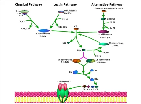
The complement system has traditionally been con-sidered a branch of the innate immune response that enhances the effects of antibodies and eliminates cellular debris and foreign intruders [10]. There are three main complement activation pathways: the classical pathway (CP), the lectin pathway (LP), and the alternative pathway (AP). All three pathways merge into a common terminal pathway that includes the activation of complement com-ponent 5 (C5) into C5a and C5b. C5b binds to C6 and C7 to form the C5b–C6–C7 complex, which is anchored to cell membranes and interacts with C8 and C9 to form the membrane attack complex (MAC), leading to antibody-mediated complement-dependent cytotoxicity (CDC). After this activation, complement proteins are activated and cleaved, and some of the resultant products are deposited on cell surfaces or released into body fluids to interact with specific receptors. The complement sys-tem acts as an efficient immune surveillance syssys-tem and contributes substantially to homeostasis [10]. However, recent studies provide new perspectives on the immuno-suppressive functions of complement components. Stud-ies over the last decade have demonstrated that these
Open Access
*Correspondence: [email protected]; [email protected]
complement components could contribute to regulating the function of the TME as a bridge between tumor-pro-moting and tumor-suppressing immune responses. This review discusses complement system activation in can-cer and interactions between the complement and the TME to provide a framework in which to understand the role of the complement system in cancer and discuss the potential of therapies targeting complement activation in the TME.
Complement activation in the TME
The complement system is important in regulating humoral immunity and complement proteins are abun-dant in the immune microenvironment [11]. The com-plement system is composed of more 50 serum proteins and membrane-bound regulators and receptors that
interact with various cells and mediators of the immune system [10, 12]. The complement cascade is summa-rized in Fig. 1. However, in the presence of malignancy, the balance between the concentrations and proportions of complement components in body fluids was observed to be lost [13, 14]. The expression of complement pro-teins is increased in malignant tumors, and complement activation in the TME promotes tumorigenesis and pro-gression. The main pathway involved in complement activation in the TME remains unclear. The CP was iden-tified as the main contributor to complement activation in a model of cervical cancer [15]. The LP was found to be significantly increased in colorectal cancer patients compared with healthy persons [16]. The complement system has been reported to be activated in tumor cells and tumor tissues, and these findings are summarized in
Fig. 1 The three pathways of the complement cascade. The complement system has three main complement activation pathways: the classical
Table 1. In addition to host cells, tumor cells can produce complement proteins. Increases in C3 and C5a concen-trations were observed in the plasma of a mouse model of metastatic breast cancer [17]. C3 cleavage products were extensively deposited along the tumor vasculature in a mouse model of cervical cancer [15]. Tumor cells were shown to secrete C3 in a syngeneic mouse model of ovarian cancer and cancer cell lines, and C3 deposition was found in tumors resected from C3-deficient mice [18]. C4d, a degradation product of complement activa-tion, was found to be elevated in malignant lung tissues, bronchoalveolar lavage fluid, and plasma from lung can-cer patients and C4d levels were associated with disease prognosis [19]. C4d fragments were also detected in oral squamous cell carcinomas, and C4d levels in saliva from patients were increased [20]. Deposition of the comple-ment proteins including C1q and C5b-9 was also dem-onstrated in melanoma and breast, colon, lung, and pancreatic cancer [21–23]. While tumor cells and stromal cells produce aberrant complement proteins, the com-plement system is pathologically activated in the TME, which reciprocally promotes tumor growth by regulating inflammation; stromal cell immunity; and the prolifera-tion, epithelial-mesenchymal transition (EMT), migra-tion and invasiveness of tumor cells.
Complement components interact with stromal cells in the TME
TAMs
TAMs have been reported to contribute to tumor pro-gression [24]. It has been reported that complement proteins could activate and recruit macrophages into tumor tissues. C1q could induce macrophage polariza-tion and suppress macrophage NLRP3 inflammasome activation [25]. Pentraxin 3 (PTX3) could regulate the complement cascade by interacting with C1q and factor H (FH), and PTX3 deficiency resulted in complement activation and the recruitment of tumor-promoting mac-rophages [26]. Recent studies showed that C1q‐polarized macrophages expressed elevated levels of programmed death-ligand 1 (PD-L1) and PD-L2 and suppressed the proliferation of human allogeneic inflammatory T cells, resulting in Treg proliferation [27]. Tumor cell-derived C3a modulated TAMs by promoting the accumulation and immunosuppressive activity via C3a–C3a recep-tor (C3aR)-PI3Kγ signaling [28]. C5a was demonstrated to inhibit the production of IL-12 in macrophages [29]. C5a mediated macrophage polarization by activation of the nuclear factor-κB (NF-κB) pathway and C5a receptor (C5aR) expressed on TAMs exhibited a tumor-promoting functional profile in colon cancer liver metastatic lesions [30]. Complement proteins are also expressed by mac-rophages. C3 produced by macrophages promoted renal
fibrosis via IL-17A secretion [31]. C9 played a crucial role in CDC-mediated tumoricidal activity, and hypoxia could downregulate C9 in TAMs, promoting non-small cell lung cancer progression [32]. Macrophages could also regulate the production of complement components. Medler et al. found that urokinase (uPA) -expressing macrophages were critical regulators of C3-independent C5a generation in squamous cell carcinomas and the C5a could foster an immunosuppressive TME during car-cinogenesis by activating C5aR1+ macrophages [33]. The cytokine production such as IL-17A, IL-17F, and IL-23 of TAMs could also be modulated by the complement pro-teins via the PI3K/Akt signaling cascade [34], suggesting dual regulation by TAMs and the complement system.
TANs
Neutrophils are the first responders among inflamma-tory cells during the acute phase of damage and infection. Recent studies showed that neutrophils in the TME, also called TANs, are associated with cancer progression [35]. Activation of the complement system could also induce the accumulation and differentiation of TANs within tumors. Allendorf et al. found that C5a could facilitate neutrophil recruitment by stimulating epithelial and endothelial cells to release leukotriene B4 (LTB4) [36], and Dick et al. found that C5aR induced neutrophil dys-function [37]. C5a generated upon complement activa-tion increased neutrophil recruitment by promoting IL-1 production [38]. C5aR deficiency could inhibit the tumor metastasis of colon cancer by reducing neutrophil infil-tration in metastatic foci in the liver [39]. C5a has also been shown to stimulate the production of functionally active tissue factor (TF) in peripheral blood neutrophils, which resulted in enhanced tumor growth and metas-tasis formation [40]. It was also shown that coagulation induced by C3aR-dependent neutrophil extracellular traps (NETs) could cause the accumulation of neutrophils with a pro-tumorigenic N2 phenotype during intestinal tumorigenesis [41]. In a model of intestinal ischemia– reperfusion injury, C3aR constrained neutrophil mobi-lization [42]. Therefore, imbalanced complement activation in the TME could reduce neutrophil proin-flammatory function, resulting in a pro-tumorigenic TME, via activation and polarization of TANs to N2-type TANs.
MDSCs
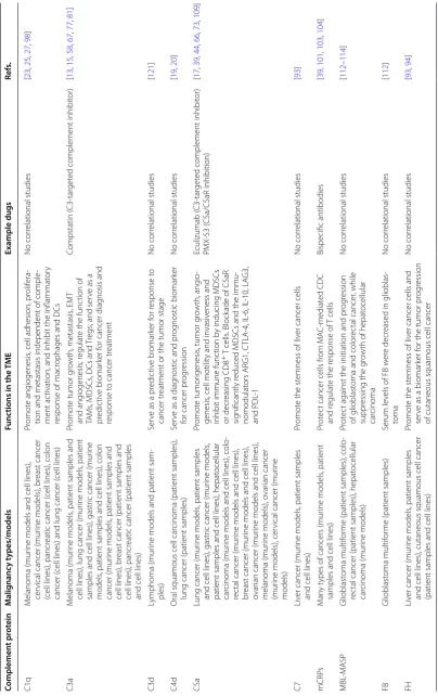
Table 1 E ff ec ts of c omplemen t sy st em on the
TME and their ther
ap eutic p ot en tial f or c anc er tr ea tmen t Complemen t pr ot ein M alig nanc y t ypes/models Func tions in the TME Example dugs Re fs. C1q M elanoma (mur
ine models and cell lines),
cer
vical cancer (mur
ine models), br
east cancer
(cell lines), pancr
eatic cancer (
cell lines), colon
cancer (
cell lines) and lung cancer (
cell lines)
Pr
omot
e ang
iogenesis
, cell adhesion, pr
olif
era
-tion and metastasis independent of comple
-ment ac
tivation, and inhibit the inflammat
or
y
response of macr
ophages and DCs
No cor relational studies [ 23 , 25 , 27 , 98 ] C3a M elanoma (mur ine models
, patient samples and
cell lines), lung cancer (mur
ine models
, patient
samples and cell lines), gastr
ic cancer (mur
ine
models
, patient samples and cell lines), colon
cancer (mur
ine models
, patient samples and
cell lines), br
east cancer (patient samples and
cell lines), pancr
eatic cancer (patient samples
and cell lines)
Pr
omot
e tumor g
ro wth, metastasis , EM T and ang iogenesis; r egulat
e the func
tion of
TA
Ms
, MDSCs
, DCs and
Tr
egs; and ser
ve as a
pr
edic
tiv
e biomar
ker f
or cancer diag
nosis and
response t
o cancer tr
eatment
Compstatin (
C3-tar
get
ed complement inhibit
or) [ 13 , 15 , 58 , 67 , 77 , 81 ] C3d Lymphoma (mur
ine models and patient sam
-ples)
Ser
ve as a pr
edic tiv e biomar ker f or r esponse t o cancer tr
eatment or the tumor stage
No cor relational studies [ 121 ] C4d
Oral squamous cell car
cinoma (patient samples),
lung cancer (patient samples)
Ser
ve as a diag
nostic and pr
og
nostic biomar
ker
for cancer pr
og ression No cor relational studies [ 19 , 20 ] C5a
Lung cancer (mur
ine models
, patient samples
and cell lines), gastr
ic cancer (mur
ine models
,
patient samples and cell lines), hepat
ocellular
car
cinoma (mur
ine models and cell lines), colo
-rec
tal cancer (mur
ine models and cell lines),
br
east cancer (mur
ine models and cell lines),
ovar
ian cancer (mur
ine models and cell lines),
melanoma (mur
ine models), o
var
ian cancer
(mur
ine models), cer
vical cancer (mur
ine models) Pr omot e tumor igenesis
, tumor g
ro
wth, ang
io
-genesis
, cell motilit
y and in
vasiv
eness and
inhibit immune func
tion b
y inducing MDSCs
or decr
easing
CD8
+ T cells
. Block
ade of C5aR
sig
nificantly r
educed MDSCs and the immu
-nomodulat ors AR G1, C TLA-4, IL -6, IL -10, LA G3, and PDL -1 Eculizumab ( C3-tar get
ed complement inhibit
or) PMX -53 ( C5a/C5aR inhibition) [ 17 , 39 , 44 , 66 , 73 , 109 ] C7 Liv
er cancer (mur
ine models
, patient samples
and cell lines)
Pr
omot
e the st
emness of liv
er cancer cells
No cor relational studies [ 93 ] mCRP s M an y t
ypes of cancers (mur
ine models
, patient
samples and cell lines)
Pr
ot
ec
t cancer cells fr
om M A C-mediat ed CDC and r egulat
e the r
esponse of T cells Bispecific antibodies [ 39 , 101 , 103 , 104 ] MBL -M ASP Glioblast oma multif or
me (patient samples), colo
-rec
tal cancer (patient samples), hepat
ocellular car cinoma (mur ine models) Pr ot ec
t against the initiation and pr
og
ression
of glioblast
oma and color
ec
tal cancer
, while
suppr
essing the g
ro
wth of hepat
ocellular car cinoma No cor relational studies [ 112 – 114 ] FB Glioblast oma multif or
me (patient samples)
Serum le
vels of FB w
er
e decr
eased in glioblas
-toma No cor relational studies [ 112 ] FH Liv
er cancer (mur
ine models
, patient samples
and cell lines), cutaneous squamous cell cancer (patient samples and cell lines)
Pr
omot
e the st
emness of liv
er cancer cells and
ser
ve as a biomar
ker f
or the tumor pr
og
ression
of cutaneous squamous cell cancer
of C5aR significantly reduced MDSCs [44]. When MDSCs enter the TME, they can suppress the function of T cells via C5a/C5aR pathways. Studies of mouse mod-els of melanoma and breast and lung cancer showed that C5aR-mediated pathways are linked to the differentiation of MDSCs and activation of these pathways is associated with the production of immunomodulators such as argi-nase-1 (Arg-1), IL-10, TGF-β1, cytotoxic T lymphocyte antigen 4 (CTLA4) and PDL-1 [17, 44, 45]. Then, immu-nomodulators induced by C5a/C5aR facilitated cancer metastasis by the suppressing T cell responses. It was also shown that C5a/C5aR signaling regulated synthesis of reactive oxygen in MDSCs and inhibited the antigen-specific responses of CD8+ T cells [46]. These studies demonstrated that tumor cell-derived C5a induced the recruitment and differentiation of MDSCs into the TME and that MDSCs exerted immunosuppressive effects by altering T cell responses, resulting in tumor progres-sion. MDSCs, the cornerstone of the immunosuppres-sive shield in the TME, can promote the formation of Tregs and TAMs to protect tumor cells from the immune system and immunotherapy [43]. Therefore, the link between MDSCs and complement activation is impor-tant for the formation of an immunosuppressive TME.
Tregs
Tregs are the immunosuppressive subsets of CD4+ T cells that can suppress antitumor immune responses in multiple ways [47]. Tregs are recruited by tumor cells and other stromal cells into the TME, where they play immunosuppressive roles in tumor progression. Impor-tantly, MDSCs play critical roles in the generation of Tregs in the TME, and the connection between comple-ment activation and Tregs is therefore arguably impor-tant. Complement activation via a C3aR pathway altered CD4+ T lymphocytes and mediated cancer progression in mouse models of lung cancer [48]. The protumor effect of the C5a–C5aR signaling axis was also demonstrated in mouse breast cancer models. In a model of metastatic breast cancer, C5aR inhibited the recruitment and func-tions of CD4+ and CD8+ effector T cells in the lung and liver, and the numbers of Tregs in the lungs of C5aR-defi-cient mice was reduced [17]. C5aR in MDSCs was also found to contribute to the polarization of CD4+ T cells in the lungs to Th2 type T cells. The reduced generation of Tregs in the absence of C5aR signaling was also asso-ciated with the reduced production of TGF-β1 [49]. C5a inhibition in combination with chemotherapy fostered TME reprogramming, resulting in CD8+ T cell-depend-ent antitumor immune responses [33]. Therefore, the combination of complement inhibition with other thera-peutic interventions is more likely to substantially benefit cancer patients.
DCs
DCs, major players in the control of cancer by adaptive immunity, are traditionally divided into plasmacytoid DCs (pDCs) and conventional DCs (cDCs) [50]. Some studies have demonstrated that DCs could be pro- or anti-tumorigenic depending on the status of the TME [51]. In a p53/KRAS-inducible mouse model of ovar-ian cancer progression, the depletion of DCs early in the disease course accelerated tumor expansion, but DC depletion at advanced stages of disease significantly delayed aggressive malignant progression. Phenotypi-cally divergent DCs both drove immunosurveillance and accelerated malignant growth [52]. Activation of the complement system exerted both protective and immunosuppressive functions in immune-inflammatory responses against injury and cancer. The complement inhibitors C4b-binding protein (C4BP) and FH were reported to play a critical role in modulating adaptive immune responses by generating an anti-inflammatory state in monocyte-derived DCs [53]. Generation of this type of DCs was accompanied by impaired CD4+ T cell proliferation and inhibited IFN-γ secretion. In a plant virus-infected model, C3 depletion in mice increased IFN-α production and the immunotherapeutic properties of immune cells [54]. In an HIV-infected model, com-plement activation could inhibit the pro-inflammatory functions of DCs [55], and this opsonization function of complement on DCs was also demonstrated in a herpes simplex virus 2 (HSV-2)-infected model [56]. In addition, the production of complement components by DCs was shown to affect their ability to regulate T cell responses. FH produced by DCs could inhibit CD4+ T cell prolifera-tion [57], and C1q-polarized DCs expressed higher lev-els of surface PD-L2 and exhibited decreased autologous Th17 and Th1 cell proliferation [27]. C3 production in DCs was increased by lithium via GSK-3 inhibition and regulated interactions between microglia and neurons [58]. C3a and C5a were also shown to be central media-tors of radiotherapy-induced, tumor-specific immunity and clinical response [59]. Therefore, it is tempting to speculate that the antigen-presenting function of DCs is limited in the presence of complement activation and that these DCs carry out their anti-inflammatory func-tion by inhibiting T cell anti-tumor responses.
CAFs
effect of immunotherapy by inducing the infiltration of MDSCs in tumors [62]. Combined depletion of CAFs in the TME and anti-CTLA4 immunotherapy improved therapeutic effects and prolonged animal survival [63]. Recent studies have shown that CD10 and GPR77 expres-sion could specifically define a CAF subset correlated with chemoresistance and poor survival in breast and lung cancer patients [64]. GPR77 has been regarded as a C5aR in the complement signaling pathway [65]. CD10+GPR77+ CAFs could induce cancer stem cell (CSC) enrichment and chemoresistance by secreting IL-6 and IL-8, and CAFs also produced complement for self-sustained GPR77 signaling. Combined chemotherapy and anti-GPR77 neutralizing antibody could inhibit tum-origenesis and enhance chemotherapeutic effects. Thus, CD10+GPR77+CAF infiltration may serve as a promising clinical biomarker to predict chemotherapy response.
Effects of the complement system on tumor progression
Proliferation
Imbalanced tumor cell proliferation and apoptosis is a distinct characteristic of carcinogenesis. There is some evidence that complement activation in the TME directly or indirectly enhanced tumor cell proliferation. Min et al. [18] showed that tumor-derived C3a and C5a played distinctly important roles in promoting tumor prolifera-tion and that C3 or C5 silencing reduced tumor growth in vivo. In this study, C3aR and C5aR agonists increased the proliferation of ovarian cancer cells, while C3aR and C5aR antagonists decreased the proliferation of these cells. There was also no significant difference in comple-ment effects on tumor proliferation in CD8−/− and WT mouse models, which indicated that complement effects on tumor proliferation were independent of T cells. Other studies have demonstrated the complement-induced pro-motion of cancer proliferation, in which complement components exerted an indirect effect via regulating the immune response of immune cells [44, 66, 67]. Corrales and colleagues found that C5a promoted cell prolifera-tion and tumor growth in the Lewis lung cancer model by creating an immunosuppressive microenvironment. The blockade of C5aR significantly reduced MDSCs and immunomodulators, inhibiting tumor growth [44]. In ovarian tumor-bearing mice, C5a-expressing tumor cells in an overall immunosuppressive state exhibited accel-erated growth, and significantly lower percentages of infiltrating CD4+ and CD8+ T cells were observed in the spleen and tumors [66]. C3a–C3aR signaling was also reported to participate in promoting tumor proliferation. Jamileh et al. showed that C3a–C3aR signaling contrib-uted to melanoma growth by inhibiting neutrophil and CD4+ T cell responses [67]. In conclusion, cancer cells
have the capacity to generate C3a and C5a, which can promote cancer cell proliferation and create an immu-nosuppressive TME for cancer progression. In addition, Agostinis et al. showed that high levels of C1q expres-sion in malignant pleural mesothelioma enhanced tumor adhesion and proliferation via enhanced ERK1/2, SAPK/ JNK, and p38 phosphorylation [68]. These results provide a new understanding of the role of the complement sys-tem in cancer proliferation and have significant implica-tions for innovative therapeutic and biomarker strategies for some cancers.
EMT
EMT, the transition between epithelial and mesenchymal phenotypes, contributes to embryonic development and carcinoma progression [69, 70]. The complement system participates in mediating EMT in multiple tumor mod-els. C3a secreted by ovarian cancer cells could induce a reduction in E-cadherin expression and promote EMT in cancer cells, which was regulated by the transcription factor TWIST1 [71]. The contribution of C3 to EMT in renal fibrosis and injury has also been investigated [72]. In addition, activation of C5aR by C5a in hepatocellular carcinoma could induce EMT by downregulating E-cad-herin and claudin-1 expression, and upregulating Snail expression via activation of the ERK1/2 pathway [73]. Furthermore, C5aR blockade could impair the migration of lung cancer cells and up-regulate E-cadherin protein expression [74]. TGF-β is a major driving force of EMT. TGF-β-induced EMT in lung cancer cells was reported to confer resistance to CDC by upregulation in the CD59 expression on the surface of cancer cells [75]. In this study, CD59 inhibition was demonstrated to enhance the efficacy of antibody-mediated CDC and inhibit metas-tasis in lung cancer. Stromal cells stimulated by com-plement components in the TME, such as TAMs and MDSCs, could also produce TGF-β1 and promote EMT. Therapeutic strategies that inhibit complement pathways may be a promising method to inhibit EMT and limit dis-tant metastasis.
Metastasis
patients enhanced the motility of malignant cells by downregulating the expression of HO-1 [78]. C5a–C5aR signaling facilitated breast cancer metastasis by promot-ing Treg generation and suppresspromot-ing T cell responses in the lungs [17]. C5a generated by colon cancer cells con-tributed to tumor metastasis by increasing the expression of monocyte chemoattractant protein-1 (MCP-1), IL-10, Arg-1 and TGF-β1 [39]. C3 and C4 could bind to colla-gen and elastin in the vascular wall, leading to increased vascular stiffness [79]. The C5a–C5aR interaction could induce the expression of MMP-1 and MMP-9, which were important for degradation of the ECM, by the acti-vation of NF-κB and AP-1 [80]. C3a–C3aR signaling in the choroid plexus epithelium has been further demon-strated to disrupt the blood-cerebrospinal fluid barrier and promote cancer cell leptomeningeal metastasis [81]. In addition, complement-regulated TAMs and MDSCs could also promote cancer metastasis by modulating the TME, as mentioned above. Surprisingly, some comple-ment components were shown to carry out functions to promote cancer metastasis independent of complement activation. In a mouse model of melanoma, more lung metastases were observed in WT mice than in C1q-defi-cient mice, suggesting that C1q promoted cancer metas-tasis [23]. Further investigations to define the underlying mechanisms of these C1q-mediated effects should be carried out.
Angiogenesis
Tumor angiogenesis is a key step in cancer progression. Tumor cells secrete pro-angiogenic factors to promote the development of abnormal vascular networks and normalization of the tumor vasculature has emerged as a new strategy for therapeutic cancer management [82]. The role of the complement system in angiogenesis is controversial. There is some evidence of the proangio-genic effects of complement components. In a transproangio-genic mouse model of ovarian cancer, complement inhibi-tion by C3 or C5aR knockout inhibited tumor growth by altering endothelial cell function and vascular endothe-lial growth factor (VEGF) expression [83]. VEGF was shown to carry out a critical function in vascularization under physiological and pathological conditions [84]. Factors promoting angiogenesis could also be secreted from TAMs or MDSCs, which could be recruited by C3a or C5a to the premetastatic niche [85, 86]. There-fore, the complement system may indirectly contribute to angiogenesis by regulating stromal cells in the TME. However, some findings have provided opposite evidence that C3a and C5a might exert an antiangiogenic effect on the course of pathological postnatal neovascularization in the retina [87]. In addition, evidence has shown that the genetic deletion of C3 or C5aR and pharmacological
blockade of C5aR impaired the ability of T cells to over-come the endothelial barrier, infiltrate tumors, and con-trol tumor progression in vivo [88]. In genetic chimera mouse models, local complement activation was dem-onstrated to disrupt the tumor endothelial barrier, which promoted the successful homing of T cells. Neverthe-less, complement activation of C3a or C5a did not con-tribute to tumor angiogenesis in murine models of lung and cervical cancer or epithelial carcinogenesis [15, 44, 89]. During would healing, C1q was shown to be very effective in inducing an angiogenic phenotype in cul-tured endothelial cells in vitro and forming new vessels in mice or rats [90]. The effects of complement on tumor angiogenesis are complicated, and the roles of comple-ment components in tumor angiogenesis require further investigation.
Stemness
CSCs are a subset of cells that possess the capacity to self-renew, differentiate, and give rise to cancer recur-rence [91, 92]. There is some evidence supporting the possible effects of the complement system on stemness. A recent report showed that the complement proteins C7 and FH were upregulated in liver tumor-initiating cells, and these proteins were needed to control the stemness of liver cancer cells via LSF-1 [93]. CSF was also reported to be upregulated in cutaneous squamous cell carcinoma and knockdown of CFH expression inhibited the prolif-eration and migration of these cancer cells by inhibiting basal ERK1/2 activation [94]. In contrast, the role of C7 is not well understood. The complement regulator CD59 was also reported to be upregulated by SOX2 to protect epithelial CSCs to evade complement surveillance [95]. In addition, Lee and colleagues found that the mobili-zation of hematopoietic stem cells in C5-deficient mice was impaired and that C5a-mediated pro-mobilization effects were mediated by the stimulation of granulo-cytes rather than hematopoietic stem cells [96]. Another study showed that the contribution of C5a to hemat-opoietic stem cell mobilization was mediated by C5aR, and C5aR antagonists diminished the pro-mobilization effects of C5a [97]. There are also some studies on the regulation of the stemness-associated signaling pathway by complement activation. Naito and colleagues found that the complement C1q could bind to Frizzled recep-tors and activate canonical Wnt signaling to promote the aging-associated impairment of muscle regeneration [98] and that Wnt signaling pathways could regulate cancer stemness in various manners [99, 100].
Immunosuppression
MAC. Products of complement activation have been shown to participate in immunosuppression by upregu-lating the expression of molecules such as PDL-1, IL-10, Arg-1, and TGF-β1 and regulating immune cell differen-tiation. In addition, the expression of membrane-bound complement regulatory proteins (mCRPs), including CD46, CD55 and CD59, is upregulated in many types of cancer cells, which can dwarf antitumor therapeu-tic efficacy. It has been reported that CD46, CD55 and CD59 could protect cancer cells from MAC-mediated CDC [101–103]. In addition, mCRPs have been shown to regulate T cell responses. John and colleagues showed that CD46 participated in switching T cells towards a regulatory phenotype by attenuating IL-2 production via the transcriptional regulator ICER/CREM and upregu-lating IL-10 expression via the serine-threonine kinase SPAK [104]. CD59 was demonstrated to downmodu-late CD4+ T cell activity and CD59 blockade enhanced
antigen-specific CD4+ T cell responses [105]. Neu-tralization or blockade of mCRPs in cancer cells could increase the efficacy of antibody-based immunotherapy [106, 107]. Strategies involving regulating the function of mCRPs and the status of immune cells may provide new insights into cancer immunotherapy. However, consid-ering the wide distribution of mCRPs on somatic cells, more studies are needed to further validate the specificity of these treatments.
Therapeutic potential of targeting complement activation in the TME
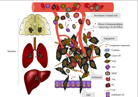
The recent clinical success of immune checkpoint block-ade suggests that treatment targeting the immune system is the most promising approach to eliminate cancer cells [108]. The multiple roles of the complement system in cancer progression, which is summarized in Fig. 2, have
Fig. 2 Effects of complement activation on the TME. Activation of the complement system inside tumors releases complement components, such
unveiled novel opportunities for the improved man-agement of cancer patients. There is some evidence indicating the therapeutic possibility of complement components as biomarkers or targets for immunothera-pies. Serum C3a and C5a have been found to be elevated in patients with lung, colorectal and gastric cancers com-pared to healthy individuals [109–111]. C4d deposition in tumors was also suggested to serve as a biomarker for the early diagnosis and prognosis of lung cancer [19]. Serum levels of factor B (FB) were decreased and serum levels of mannose-binding lectin (MBL) were elevated in patients with glial tumor, suggesting that low levels of MBL might protect against the initiation and progres-sion of glioblastoma multiforme [112]; furthermore, high serum levels of mannan-binding lectin-associated serine protease 2 (MASP-2) predicted recurrence and poor sur-vival in colorectal cancer patients [113]. However, MBL was reported to suppress tumor growth by regulating hepatic stellate cell activation in a mouse model of hepa-tocellular carcinoma [114]. Some studies have suggested that the levels of complement proteins served as predic-tive biomarkers for the response to cancer treatment. Zhang et al. have suggested that C3a was at higher level in samples after neoadjuvant chemotherapy than in that before treatment indicating that C3a might serve as a bio-marker to predict the sensitivity of breast cancer to neo-adjuvant chemotherapy [115]. Maher et al. demonstrated that serum levels of C4a and C3a might act as predictive biomarkers of the response of esophageal cancer patients to chemoradiation [116]. They found that serum C4a and C3a levels were significantly higher in poor responders versus good responders and these proteins could pre-dict response to neoadjuvant chemoradiotherapy with a sensitivity and specificity of 78.6% and 83.3% in esopha-geal cancer. Surace et al. found that radiotherapy induced intratumoral complement activation in melanoma and colon carcinoma and C3aR or C5aR blockade before applying radiotherapy could affect antitumor effect of radiotherapy by disturbing DC and CD8+ T cell activa-tion. Various studies have suggested the potential appli-cation of complement components as cancer biomarkers for the diagnosis, prognosis or response to cancer treat-ment. However, evidence of their specificity and sensitiv-ity remains insufficient, and the exact mechanisms are still unclear.
PD-1/PD-L1 checkpoint blockades have been shown to be remarkably clinically efficient strategies for various malignancies [117]. C5a blockade was demonstrated to work synergistically with anti-PD-1 inhibition in mela-noma and colon and lung cancer growth associated with the activation of CD8+ T cells and inhibition of MDSCs [118, 119]. In addition, the targeting of mCRPs, such as
CD46 and CD59, for cancer immunotherapy has recently been explored. An antibody–drug conjugate targeting CD46 was shown to eliminate myeloma growth [120], and bispecific antibodies targeting tumor-associated antigens and CD59 increased the efficacy of immuno-therapy in a lymphoma mouse model [106]. However, these studies were limited to animal experiments, and differences between the mouse and human complement systems should be considered. There are many challenges and constraints that could hinder development and application in this area. A more detailed understanding of the complex network established between the com-plement system and cancer is essential to bridge the gap between promising preclinical trials and effective clinical treatments.
Conclusions and perspective
Recent studies have shown the complex and multifac-eted role of complement proteins in immune regulation and cancer. Complement components have been shown to contribute to regulating the functions of the TME and exert immunoregulatory effects under certain conditions. Although we have gained knowledge about the role of the complement system in cancer, molecules that activate the complement cascade in cancer cells are essentially unknown. Due to the high heterogeneity of human can-cer, different complement activation pathways and mech-anisms may be involved, and different strategies to treat different tumor types could be combined with traditional chemotherapies or immunotherapies. A better under-standing of the mechanistic interaction between the complement system and TME will provide a new break-through in cancer immunotherapy. In conclusion, target-ing complement reagents might be a promistarget-ing challenge in cancer immunotherapy, and we hope that more effi-cient therapeutic strategies are developed to improve the efficacy of complement-related anticancer therapies.
Abbreviations
AP: alternative pathway; Arg-1: arginase-1; C4BP: C4b-binding protein; C5aR: C5a receptor; CAFs: cancer-associated fibroblasts; CDC: complement-depend-ent cytotoxicity; cDCs: convcomplement-depend-entional DCs; CP: classical pathway; CSC: cancer stem cell; CTLA4: cytotoxic T lymphocyte antigen 4; DCs: dendritic cells; ECM: extracellular matrix; EMT: epithelial–mesenchymal transition; FB: factor B; FH: factor H; HSV-2: herpes simplex virus 2; LP: lectin pathway; LTB4: leukotriene B4; MAC: membrane attack complex; MASP: mannan-binding lectin-associated serine protease; MBL: mannan-binding lectin; MCP-1: monocyte chemoat-tractant protein-1; mCRPs: membrane-bound complement regulatory pro-teins; MDSCs: myeloid-derived suppressor cells; NETs: neutrophil extracellular traps; NF-κB: nuclear factor-κB; pDCs: plasmacytoid DCs; PD-L1: programmed death-ligand 1; PTX3: pentraxin 3; TAMs: tumor-associated macrophages; TANs: tumor-associated neutrophils; TF: tissue factor; TME: tumor microenvironment; Tregs: regulatory T cells; VEGF: vascular endothelial growth factor.
Authors’ contributions
QL, YZ and RZ designed the study. RZ and QL discussed and wrote the manu-script. LQ and YZ revised the manumanu-script. All authors read and approved the final manuscript.
Funding
This work was supported by Grants from National Natural Science Foundation of China (81502068, 81673023, 81272573 and 81872501), Beijing Natural Sci-ence Foundation (7172177) the Non-profit Central Research Institute Fund of Chinese Academy of Medical Sciences (No. 2018PT32014).
Ethics approval and consent to participate Not applicable.
Consent for publication Not applicable.
Competing interests
The authors declare that they have no competing interests.
Received: 27 August 2019 Accepted: 11 November 2019
References
1. Siegel RL, Miller KD. Cancer statistics, 2019. CA Cancer J Clin. 2019;69(1):7–34.
2. Torre LA, Bray F, Siegel RL, et al. Global cancer statistics, 2012. CA Cancer J Clin. 2015;65(2):87–108.
3. Guo S, Deng CX. Effect of stromal cells in tumor microenvironment on metastasis initiation. Int J Biol Sci. 2018;14(14):2083–93.
4. Balkwill FR, Capasso M, Hagemann T. The tumor microenvironment at a glance. J Cell Sci. 2012;125(Pt 23):5591–6.
5. Liu Q, Liao Q, Zhao Y. Chemotherapy and tumor microenvironment of pancreatic cancer. Cancer Cell Int. 2017;17:68.
6. Albini A, Bruno A, Noonan DM, et al. Contribution to tumor angiogen-esis from innate immune cells within the tumor microenvironment: implications for immunotherapy. Front Immunol. 2018;9:527. 7. Munn DH, Sharma MD, Johnson TS. Treg destabilization and
repro-gramming: implications for cancer immunotherapy. Cancer Res. 2018;78(18):5191–9.
8. Sun Q, Zhang B, Hu Q, et al. The impact of cancer-associated fibroblasts on major hallmarks of pancreatic cancer. Theranostics. 2018;8(18):5072–87.
9. Labidi-Galy SI, Treilleux I, Goddard-Leon S, et al. Plasmacytoid dendritic cells infiltrating ovarian cancer are associated with poor prognosis. Oncoimmunology. 2012;1(3):380–2.
10. Ricklin D, Hajishengallis G, Yang K, et al. Complement: a key sys-tem for immune surveillance and homeostasis. Nat Immunol. 2010;11(9):785–97.
11. Holers VM. Complement and its receptors: new insights into human disease. Annu Rev Immunol. 2014;32:433–59.
12. Nesargikar PN, Spiller B, Chavez R. The complement system: history, pathways, cascade and inhibitors. Eur J Microbiol Immunol (Bp). 2012;2(2):103–11.
13. Ajona D, Ortiz-Espinosa S, Pio R. Complement anaphylatoxins C3a and C5a: emerging roles in cancer progression and treatment. Semin Cell Dev Biol. 2019;85:153–63.
14. Bajic G, Degn SE, Thiel S, et al. Complement activation, regulation, and molecular basis for complement-related diseases. EMBO J. 2015;34(22):2735–57.
15. Markiewski MM, DeAngelis RA, Benencia F, et al. Modulation of the antitumor immune response by complement. Nat Immunol. 2008;9(11):1225–35.
16. Ytting H, Jensenius JC, Christensen IJ, et al. Increased activity of the mannan-binding lectin complement activation pathway in patients with colorectal cancer. Scand J Gastroenterol. 2004;39(7):674–9. 17. Vadrevu SK, Chintala NK, Sharma SK, et al. Complement c5a receptor
facilitates cancer metastasis by altering T-cell responses in the meta-static niche. Cancer Res. 2014;74(13):3454–65.
18. Cho MS, Vasquez HG, Rupaimoole R, et al. Autocrine effects of tumor-derived complement. Cell Rep. 2014;6(6):1085–95.
19. Ajona D, Pajares MJ, Corrales L, et al. Investigation of complement activation product c4d as a diagnostic and prognostic biomarker for lung cancer. J Natl Cancer Inst. 2013;105(18):1385–93.
20. Ajona D, Pajares MJ, Chiara MD, et al. Complement activation product C4d in oral and oropharyngeal squamous cell carcinoma. Oral Dis. 2015;21(7):899–904.
21. Bandini S, Macagno M, Hysi A, et al. The non-inflammatory role of C1q during Her2/neu-driven mammary carcinogenesis. Oncoimmu-nology. 2016;5(12):e1253653.
22. Bjorge L, Hakulinen J, Vintermyr OK, et al. Ascitic complement system in ovarian cancer. Br J Cancer. 2005;92(5):895–905.
23. Bulla R, Tripodo C, Rami D, et al. C1q acts in the tumour microenvi-ronment as a cancer-promoting factor independently of comple-ment activation. Nat Commun. 2016;7:10346.
24. Mantovani A, Marchesi F, Malesci A, et al. Tumour-associated macrophages as treatment targets in oncology. Nat Rev Clin Oncol. 2017;14(7):399–416.
25. Benoit ME, Clarke EV, Morgado P, et al. Complement protein C1q directs macrophage polarization and limits inflamma-some activity during the uptake of apoptotic cells. J Immunol. 2012;188(11):5682–93.
26. Bonavita E, Gentile S, Rubino M, et al. PTX3 is an extrinsic oncosup-pressor regulating complement-dependent inflammation in cancer. Cell. 2015;160(4):700–14.
27. Clarke EV, Weist BM, Walsh CM, et al. Complement protein C1q bound to apoptotic cells suppresses human macrophage and dendritic cell-mediated Th17 and Th1 T cell subset proliferation. J Leukoc Biol. 2015;97(1):147–60.
28. Zha H, Wang X, Zhu Y, et al. Intracellular activation of complement C3 leads to PD-L1 antibody treatment resistance by modulating tumor-associated macrophages. Cancer Immunol Res. 2019;7(2):193–207. 29. Hawlisch H, Belkaid Y, Baelder R, et al. C5a negatively regulates
toll-like receptor 4-induced immune responses. Immunity. 2005;22(4):415–26.
30. Piao C, Zhang WM, Li TT, et al. Complement 5a stimulates macrophage polarization and contributes to tumor metastases of colon cancer. Exp Cell Res. 2018;366(2):127–38.
31. Liu Y, Wang K, Liang X, et al. Complement C3 produced by mac-rophages promotes renal fibrosis via IL-17A secretion. Front Immunol. 2018;9:2385.
32. Li L, Yang H, Li Y, et al. Hypoxia restrains the expression of complement component 9 in tumor-associated macrophages promoting non-small cell lung cancer progression. Cell Death Discov. 2018;4:63.
33. Medler TR, Murugan D, Horton W, et al. Complement C5a fosters squamous carcinogenesis and limits T cell response to chemotherapy. Cancer Cell. 2018;34(4):561–578.e566.
34. Grailer JJ, Bosmann M, Ward PA. Regulatory effects of C5a on IL-17A, IL-17F, and IL-23. Front Immunol. 2012;3:387.
35. Powell DR, Huttenlocher A. Neutrophils in the tumor microenviron-ment. Trends Immunol. 2016;37(1):41–52.
36. Allendorf DJ, Yan J, Ross GD, et al. C5a-mediated leukotriene B4-ampli-fied neutrophil chemotaxis is essential in tumor immunotherapy facili-tated by anti-tumor monoclonal antibody and beta-glucan. J Immunol. 2005;174(11):7050–6.
37. Dick J, Gan PY, Ford SL, et al. C5a receptor 1 promotes autoimmunity, neutrophil dysfunction and injury in experimental anti-myeloperoxi-dase glomerulonephritis. Kidney Int. 2018;93(3):615–25.
38. Khameneh HJ, Ho AW, Laudisi F, et al. C5a regulates IL-1beta production and leukocyte recruitment in a murine model of monosodium urate crystal-induced peritonitis. Front Pharmacol. 2017;8:10.
39. Piao C, Cai L, Qiu S, et al. Complement 5a enhances hepatic metastases of colon cancer via monocyte chemoattractant protein-1-mediated inflammatory cell infiltration. J Biol Chem. 2015;290(17):10667–76. 40. Kourtzelis I, Markiewski MM, Doumas M, et al. Complement
anaphyla-toxin C5a contributes to hemodialysis-associated thrombosis. Blood. 2010;116(4):631–9.
42. Wu MC, Brennan FH, Lynch JP, et al. The receptor for complement component C3a mediates protection from intestinal ischemia-reper-fusion injuries by inhibiting neutrophil mobilization. Proc Natl Acad Sci USA. 2013;110(23):9439–44.
43. Tesi RJ. MDSC; the most important cell you have never heard of. Trends Pharmacol Sci. 2019;40(1):4–7.
44. Corrales L, Ajona D, Rafail S, et al. Anaphylatoxin C5a creates a favorable microenvironment for lung cancer progression. J Immunol. 2012;189(9):4674–83.
45. Ning C, Li YY, Wang Y, et al. Complement activation promotes colitis-associated carcinogenesis through activating intestinal IL-1beta/ IL-17A axis. Mucosal Immunol. 2015;8(6):1275–84.
46. Kusmartsev S, Nefedova Y, Yoder D, et al. Antigen-specific inhibition of CD8+ T cell response by immature myeloid cells in cancer is medi-ated by reactive oxygen species. J Immunol. 2004;172(2):989–99. 47. Togashi Y, Shitara K, Nishikawa H. Regulatory T cells in cancer
immu-nosuppression—implications for anticancer therapy. Nat Rev Clin Oncol. 2019;16(6):356–71.
48. Kwak JW, Laskowski J, Li HY, et al. Complement activation via a C3a receptor pathway alters CD4(+) T lymphocytes and mediates lung cancer progression. Cancer Res. 2018;78(1):143–56.
49. Markiewski MM, Vadrevu SK, Sharma SK. The ribosomal protein S19 suppresses antitumor immune responses via the complement C5a receptor 1. J Immunol. 2017;198(7):2989–99.
50. Villadangos JA, Schnorrer P. Intrinsic and cooperative antigen-pre-senting functions of dendritic-cell subsets in vivo. Nat Rev Immunol. 2007;7(7):543–55.
51. Hansen M, Andersen MH. The role of dendritic cells in cancer. Semin Immunopathol. 2017;39(3):307–16.
52. Scarlett UK, Rutkowski MR, Rauwerdink AM, et al. Ovarian cancer progression is controlled by phenotypic changes in dendritic cells. J Exp Med. 2012;209(3):495–506.
53. Olivar R, Luque A, Cardenas-Brito S, et al. The complement inhibitor factor H generates an anti-inflammatory and tolerogenic state in monocyte-derived dendritic cells. J Immunol. 2016;196(10):4274–90. 54. Lebel ME, Langlois MP. Complement component 3 regulates
IFN-alpha production by plasmacytoid dendritic cells follow-ing TLR7 activation by a plant virus-like nanoparticle. J Immunol. 2017;198(1):292–9.
55. Posch W, Steger M, Knackmuss U, et al. Complement-opsonized HIV-1 overcomes restriction in dendritic cells. PLoS Pathog. 2015;11(6):e1005005.
56. Crisci E, Ellegard R, Nystrom S, et al. Complement opsonization promotes herpes simplex virus 2 infection of human dendritic cells. J Virol. 2016;90(10):4939–50.
57. Dixon KO, O’Flynn J, Klar-Mohamad N, et al. Properdin and factor H production by human dendritic cells modulates their T-cell stimulatory capacity and is regulated by IFN-gamma. Eur J Immunol. 2017;47(3):470–80.
58. Yu Z, Ono C, Aiba S, et al. Therapeutic concentration of lithium stimu-lates complement C3 production in dendritic cells and microglia via GSK-3 inhibition. Glia. 2015;63(2):257–70.
59. Surace L, Lysenko V, Fontana AO, et al. Complement is a central mediator of radiotherapy-induced tumor-specific immunity and clini-cal response. Immunity. 2015;42(4):767–77.
60. Gascard P, Tlsty TD. Carcinoma-associated fibroblasts: orchestrating the composition of malignancy. Genes Dev. 2016;30(9):1002–19. 61. Ohlund D, Handly-Santana A, Biffi G, et al. Distinct populations of
inflammatory fibroblasts and myofibroblasts in pancreatic cancer. J Exp Med. 2017;214(3):579–96.
62. Kumar V, Donthireddy L, Marvel D, et al. Cancer-associated fibroblasts neutralize the anti-tumor effect of CSF1 receptor blockade by induc-ing PMN-MDSC infiltration of tumors. Cancer Cell. 2017;32(5):654– 668.e655.
63. Ozdemir BC, Pentcheva-Hoang T, Carstens JL, et al. Depletion of carcinoma-associated fibroblasts and fibrosis induces immunosuppres-sion and accelerates pancreas cancer with reduced survival. Cancer Cell. 2014;25(6):719–34.
64. Su S, Chen J, Yao H, et al. CD10(+)GPR77(+) cancer-associated fibro-blasts promote cancer formation and chemoresistance by sustaining cancer stemness. Cell. 2018;172(4):841–856.e816.
65. Klos A, Wende E, Wareham KJ, et al. International union of basic and clinical pharmacology. [corrected]. LXXXVII. Complement peptide C5a, C4a, and C3a receptors. Pharmacol Rev. 2013;65(1):500–43.
66. Gunn L, Ding C, Liu M, et al. Opposing roles for complement compo-nent C5a in tumor progression and the tumor microenvironment. J Immunol. 2012;189(6):2985–94.
67. Nabizadeh JA, Manthey HD. The complement C3a receptor contributes to melanoma tumorigenesis by inhibiting neutrophil and CD4+ T cell responses. J Immunol. 2016;196(11):4783–92.
68. Agostinis C, Vidergar R, Belmonte B, et al. Complement protein C1q binds to hyaluronic acid in the malignant pleural mesothelioma microenvironment and promotes tumor growth. Front Immunol. 2017;8:1559.
69. Lamouille S, Xu J, Derynck R. Molecular mechanisms of epithelial–mes-enchymal transition. Nat Rev Mol Cell Biol. 2014;15(3):178–96. 70. Nieto MA, Huang RY, Jackson RA, et al. EMT: 2016. Cell.
2016;166(1):21–45.
71. Cho MS, Rupaimoole R, Choi HJ, et al. Complement component 3 is regulated by TWIST1 and mediates epithelial–mesenchymal transition. J Immunol. 2016;196(3):1412–8.
72. Zhou X, Fukuda N, Matsuda H, et al. Complement 3 activates the renal renin-angiotensin system by induction of epithelial-to-mesenchymal transition of the nephrotubulus in mice. Am J Physiol Renal Physiol. 2013;305(7):F957–67.
73. Hu WH, Hu Z, Shen X, et al. C5a receptor enhances hepatocellular carcinoma cell invasiveness via activating ERK1/2-mediated epithelial– mesenchymal transition. Exp Mol Pathol. 2016;100(1):101–8. 74. Gu J, Ding JY, Lu CL, et al. Overexpression of CD88 predicts poor
prog-nosis in non-small-cell lung cancer. Lung Cancer. 2013;81(2):259–65. 75. Goswami MT, Reka AK, Kurapati H, et al. Regulation of
complement-dependent cytotoxicity by TGF-beta-induced epithelial–mesenchymal transition. Oncogene. 2016;35(15):1888–98.
76. Steeg PS. Targeting metastasis. Nat Rev Cancer. 2016;16(4):201–18. 77. Carmona-Fontaine C, Theveneau E, Tzekou A, et al. Complement
frag-ment C3a controls mutual cell attraction during collective cell migra-tion. Dev Cell. 2011;21(6):1026–37.
78. Abdelbaset-Ismail A, Borkowska-Rzeszotek S, Kubis E, et al. Activation of the complement cascade enhances motility of leukemic cells by downregulating expression of HO-1. Leukemia. 2017;31(2):446–58. 79. Shields KJ, Stolz D, Watkins SC, et al. Complement proteins C3 and C4
bind to collagen and elastin in the vascular wall: a potential role in vascular stiffness and atherosclerosis. Clin Transl Sci. 2011;4(3):146–52. 80. Speidl WS, Kastl SP, Hutter R, et al. The complement component C5a is
present in human coronary lesions in vivo and induces the expres-sion of MMP-1 and MMP-9 in human macrophages in vitro. Faseb j. 2011;25(1):35–44.
81. Boire A, Zou Y, Shieh J, et al. Complement component 3 adapts the cer-ebrospinal fluid for leptomeningeal metastasis. Cell. 2017;168(6):1101– 1113.e1113.
82. Viallard C, Larrivee B. Tumor angiogenesis and vascular normalization: alternative therapeutic targets. Angiogenesis. 2017;20(4):409–26. 83. Nunez-Cruz S, Gimotty PA, Guerra MW, et al. Genetic and
pharma-cologic inhibition of complement impairs endothelial cell func-tion and ablates ovarian cancer neovascularizafunc-tion. Neoplasia. 2012;14(11):994–1004.
84. Vempati P, Popel AS, Mac Gabhann F. Extracellular regulation of VEGF: isoforms, proteolysis, and vascular patterning. Cytokine Growth Factor Rev. 2014;25(1):1–19.
85. Zhang T, Zhou J, Man GCW, et al. MDSCs drive the process of endome-triosis by enhancing angiogenesis and are a new potential therapeutic target. Eur J Immunol. 2018;48(6):1059–73.
86. Zhu C, Kros JM, Cheng C, et al. The contribution of tumor-associated macrophages in glioma neo-angiogenesis and implications for anti-angiogenic strategies. Neuro Oncol. 2017;19(11):1435–46.
87. Langer HF, Chung KJ, Orlova VV, et al. Complement-mediated inhibition of neovascularization reveals a point of convergence between innate immunity and angiogenesis. Blood. 2010;116(22):4395–403. 88. Facciabene A, De Sanctis F, Pierini S, et al. Local endothelial
•fast, convenient online submission
•
thorough peer review by experienced researchers in your field
• rapid publication on acceptance
• support for research data, including large and complex data types
•
gold Open Access which fosters wider collaboration and increased citations maximum visibility for your research: over 100M website views per year
•
At BMC, research is always in progress.
Learn more biomedcentral.com/submissions
Ready to submit your research? Choose BMC and benefit from:
89. de Visser KE, Korets LV, Coussens LM. Early neoplastic progression is complement independent. Neoplasia. 2004;6(6):768–76.
90. Bossi F, Tripodo C, Rizzi L, et al. C1q as a unique player in angiogenesis with therapeutic implication in wound healing. Proc Natl Acad Sci USA. 2014;111(11):4209–14.
91. Batlle E, Clevers H. Cancer stem cells revisited. Nat Med. 2017;23(10):1124–34.
92. Vlashi E, Pajonk F. Cancer stem cells, cancer cell plasticity and radiation therapy. Semin Cancer Biol. 2015;31:28–35.
93. Seol HS, Lee SE, Song JS, et al. Complement proteins C7 and CFH control the stemness of liver cancer cells via LSF-1. Cancer Lett. 2016;372(1):24–35.
94. Riihila PM, Nissinen LM, Ala-Aho R, et al. Complement factor H: a bio-marker for progression of cutaneous squamous cell carcinoma. J Invest Dermatol. 2014;134(2):498–506.
95. Chen J, Ding P, Li L, et al. CD59 regulation by SOX2 is required for epi-thelial cancer stem cells to evade complement surveillance. Stem Cell Reports. 2017;8(1):140–51.
96. Lee HM, Wu W, Wysoczynski M, et al. Impaired mobilization of hematopoietic stem/progenitor cells in C5-deficient mice supports the pivotal involvement of innate immunity in this process and reveals novel promobilization effects of granulocytes. Leukemia. 2009;23(11):2052–62.
97. Bujko K, Rzeszotek S, Hoehlig K, et al. Signaling of the complement cleavage product anaphylatoxin C5a through C5aR (CD88) contributes to pharmacological hematopoietic stem cell mobilization. Stem Cell Rev. 2017;13(6):793–800.
98. Naito AT, Sumida T, Nomura S, et al. Complement C1q activates canonical Wnt signaling and promotes aging-related phenotypes. Cell. 2012;149(6):1298–313.
99. Kahn M. Wnt Signaling in Stem Cells and Cancer Stem Cells: a Tale of Two Coactivators. Prog Mol Biol Transl Sci. 2018;153:209–44. 100. Regan JL, Schumacher D, Staudte S, et al. Non-canonical hedgehog
signaling is a positive regulator of the WNT pathway and is required for the survival of colon cancer stem cells. Cell Rep. 2017;21(10):2813–28. 101. Cui W, Zhao Y, Shan C, et al. HBXIP upregulates CD46, CD55 and CD59 through ERK1/2/NF-kappaB signaling to protect breast cancer cells from complement attack. FEBS Lett. 2012;586(6):766–71. 102. Yan J, Allendorf DJ, Li B, et al. The role of membrane complement
regulatory proteins in cancer immunotherapy. Adv Exp Med Biol. 2008;632:159–74.
103. Zhang R, Liu Q, Liao Q, et al. CD59: a promising target for tumor immu-notherapy. Futur Oncol. 2018;14(8):781–91.
104. Cardone J, Le Friec G, Vantourout P, et al. Complement regulator CD46 temporally regulates cytokine production by conventional and uncon-ventional T cells. Nat Immunol. 2010;11(9):862–71.
105. Sivasankar B, Longhi MP, Gallagher KM, et al. CD59 blockade enhances antigen-specific CD4+ T cell responses in humans: a new target for cancer immunotherapy? J Immunol. 2009;182(9):5203–7. 106. Macor P, Secco E, Mezzaroba N, et al. Bispecific antibodies targeting
tumor-associated antigens and neutralizing complement regulators increase the efficacy of antibody-based immunotherapy in mice. Leukemia. 2015;29(2):406–14.
107. Macor P, Tripodo C, Zorzet S, et al. In vivo targeting of human neutral-izing antibodies against CD55 and CD59 to lymphoma cells increases the antitumor activity of rituximab. Cancer Res. 2007;67(21):10556–63. 108. Yang Y. Cancer immunotherapy: harnessing the immune system to
battle cancer. J Clin Invest. 2015;125(9):3335–7.
109. Chen J, Li GQ, Zhang L, et al. Complement C5a/C5aR pathway potenti-ates the pathogenesis of gastric cancer by down-regulating p21 expression. Cancer Lett. 2018;412:30–6.
110. Fentz AK, Sporl M, Spangenberg J, et al. Detection of colorectal adenoma and cancer based on transthyretin and C3a-desArg serum levels. Proteomics Clin Appl. 2007;1(6):536–44.
111. Habermann JK, Roblick UJ, Luke BT, et al. Increased serum levels of complement C3a anaphylatoxin indicate the presence of colorectal tumors. Gastroenterology. 2006;131(4):1020–9.
112. Bouwens TA, Trouw LA, Veerhuis R, et al. Complement activation in Glioblastoma multiforme pathophysiology: evidence from serum levels and presence of complement activation products in tumor tissue. J Neuroimmunol. 2015;278:271–6.
113. Ytting H, Christensen IJ, Thiel S, et al. Serum mannan-binding lectin-associated serine protease 2 levels in colorectal cancer: relation to recurrence and mortality. Clin Cancer Res. 2005;11(4):1441–6. 114. Li J, Li H, Yu Y, et al. Mannan-binding lectin suppresses growth of
hepa-tocellular carcinoma by regulating hepatic stellate cell activation via the ERK/COX-2/PGE2 pathway. Oncoimmunology. 2019;8(2):e1527650. 115. Zhang K, Yuan K, Wu H, et al. Identification of potential markers related to neoadjuvant chemotherapy sensitivity of breast cancer by SELDI-TOF MS. Appl Biochem Biotechnol. 2012;166(3):753–63.
116. Maher SG, McDowell DT, Collins BC, et al. Serum proteomic profiling reveals that pretreatment complement protein levels are predictive of esophageal cancer patient response to neoadjuvant chemoradiation. Ann Surg. 2011;254(5):809–16 (discussion 816–807).
117. Meng X, Huang Z, Teng F, et al. Predictive biomarkers in PD-1/ PD-L1 checkpoint blockade immunotherapy. Cancer Treat Rev. 2015;41(10):868–76.
118. Ajona D, Ortiz-Espinosa S, Moreno H, et al. A combined PD-1/C5a block-ade synergistically protects against lung cancer growth and metastasis. Cancer Discov. 2017;7(7):694–703.
119. Zha H, Han X, Zhu Y, et al. Blocking C5aR signaling promotes the anti-tumor efficacy of PD-1/PD-L1 blockade. Oncoimmunology. 2017;6(10):e1349587.
120. Sherbenou DW, Aftab BT, Su Y, et al. Antibody-drug conjugate targeting CD46 eliminates multiple myeloma cells. J Clin Invest. 2016;126(12):4640–53.
121. Elvington M, Scheiber M, Yang X, et al. Complement-dependent modu-lation of antitumor immunity following radiation therapy. Cell Rep. 2014;8(3):818–30.
Publisher’s Note