R E S E A R C H
Open Access
Co-option of a coordinate system defined by
the EGFr and Dpp pathways in the evolution of
a morphological novelty
Barbara MI Vreede
1*, Jeremy A Lynch
3,4, Siegfried Roth
3and ´Elio Sucena
1,2*Abstract
Background: Morphological innovation is an elusive and fascinating concept in evolutionary biology. A novel structure may open up an array of possibilities for adaptation, and thus is fundamental to the evolution of complex multicellular life. We use the respiratory appendages on the dorsal-anterior side of theDrosophilaeggshell as a model system for morphological novelty. To study the co-option of genetic pathways in the evolution of this novelty we have compared oogenesis and eggshell patterning inDrosophila melanogasterwithCeratitis capitata, a dipteran whose eggs do not bear dorsal appendages.
Results: During the final stages of oogenesis, the appendages are formed by specific groups of cells in the follicular epithelium of the egg chamber. These cells are defined via signaling activity of the Dpp and EGFr pathways, and we find that both pathways are active inC. capitataoogenesis. The transcription factor genemirroris expressed downstream of EGFr activation in a dorsolateral domain in theD. melanogasteregg chamber, but could not be detected duringC. capitataoogenesis. InD. melanogaster, Mirror regulates the expression of two important genes: broad, which defines the appendage primordia, andpipe, involved in embryonic dorsoventral polarity. InC. capitata, broadremains expressed ubiquitously throughout the follicular epithelium, and is not restricted to the appendage primordia. Interestinglypipeexpression did not differ between the two species.
Conclusions: Our analysis identifies bothbroadandmirroras important nodes that have been redeployed in the Drosophilaegg chamber patterning network in the evolution of a morphologically novel feature. Further, our results show how pre-existing signals can provide an epithelium with a spatial coordinate system, which can be co-opted for novel patterns.
Keywords: Evolutionary novelty,Drosophila melanogaster,Ceratitis capitata, Oogenesis, Pattern formation, Dorsal appendages, Genetic network
Background
Classically, the concept of evolutionary novelty is that of a new trait, usually an anatomical or morphological one, that opens up the possibility of a wide adaptive radiation into new niches [1]. This definition places an emphasis on adaptation and is thus illustrative of the central role novel traits may have on shaping life on earth. Yet, it is a restrictive definition in that it implies
*Correspondence: [email protected]; [email protected] 1Instituto Gulbenkian de Ciˆencia, Rua da Quinta Grande 6, Oeiras, Portugal 2Universidade de Lisboa, Faculdade de Ciˆencias, Departamento da Biologia Animal, Lisbon, Portugal
Full list of author information is available at the end of the article
knowledge of the adaptive value of the trait, eliminating traits that have been phylogenetically validated as novel-ties but lack ecological context. Moreover, this definition disregards the ontogenic aspects of the new trait, par-ticularly of novel morphologies, the most prevalent type of novelty reported. An alternative definition has been proposed by M¨uller and Wagner [2] to a great extent cir-cumventing the limitations described above. They define a morphological novelty as ‘a structure that is neither homologous to any structure in the ancestral species nor homonomous to any other structure of the same organ-ism’ [2]. Still, this definition is not without problems, as it dislocates the problem of defining novelty to the definition
of homology, which is another particularly elusive con-cept in biology [3-5]. Or, as phrased by Moczek [6]: ‘our definition of novelty now only becomes as strong as our definition of homology’. Nonetheless, and as new perspec-tives and conceptual contributions to this debate arise [7], in the confined context of this paper we will adopt this latter, more operational definition of a morphological novelty.
At the mechanistic level, one of the most important contributions of evo-devo to our understanding of the evolutionary process has been the refinement and exper-imental validation of the gene recruitment concept (co-option). These are key innovations at the genetic level that may underlie differences in cellular growth and mor-phogenetic processes between related organisms, which have diverged morphologically [8]. In recent years many examples have demonstrated that evolution largely relies on recycling old genes and pathways to generate novel patterns and morphologies [9,10].
The model Drosophila melanogaster has often been
criticized for being extremely derived, and therefore a poor reference in understanding the prototypical insect. Here, we turn this argument around and use D. melanogaster as a source of novelty by identifying a novel morphological feature acquired in the evolution of the Drosophilidae family: the egg dorsal appendages (Figure 1B). The formation of these dorsal-anterior
chori-onic filaments during Drosophila oogenesis has already
been used as a model system for the study of many developmental mechanisms, such as epithelial patterning [11,12], and tube formation [13-15].
Most (though not all) eggs of Drosophilidae bear dor-sal appendages, which are thought to have a single origin in their last common ancestor [16]. The appendages are hollow tubes protruding from the dorsal-anterior end of the chorion, and provide an oxygen supply to the immersed egg [16,17]. They portray a striking diversity within the Drosophilidae family [18-20], which makes
the appendages an interesting subject from an evolution-ary perspective. The adaptive advantage of respiratory appendages is emphasized by Hinton [16]: they allow the egg to increase its oxygen-absorbing surface without risking desiccation. Indeed, similar eggshell structures have evolved independently at least 11 more times within Diptera, and at seven more instances in other insects [16,17]. Nonetheless, and despite their assumed evolu-tionary advantage, they are not so prevalent that a single origin of these structures in all Diptera seems likely.
In addition to the dorsal appendages, theDrosophilaegg carries an operculum and a micropyle: structures relevant for hatching and fertilization, respectively (Figure 1B). These structures are formed during the last stage of ooge-nesis by designated cells in the follicular epithelium that change shape prior to the deposition of chorionic pro-teins [13,21]. Specification of the appendage primordia occurs chiefly through the activity of two main signaling pathways: EGFr and Dpp [13,22].
EGFr and Dpp signaling define appendage primordia
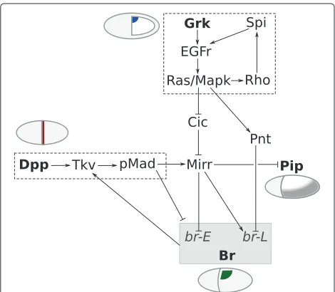
Pattern formation on the follicular epithelium occurs through the activation of a genetic network by two main input pathways: EGFr and Dpp signaling (Figure 2).
Around stage 8 of Drosophila oogenesis, dorsal
pat-terning is initiated when the TGF-α-like ligand Gurken (Grk) localizes to the dorsal-anterior corner of the oocyte (Figure 3A). Grk associates with the oocyte nucleus, which is pushed by microtubules to a dorsal-anterior position
Figure 2A simplified representation of the genetic network underlying dorsoventral polarity (pip) and DA-formation (br) duringD. melanogasteroogenesis.Input comes from two main signaling pathways, EGFr and Dpp, providing dorsoventral and anteroposterior information, respectively, and results in the specification of domains on the epithelium expressingpipandbr.
[23], breaking dorsoventral symmetry in the egg chamber [24]. The Grk signal then activates EGFr in the adjacent follicle cells, leading (directly and indirectly) to the expres-sion of several transcriptional targets, among which are mirror (mirr) [25,26], rhomboid (rho) [27], and pointed (pnt) [28] (Figure 2).
Meanwhile, Dpp signaling starts at stage 8 with the expression ofdppin a subset of anterior follicle cells [29] (Figure 3C). Dpp protein diffuses to more posterior fol-licle cells, forming a morphogen gradient. It acts via the receptor Thickveins (Tkv) in the follicular epithelium to phosphorylate Mothers Against Dpp (Mad), activating the pathway in a graded manner [30]. Dpp has also been sug-gested to be required for the expression of mirr [31], which starts at stage 10A in a wide dorsoanterior domain (Figure 4A). Recent work by Fuchset al.[32] shows how the transcription factor Mirr, regulated by both Dpp and EGFr activity, and the ETS-domain transcription factor Pnt, expressed in a more narrow stripe along the mid-line, subsequently establish two groups of cells expressing broad (br) through two rounds of signaling. First, Mirr repressesbr, which has been expressed in all follicle cells up to this point, in a wide dorsoanterior region through thebrEenhancer. Then,brexpression is upregulated again by Mirr, but repressed by Pnt, through thebrLenhancer (Figure 2). The two resulting patches of Br-positive cells on either side of the midline are identified as ‘roof cells’: they will later constrict apically and shape the roof of the appendage tube [33]. Adjacent to the Br-positive patches is a single L-shaped row of cells, bordering the ante-rior and the central edge of the roof domain. These cells express high levels of rho, and elongate directionally to form the floor of the tube [33]. rho expression is regu-lated mainly by activation of the EGFr pathway, which is highly dynamic throughout oogenesis, and shows the same L-shaped pattern at the definition of the floor cells [34]. Rho itself is involved in the dynamic EGFr activa-tion as it cleaves the EGFr ligand Spitz (Spi) into its active form, thereby providing a positive feedback loop for EGFr signaling [27,35,36] (Figure 2).
Importantly, EGFr signaling also determines the dorsoventral axis of the future embryo [37]. Via Mirr,pipe (pip) expression is restricted to the ventral follicle cells (Figure 2), leaving an asymmetric distribution of Pip pro-tein at the end of oogenesis [32,38-40]. Pip is upstream of a proteolytic cascade in the embryo, leading to the well-known gradient of nuclear Dorsal that regulates the germ layers of the early embryo [41].
Figure 3Dpp and EGFr activity inC. capitataandD. melanogasteroogenesis.Posterior is to the right, ventral to the bottom.(AtoB’)Thegrk
transcript localizes in the dorsal-anterior corner of the oocyte, both inD. melanogaster(A,A’)andC. capitata(B,B’). A and B are stage 8;A’andB’are stage 10B. (CtoD’) Expression ofdppdiffers between the two species:(C)D. melanogaster dppexpression starts in a subset of anterior follicle cells at stage 8 (arrowhead). (C’) At stage 10Adppis only seen in a ring of follicle cells at the border between the nurse cells and oocyte (arrowhead).(D)In a stage 8C. capitataegg chamber, thedpptranscript is seen in the border cell cluster (empty arrowhead), the nurse cells, and localized anteriorly in the oocyte (black arrowhead).(D’)At stage 10, the transcript localizes in a ring at the anterior-outer edge of the oocyte (arrowhead), see also(I).(E toF’)Expression ofthickveinsin all follicle cells of stage 9(E,F)and early stage 10(E’,F’)egg chambers of both species.(GtoH)In bothD. melanogaster(G)andC. capitata(H)stage 10A egg chambers, activation of the Dpp pathway, visualized with immunohistochemistry against pMad, occurs in the stretched follicle cells overlying the nurse cells, and a few anterior rows of columnar follicle cells.(I,I’)FISH ofdppinC. capitata.(I)In a stage 10A egg chamber thedpptranscript localizes just underneath the follicle cells (arrowhead; dashed line indicates border of follicle cells).(I’)In stage 11 expression can be seen in migrated follicle cells between the nurse cells and oocyte (arrowhead).
required furthermore for the formation of the operculum [29,42].
In summary, EGFr and Dpp activity specify dorsoventral and anteroposterior polarity in the epithelium, respec-tively, and their signaling information is integrated by Br and Rho, which together specify the appendage pri-mordia. In addition, both signaling pathways are cru-cial for proper egg formation and further embryonic development, linking the formation of secondary (novel) structures to essential (thus presumably ancestral) devel-opmental events.
Ceratitis capitata
Considering the relatively novel acquisition of eggshell appendages in the family Drosophilidae, it is interesting to examine the underlying patterning network in the context
of a fly species that does not possess these specialized structures. Tephritidae are estimated to be separated by about 65 million years of evolution from Drosophili-dae [43]. For our comparison we chose a Tephritid fly that has been established as a laboratory organism: the Mediterranean fruit flyCeratitis capitata.C. capitatais an agricultural pest, which has motivated widespread inter-national research, including a genome project and the development of genetic tools [44-46].
In this study, we have examined both EGFr and Dpp sig-naling as well as their downstream targets inC. capitata oogenesis, in order to understand the genetic network patterning the follicular epithelium prior to the evolution of dorsal appendages. Determining which genes behave
differently in the formation of appendage-bearing (D.
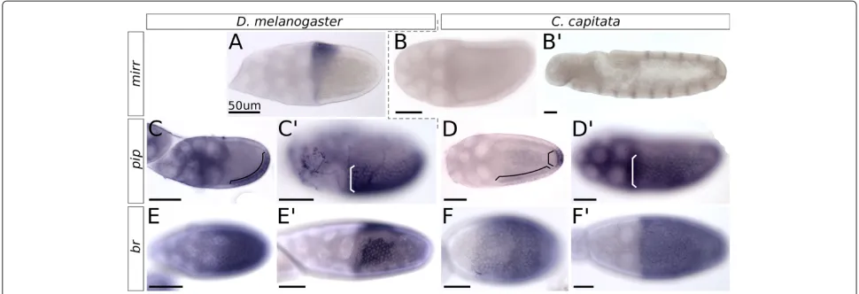
Figure 4Expression ofmirr,pipandbrinC. capitataandD. melanogasteroogenesis.All images arein situhybridizations; posterior is to the right, and ventral to the bottom. The scale bar is always 50μm.(A)mirrexpression in a stage 10 egg chamber ofD. melanogaster.(B)mirr
expression in a stage 10 egg chamber ofC. capitata, with(B’)a positive control for the probe in the embryo.(C)Expression ofpipin a stage 9 egg chamber ofD. melanogastershows dorsal and anterior repression of the gene, and an equal expression strength in ventral and posterior follicle cells (marked by bracket).(C’)Stage 10B shows the final stabilizedpippattern.(D)InC. capitata, ventralpipexpression starts only at stage 10A, and is visibly lighter than the posterior domain (domains marked by separate brackets).(D’)At stage 10B the pattern has stabilized and shows the same sharp on-off boundary between cells expressing and not-expressingpipas seen inD. melanogaster(C’).(E)Expression ofbris visible in all follicle cells of theD. melanogasterstage 9 egg chamber.(E’)Stage 10B showsbrexpressed in the roof cells of the appendage primordia. (F) InC. capitata, stage 9 expression is similar with all cells expressingbr.(F’)AC. capitatastage 10B egg chamber shows how all follicle cells continue expressingbr, and a pattern such as inD. melanogaster(E’)is not formed.
can help us understand the co-option of genes and the genetic network in the evolution of this novel feature. Our analysis points to a key role for the transcription factor Mirr, both in its regulation and in its transcriptional tar-gets. Furthermore, the activity of both the EGFr and the Dpp pathway inC. capitataoogenesis leads us to hypoth-esize that these pathways provided positional information to the ancestral follicular epithelium, which could have facilitated further downstream patterning required for the development of the dorsal appendages.
Material and methods
Fly maintenance
Our initial Ceratitis capitata culture was kindly (and repeatedly) provided by Andrew Jessup (IAEA Seibers-dorf, Austria), originating from flies captured in Argentina. Adult flies were maintained on a diet of sugar and hydrolyzed yeast protein, and larvae were reared on a mixture of bran, sugar and yeast. All stages were
main-tained at room temperature. Drosophila melanogaster
Oregon R. was maintained on regular fly food at room temperature.
Cloning
Gene-specific sequences were isolated fromC. capitata
cDNA by PCR using degenerate primers (fordpp, mirr, rhoandtkv), as well asC. capitataspecific primers (for Cc-br, Cc-cic (capicua), Cc-grk, Cc-pnt, Cc-pip and Cc-slbo). Specific primers were designed using contigs from
the C. capitatagenome project, provided by the Med-fly Whole Genome Sequencing Consortium (led by Drs Alfred Handler and Marc Schetelig, USDA, Agricultural Research Service, Gainesville, Florida; Giuliano Gasperi and Ludvik Gomulski, Department of Biology & Biotech-nology, University of Pavia, Italy; and Stephen Richards and Steven Scherer, Baylor College of Medicine Human Genome Sequencing Center).
ForCc-piptwo primer combinations were used, gener-ating two separate probes forin situhybridization. These probes were (1) against the common part of allpip iso-forms, and (2) againstCc-pip-ST2, the homologue of
Dm-pip-ST2 (isoform A). Corresponding probes were made
for the positive controls inD. melanogaster.
The (partial) nucleotide sequences of all C.
capi-tata genes used in this study have been deposited
with GenBank, and are available under the following
accession numbers: KC150010 (Cc-br), KC150011 (
Cc-cic), KC150006 (Cc-dpp), KC150012 (Cc-grk), KC150007 (Cc-mirr), KC150013 (Cc-pip, common part), KC150014 (Cc-pip, specific to isoform ST2), KC150015 (Cc-pnt), KC150008 (Cc-rho), KC150016 (Cc-slbo) and KC150009 (Cc-tkv).
Immunohistochemistry
room temperature for 1 hour. Antibody incubation was done overnight at 4°C. The rabbit anti-pMad antibody was kindly provided by the laboratory of Gin´es Morata (Cen-tro de Biolog´ıa Molecular Severo Ochoa, Autonomous University of Madrid, Spain), and was used at a con-centration of 1 : 100 in PBTx-B. Anti-Fasciclin II (1D4) was obtained from the Developmental Studies Hybridoma Bank (maintained at the University of Iowa, Department of Biology, Iowa), and was used at a concentration of 1 : 50. Secondary antibodies (Alexa fluor 488/546 goat-anti-rabbit or anti-mouse IgG (H+L), Molecular Probes) were used at a concentration of 1 : 2000, overnight at 4°C. Nuclear staining was done with Dapi.
In situhybridization (ISH)
Ovaries were dissected in cold PBT (0.1% Tween-20 in PBS) and transferred to 4% paraformaldehyde in PBS, where they were fixed overnight. They were subsequently washed in PBS, dehydrated and stored in 100% MeOH at
−20◦C. The protocol for ISH was taken from Tautz and Pfeifle [47] and modified for oogenesis. The main change concerned the adjustment of the proteinase K digestion to 10 minutes 50μg/mL at room temperature.
To ensure identical conditions during the experiment, the positive controls with embryos were done in the same well as the ovaries, starting at the pre-hybridization incu-bation in hybridization buffer at hybridization tempera-ture. This was done because the proteinase K treatment for ovaries is much harsher than the one we used for embryos (10 minutes 50μg/mL vs. no proteinase K at all).
Results
C. capitataoogenesis is a suitable system for comparison
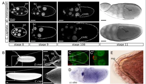
The C. capitataeggshell carries no structures that can be identified as homologues of the operculum, outward micropyle and dorsal appendages (Figure 1B). Still, it is not entirely symmetrical, both over the anteroposterior axis and the dorsoventral axis. The anterior end of the chorion shows markedly stronger imprints of (previously present) follicle cells when compared to the posterior end. While we cannot say with certainty which side is dorsal and which is ventral, it is clear that one is more convex than the other. As both late stage egg chambers (Figure 1A,D) and early embryos (data not shown) are clearly more con-vex at the ventral side, it is a reasonable assumption that the convex side of the egg is ventral.
From an initial observation of C. capitata oogenesis we can conclude first and foremost that it is a suitable
system for comparison with D. melanogaster. C.
capi-tata ovaries, like those of Drosophilidae, are meroistic polytrophic ovaries. While the egg chamber of C. capi-tata is usually larger than the corresponding stage inD. melanogaster, there is no notable difference in the number of cells that make up the follicular epithelium. Instead, the
size ofC. capitatafollicle cells is increased with respect to those ofD. melanogaster, thus contributing to a larger egg chamber as a whole (Figure 1A).
The structure of the egg chambers as well as the pro-gression of stages is nearly identical to that ofDrosophila, providing a good basis for comparison (Figure 1A). Start-ing at mid-oogenesis, we can observe the asymmetric localization of the oocyte nucleus (stage 8), as well as follicle cell migration (stage 9), and centripetal migra-tion (stage 10B). Also visible is the dumping of nurse-cell content into the oocyte, as evidenced by the increas-ing size of the oocyte relative to the nurse cells, which disappear eventually. All these are important and stage-defining steps inDrosophilaoogenesis. We will therefore refer to the stages defined inD. melanogaster[48] when describingC. capitataoogenesis.
In addition to the migration of the main body folli-cle cells, a cluster of anterior follifolli-cle cells can be seen to migrate between the nurse cells at stage 9. Their migra-tion ends at the posterior edge of the nurse cells, adja-cent to the oocyte, where they are shortly joined by the centripetally migrating follicle cells. In D. melanogaster these cells are known as border cells, and can be iden-tified by the expression ofslbo[49], as well as with the polar-cell-specific label Fasciclin II. Both markers con-firmed the identity of the border cell cluster inC. capitata (Figure 1C,D). Interestingly, as the border cells have been associated inD. melanogaster with the formation of the micropyle, no obvious external micropyle can be seen on the C. capitata egg (Figure 1B). However, upon closer examination of the newly formed eggshell we found a pore-like structure on the anterior side of the eggshell, likely homologous to the micropyle pore (Figure 1E). This is consistent with the observed border cell localization in C. capitata, as these cells are known to form the pore of the micropyle, but not the outwardly visible structure [48].
Both EGFr and Dpp pathways are active inC. capitata
oogenesis
In C. capitata ovaries, the initial activation of the dorsoventral patterning cascade by the ligand Gurken occurs similarly to D. melanogaster. In the early stages, theCc-grk transcript is visible in the oocyte at the ante-rior cortex (data not shown), and around stage 8 the pattern becomes restricted to the putative dorsoanterior side of the oocyte (Figure 3A,B). The transcript disappears around stage 11.
the dorsal repression of a known target of EGFr signaling inD. melanogaster: the genepip(Figure 4D,D).
In contrast with oogenesis inD. melanogaster,Cc-dpp is not expressed in the somatic follicle cells, but instead in the germ line. Expression ofCc-dppis first visible as early as the germarium. Once the egg chamber is formed, the dpptranscript localizes to the oocyte. When the oocyte increases in size, the mRNA seems to accumulate at the putative anterior end of the oocyte, in a ring around the edge, adjacent to the follicle cells (Figure 3D,D,I). Inter-estingly, this ring is reminiscent of theD. melanogaster pattern, wheredpp is expressed in the stretched follicle cells as well as a few anterior rows of columnar follicle cells, resulting in a similar ring ofdppexpression around the anterior end of the oocyte (Figure 3C). The main difference, of course, is that the transcript is located in different cell types.
One exception to the exclusive germ line expression of Cc-dppis the border cell cluster. This migrating group of anterior follicle cells is not known to expressdpp in D. melanogaster, but is the only group of somatic cells during oogenesis to expressCc-dpp. Expression is visible around stage 8, when the cell cluster is defined (Figure 3D, empty arrowhead), and persists through migration until the edge of the nurse cells is reached.
A possible second group ofCc-dppexpressing follicle cells was identified using fluorescentin situhybridization (FISH). This group of cells is centrally located between the nurse cells and the oocyte in late stage 11 (Figure 3I). Due to the very small sample size we cannot say with certainty whether these cells are the border cells or part of the fol-licle cells that have centripetally migrated inwards. As the signal ofCc-dppexpression does not persist in the border cell cluster after migration is completed, the observation could either indicate a new round ofCc-dppexpression in this cluster should these cells indeed be border cells, or it could point to conservation ofdppexpression in the leading edge of centripetally migrating follicle cells.
While expression of the ligand may differ somewhat between the two species, downstream signaling is remark-ably similar. The expression of the homologue of the Dpp pathway type I receptor tkvis not visibly different inC. capitatafromD. melanogaster:Cc-tkvis expressed in the follicular epithelium (Figure 3E,F), and disappears around stage 11 or 12. More importantly, the activity of the path-way, shown through immunohistochemistry for the phos-phorylated form of Mad (pMad), is initially not different between the two species, despite the altered localization of thedpptranscript (Figure 3G,H).
Differences in Dpp pathway activation between C.
capitata and D. melanogaster start around stage 10B,
when expression of Dm-tkv becomes restricted to the
Br-positive cells of the appendage primordia, naturally affecting pMad patterns [51,52]. These dynamics were not
observed inC. capitata, where no Br-positive domains are formed (Figure 4F).
Patterning of the follicular epithelium downstream of EGFr and Dpp
The dynamics of EGFr and Dpp signaling and subsequent epithelial patterning inD. melanogasteregg chambers are key in defining the appendage primordia. Identifying the point in the genetic network whereC. capitatano longer resemblesD. melanogasteris therefore an important step in understanding the evolution of the dorsal appendages, as it could indicate the point where the network was co-opted.
Our first candidate for co-option was found when we saw that no expression of mirr could be detected inC. capitataegg chambers (Figure 4B). The probe against Cc-mirrdid reveal clear expression in theC. capitataembryo, in a pattern familiar from expression inD. melanogaster (Figure 4B) [53].
Mirr regulates the transcription ofbrin those cells that will give rise to the dorsal appendages (Figure 4E). Unsur-prisingly, thebr-positive domains do not appear on theC. capitatastage 10B follicular epithelium (Figure 4F), nor during any other stage of oogenesis. Early expression of br could be seen uniformly in the follicular epithelium, as inD. melanogaster, but the late expression dynamics, both the dorsal-anterior repression and the appearance of the two domains, were not observed; instead, expression diminished around stage 11 and had disappeared entirely by stage 12.
Preliminary results indicate that two other genes rel-evant for D. melanogaster epithelial patterning do not play a role in the C. capitata dorsal-anterior epithe-lium: expression ofpnt, encoding the transcription factor responsible for the midline repression ofbr, could not be detected in the dorsal-anterior follicular epithelium ofC. capitata. A second known expression domain of pntat the posterior pole of the egg chamber was clearly visible from an early stage (stage 8), providing a positive control for thein situhybridization and thepntprobe (Additional file 1). Transcription of the generhowas also not detected in either the early broad dorsoanterior domain, or in the late hinge-shaped patterns adjacent to thebr expressing domains [22] (Additional file 1). However, as both early rhoexpression and the dorsoanterior domain ofpntcan be difficult to detect inD. melanogasteregg chambers as well, we cannot be completely certain of the absence ofpntand rhotranscripts in the dorsoanterior follicular epithelium ofC. capitata.
Conserved expression ofpip
clearly localizes to the ventral follicular epithelium. In a
similar dynamic—though not precisely identical—to D.
melanogaster,Cc-pipexpression starts at stage 8 in follicle cells at the posterior pole of the egg chamber (Figure 4D). This posterior expression domain during stages 8 and 9 is well known in D. melanogaster [38,40]. During early stage 10, ventral follicle cells start expressingCc-pip, and by late stage 10 expression in ventral and posterior folli-cle cells is of equal strength (Figure 4D). The pattern at this stage is identical to the expression pattern ofDm-pip (Figure 4C), including the sharp on-off boundary between ventral and dorsal cells. These results were obtained using two separate probes: one against the common part of all pipisoforms, and one specific to the homologue of iso-form A (orpipe-ST2), confirming that the same isoform is used inC. capitataoogenesis as is known to function in D. melanogaster[54].
Discussion
Pre-existing functional signals provide positional information
A first conclusion we can draw from the work presented is the fact that the activity during oogenesis of the two main patterning pathways, EGFr and Dpp, preceded the evo-lution of dorsal appendages and their underlying epithe-lial patterns [29]. The ancestral role of EGFr signaling lies in determining the dorsoventral axis of the future embryo [50], while Dpp is involved in various cell migra-tions required for the developmental progression of the egg chamber. Activity from these pathways provides the epithelium with positional information that may consti-tute an important facilitator for novel patterns to evolve.
A formalism for pattern formation on the Drosophila
melanogasterfollicular epithelium was developed in 2008 by Yakobyet al. [11]. They propose a combinatorial code of principle patterns from which all expression patterns at the dorsal-anterior follicular epithelium can be derived. As their formalism includes both the EGFr and Dpp input, as well as three additional primary building blocks specific to the dorsal-anterior epithelium, this system constitutes the next step of pattern formation in the evolution of Drosophilaeggshell morphology. Interestingly, this spec-ification of patterns from a system of higher order com-ponents is an emerging theme in regulatory evolution [55]. Our results therefore fit within the larger research theme of how pre-existing information may bias the future evolution of pattern formation and morphology.
Upstream differences in Dpp signaling betweenD. melanogasterandC. capitata
Despite the fact that the pattern of Dpp activity is similar betweenC. capitataandD. melanogaster, the differences in the underlying expression of its liganddppare puzzling. Not only are there differences in the expression patterns of
Dm-dppandCc-dpp, but the transcripts are produced by an altogether different cell type.Cc-dppis likely expressed by the nurse cells and transported to the oocyte, both of which are germ line, whileD. melanogasterrequiresdpp expression in the somatic follicle cells.
Several functions have been described for Dpp signal-ing inD. melanogaster[56]. In the context of oogenesis, the need for Dpp signaling in the formation of ante-rior eggshell structures has been clearly established:dpp is expressed in the cells that will form the operculum, and disruptions of Dpp signaling cause misplaced and deformed appendages [29,57-59]. Additionally, Dpp sig-naling is needed for the centripetal migration of follicle cells, to maintain structural integrity of the egg cham-ber, and for dumping of nurse cell content into the oocyte [29]. However, expression of the signaling molecule Dpp is only required in the somatic follicular epithelium: inD. melanogastergerm linedppis not required during ooge-nesis [60]. InC. capitata,dppis clearly expressed in the germ line, and the signal acts through receptors in the soma. While it cannot be ruled out that the Dpp activity in the follicular epithelium is a response to earlydpp expres-sion in the border cell cluster, the transcript in the nurse cells as well as the ring of dppin the oocyte are a likely origin for Dpp signaling in the stretched and centripetally migrating follicle cells, respectively (Figure 3D,H,I).
Although it is intriguing to observe such apparent dra-matic changes in expression patterns, conservation of phenotype in face of substantial changes in the architec-ture of developmental programs has been widely reported and discussed under the concept of developmental sys-tems drift [61]. In this particular case, it is important to remember that the functional event, the actual Dpp sig-nal, remains a cooperative act between the ligand and its receptors. Thus, the selective pressure for Dpp function will be on this signaling event, as opposed to the source of the ligand. Interestingly, a similar interaction between germ line and soma has been described regarding Dpp sig-naling in the honeybeeApis mellifera[62]. In this system, dpp mRNA is localized to a dorsal stripe in the oocyte, and signaling activity is observed in the overlying follicle cells. While the absence of data ondppexpression in other closely related dipteran species precludes a clear evolu-tionary interpretation of these patterns, it does suggest that dppexpression in the follicle cells is a recent adap-tation. A possible reason could be to prevent Dpp from remaining in the perivitelline cleft at the end of oogenesis, which could interfere with future embryonic dorsoventral patterning in which Dpp plays a large role.
Regulation ofmirr
ancestral and vital feature of embryonic development— dorsoventral polarity. One element of the network draws specific attention: the transcription factor Mirror (Mirr). Mirr regulates both the expression ofpipe(pip), the gene encoding a sulfotransferase that is pivotal in providing dorsoventral polarity to the embryo, andbroad(br), the gene that defines the dorsal appendage primordia. Our results show that pipexpression is conserved, while its upstream regulator Mirr appears to be part of the novel branch of the network inD. melanogaster. This observa-tion suggests that Mirr, rather than Br, operates as the key node of the network underlying the evolution of dorsal appendages.
Understanding the regulation ofmirrinD. melanogaster then is necessary to understand how Mirr could have been co-opted to regulatebr, and possiblypip, in a novel man-ner. The best substantiated link between EGFr activation andmirrexpression is the HMG-box transcription factor Capicua (Cic). Cic is a repressor of mirr in ventral and lateral follicle cells, and is downregulated in response to EGFr signaling [63,64]. However, global de-repression of mirr through Cic loss-of-function only results in visible expression ofmirrin anterior follicle cells [65]. This obser-vation suggests the additional involvement of Dpp activity inmirrregulation [31].
However, it has recently become clear that detectable
mirr may not fully represent Mirr activity throughout
the follicular epithelium. Indeed, local de-repression of mirr through follicle cell clones in the posterior part of the epithelium is still sufficient to repress pip [40]. Interestingly,pipexpression is unaffected when the Dpp pathway is disrupted [30]. Moreover, computational anal-yses have shown that the two-dimensional EGFr signaling profile is sufficient to explain thepipexpression pattern, without any additional requirements for Dpp or other factors [66].
Thus, the fact thatmirr expression is not seen within situhybridization does not preclude its activity in the fol-licular epithelium at a level sufficient to repress pip. In
other words, we cannot conclude that mirr expression
is absent in C. capitata from our data alone. However, although low (undetectable)mirrexpression may be suf-ficient in D. melanogaster for regulation of pip, high (detectable) expression levels are necessary for activating the late enhancer ofbrand defining the dorsal appendage fate [31,32,65]. These high levels ofmirr expression are clearly absent in C. capitata, and constitute a novel expression pattern related to the formation of a novel trait.
Expression of br also depends on input from the Dpp
pathway [22,30].
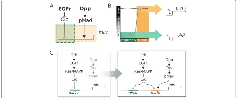
Based on this data and our observations inC. capitata we propose a model that separates the contribution of mirrto dorsoventral polarity from its function in epithe-lial patterning, using two regulatory modules inmirr to
obtain two distinct levels of expression (Figure 5A,B). One of these, responding only to EGFr activation—presumably through Cic down-regulation—is sufficient to generate the low expression levels required to repress pip (and likely also act on the early enhancer ofbr). Conversely, the other module requires Dpp signaling in addition to Cic down-regulation, and is able to regulatemirr expres-sion to the high levels observed within situhybridization in the dorsal-anterior follicle cells of wild-typeDrosophila egg chambers.
With this model we predict that dorsal de-repression of mirr through Cic is sufficient forpiprepression, and constitutes an ancestral signaling cassette linking EGFr activation to embryonic dorsoventral patterning. Due to a non-cross-reactive antibody we were unable to confirm whether the localization of Cic in theC. capitatafollicular epithelium fits our model, but we do note thatcicmRNA is expressed in the egg chambers (Additional file 1).
Alternatively,mirrexpression could be absent inC. cap-itata altogether, andpip could be regulated by another transcription factor. However, given the important role of Pip in embryonic dorsoventral axis determination, and the dramatic defects that are caused with minimal variation in factors along the anteroposterior axis [67,68], we consider it likely thatpipregulation happens through a conserved mechanism involvingmirr. Pending the completion of the C. capitatawhole genome sequencing project, it will be possible to test this hypothesis with a search for Mirr-responsive elements in aCc-pipregulatory region.
Presenting both modules as enhancers ofmirrprovides us with a hypothesis regarding their evolution (Figure 5C). The predicted ‘mirLo’ enhancer is expected to be ances-tral, as we base its existence on themirrandpip expres-sion patterns in C. capitataegg chambers.MirLowould drive mirr expression in dorsal follicle cells in a level sufficiently high to represspip, thus regulating dorsoven-tral polarity of the future embryo, downstream of EGFr signaling and independent of Dpp. The appearance of the second enhancer ‘mirHi’ would allow mirr to start responding to information from the Dpp pathway, and open up the evolutionary road to new patterns on the follicular epithelium.
Conclusions
In the evolution of dorsal appendages, several genes have been co-opted into a network that originally regulated only dorsoventral polarity, using the input from a sec-ond signaling pathway active in the tissue. The activity of these pathways, EGFr and Dpp, defined a coordi-nate system on the epithelium upon which novel gene expression patterns were built. We have shown here how this coordinate system predates the evolution of dor-sal appendages, providing positional information to the
follicular epithelium that played a crucial role in future pattern formation.
The main regulators in this novel genetic network are transcription factors Mirr, Pnt and Br. The lat-ter integrates the information from upstream Mirr and Pnt to specify the appendage primordia, and drives morphogenesis of the appendage [32,57]. Interestingly, while all three transcription factors have co-opted novel expression patterns and interactions to provide the main regulatory information for the epithelial positional cues to be translated into a novel morphology, both br, pnt and very likelymirrwere already expressed in the ances-tral non-appendage-forming epithelium. In this case it is notable that evolution may have taught ‘old genes new tricks’ [69] within the same broad spatial and develop-mental context. A novel morphological feature has been achieved by modifying the levels of existing expression patterns allocated to ancient developmental roles (brand likelymirr) with new enhancers responding to new infor-mation, or through the addition of an expression domain at another end of the same epithelium (pnt).
Most likely, the transcription factors Br, Mirr and Pnt have not been the only ones to evolve new roles. Future research could go into the EGFr feedback loop, looking at rho andaos, as well as other EGFr targets. However, while the EGFr feedback loop was long thought to be the main patterning component of the eggshell [35], it has meanwhile been shown that eggshell patterning functions normally without several elements of this feedback sys-tem [70,71]. We can therefore conclude that changes in the regulation ofmirr,pntandbrplayed important roles in the evolution of this novel morphology, and the detailed dissection of its molecular genetic basis constitutes the most important and immediate research agenda in the comprehension of the evolution of this morphological novelty.
Finally, we would like to stress that, as a derived organ-ism, the model systemD. melanogasterprovides us with an excellent handle to tackle the question of evolutionary novelties, and for the dissection of the molecular mecha-nisms that underlie the process of co-option. We propose that many other traits that defineDrosophilaas derived— be they morphological or behavioural in nature—can be amenable to study using a comparative approach with other emergent dipteran model systems.
Additional file
Additional file 1: Preliminary results: expression patterns ofCc-rho,
at the dorsal-anterior side of egg chambers could be detected. Fibers surrounding the egg chamber sometimes obscure the results, but changing the focal plane can confirm the identity of a signal as either coming from aspecific staining in fibers or staining of the cells in the follicular epithelium.
Capicua: Strong expression ofcapicuais seen in the nurse cells, but weaker expression can also be seen in the follicular epithelium (stage 10 egg chamber, below).
Abbreviations
PCR: polymerase chain reaction; PBS: phosphate-buffered saline; ISH:in situ hybridization; FISH: fluorescentin situhybridization.
Competing interests
The authors declare that they have no competing interests.
Authors’ contributions
ES conceived and designed the project, and wrote the paper; BMIV participated in the project design, performed the experiments, and wrote the paper; SR and JAL were invaluable for the execution of the experiments and the interpretation of results. All authors read and approved the final manuscript.
Acknowledgements
We are indebted to the Medfly Whole Genome Sequencing Consortium led by Alfred Handler and Marc Schetelig (USDA, FL, USA), Giuliano Gasperi and Ludvik Gomulski (University of Pavia, Italy), and Stephen Richards and Steven Scherer (Baylor College, TX, USA) for allowing access to unpublished data of theC. capitatagenome sequencing project. Further thanks go to Andrew Jessup (IAEA, Austria) forC. capitatapupae and supplies, and to Gin´es Morata (CBM Severo Ochoa, Madrid, Spain) for providing the p-Mad antibody. The anti-Fasciclin II antibody was obtained from the Developmental Studies Hybridoma Bank developed under the auspices of the NICHD and maintained by the University of Iowa, Department of Biology, Iowa City, IA 52242. The electron microscopy (EM) imaging was done under the skillful guidance of Gerda Lamers and Merijn de Bakker at the Leiden University Institute of Biology, The Netherlands. We also thank Merijn de Bakker and Alexandre Raposo for helpful suggestions during the development of anin situ hybridization protocol, Meike Knispel, Adrien Faur´e, Claudine Chaouiya, Patr´ıcia Beldade and Jos´e Pereira-Leal for invaluable discussions, and Leila Shirai, Matthew Child, Hanneke Meijer and two anonymous referees for their helpful comments on the manuscript.
This work was supported by FCT grants #410540-PFE-61-FCT-PTDC/BIA-BCM/74583/06 and #SFRH/BD/33216/2007 (BMIV), and a DFG grant of the Collaborative Research Centre 680 ‘Molecular Basis of Evolutionary Innovations’ (SR). We are very grateful to the Fundac¸˜ao Calouste Gulbenkian and the Instituto Gulbenkian de Ciˆencia for indispensable support.
Author details
1Instituto Gulbenkian de Ciˆencia, Rua da Quinta Grande 6, Oeiras, Portugal. 2Universidade de Lisboa, Faculdade de Ci ˆencias, Departamento da Biologia Animal, Lisbon, Portugal.3Institute for Developmental Biology, Biocenter, University of Cologne, Z ¨ulpicher Strasse 47b, Cologne, Germany.4Present address: Dept. of Biological Sciences, University of Illinois at Chicago, 900 S. Ashland Avenue, Chicago IL, USA.
Received: 1 October 2012 Accepted: 6 December 2012 Published: 1 March 2013
References
1. Simpson G:The major features of evolution. New York, USA: Simon and Schuster; 1953.
2. M ¨uller G, Wagner GP:Novelty in evolution: restructuring the concept.
Annu Rev Ecol Syst1991,22:229–256.
3. Abouheif E, Akam M, Dickinson WJ, Holland PW, Meyer A, Patel NH, Raff RA, Roth VL, Wray GA:Homology and developmental genes.Trends Genet1997,13(11):432–433.
4. Wagner GP:A research programme for testing the biological homology concept.Novartis Found Symp1999,222:125–34. discussion 134–40.
5. Brigandt I:Homology in comparative, molecular, and evolutionary developmental biology: the radiation of a concept.J Exp Zool B Mol Dev Evol2003,299:9–17.
6. Moczek AP:On the origins of novelty in development and evolution.
Bioessays2008,30(5):432–447.
7. Brigandt I, Love AC:Conceptualizing evolutionary novelty: moving beyond definitional debates.J Exp Zool B Mol Dev Evol2012,
318(6):417–427.
8. Wilkins A:The evolution of developmental pathways: Sinauer Associates, Inc; 2002.
9. Brakefield PM, Gates J, Keys D, Kesbeke F, Wijngaarden PJ, Monteiro A, French V, Carroll SB:Development, plasticity and evolution of butterfly eyespot patterns.Nature1996,384(6606):236–242. 10. Moczek AP, Nagy LM:Diverse developmental mechanisms contribute
to different levels of diversity in horned beetles.Evol Dev2005,
7(3):175–185.
11. Yakoby N, Bristow CA, Gong D, Schafer X, Lembong J, Zartman JJ, Halfon MS, Sch ¨upbach T, Shvartsman SY:A combinatorial code for pattern formation inDrosophilaoogenesis.Dev Cell2008,15(5):725–737. 12. Lembong J, Yakoby N, Shvartsman SY:Pattern formation by
dynamically interacting network motifs.Proc Natl Acad Sci USA2009,
106(9):3213–3218.
13. Berg CA:TheDrosophilashell game: patterning genes and morphological change.Trends Genet2005,21(6):346–355.
14. Berg CA:Tube formation inDrosophilaegg chambers.Tissue Eng Part A2008,14(9):1479–1488.
15. Boyle MJ, French RL, Cosand KA, Dorman JB, Kiehart DP, Berg CA:Division of labor: subsets of dorsal-appendage-forming cells control the shape of the entire tube.Dev Biol2010,346:68–79.
16. Hinton HE:Respiratory systems of insect egg shells.Annu Rev Entomol 1969,14:343–368.
17. Hinton HE:Biology of Insect Eggs. Oxford, UK: Pergamon Press; 1981. 18. Okada T:Systematic study of the early stages of Drosophilidae. Tokyo, Japan:
Department of Biology, Faculty of Science, Metropolitan University; 1968. 19. Nakamura Y, Matsuno K:Species-specific activation of EGF receptor
signaling underlies evolutionary diversity in the dorsal appendage number of the genusDrosophilaeggshells.Mech Dev2003,
120(8):897–907.
20. Kagesawa T, Nakamura Y, Nishikawa M, Akiyama Y, Kajiwara M, Matsuno K:
Distinct activation patterns of EGF receptor signaling in the homoplastic evolution of eggshell morphology in genusDrosophila.
Mech Dev2008,125(11-12):1020–1032.
21. Dorman JB, James KE, Fraser SE, Kiehart DP, Berg CA:bullwinkleis required for epithelial morphogenesis duringDrosophilaoogenesis.
Dev Biol2004,267(2):320–341.
22. Peri F, Roth S:Combined activities of Gurken and decapentaplegic specify dorsal chorion structures of theDrosophilaeggs.
Development2000,127(4):841–850.
23. Zhao T, Graham OS, Raposo A, St Johnston D:Growing microtubules push the oocyte nucleus to polarize theDrosophiladorsal-ventral axis.Science2012,336:999–1003.
24. Neuman-Silberberg FS, Sch ¨upbach T:TheDrosophiladorsoventral patterning genegurkenproduces a dorsally localized RNA and encodes a TGF alpha-like protein.Cell1993,75:165–174. 25. Jordan KC, Clegg NJ, Blasi JA, Morimoto AM, Sen J, Stein D, McNeill H,
Deng WM, Tworoger M, Ruohola-Baker H:The homeobox genemirror
links EGF signalling to embryonic dorso-ventral axis formation through Notch activation.Nat Genet2000,24(4):429–433. 26. Zhao D, Woolner S, Bownes M:The Mirror transcription factor links
signalling pathways inDrosophilaoogenesis.Dev Genes Evol2000,
210(8-9):449–457.
27. Ruohola-Baker H, Grell E, Chou TB, Baker D, Jan LY, Jan YN:Spatially localizedrhomboidis required for establishment of the
dorsal-ventral axis inDrosophilaoogenesis.Cell1993,73(5):953–965. 28. Morimoto AM, Jordan KC, Tietze K, Britton JS, O’Neill EM, Ruohola-Baker H:
Pointed, an ETS domain transcription factor, negatively regulates the EGF receptor pathway inDrosophilaoogenesis.Development 1996,122(12):3745–3754.
29. Twombly V, Blackman RK, Jin H, Graff JM, Padgett RW, Gelbart WM:The TGF-beta signaling pathway is essential forDrosophilaoogenesis.
30. Shravage BV, Altmann G, Technau M, Roth S:The role of Dpp and its inhibitors during eggshell patterning inDrosophila.Development 2007,134(12):2261–2271.
31. Atkey MR, Boisclair Lachance JF, Walczak M, Rebello T, Nilson LA:Capicua regulates follicle cell fate in theDrosophilaovary through repression ofmirror.Development2006,133(11):2115–2123. 32. Fuchs A, Cheung LS, Charbonnier E, Shvartsman SY, Pyrowolakis G:
Transcriptional interpretation of the EGF receptor signaling gradient.Proc Natl Acad Sci USA2012,109(5):1572–1577.
33. Ward EJ, Berg CA:Juxtaposition between two cell types is necessary for dorsal appendage tube formation.Mech Dev2005,122(2):241–255. 34. Nakamura Y, Kagesawa T, Nishikawa M, Hayashi Y, Kobayashi S, Niimi T,
Matsuno K:Soma-dependent modulations contribute to divergence ofrhomboidexpression during evolution ofDrosophilaeggshell morphology.Development2007,134(8):1529–1537.
35. Wasserman JD, Freeman M:An autoregulatory cascade of EGF receptor signaling patterns theDrosophilaegg.Cell1998,
95(3):355–364.
36. Urban S, Lee JR, Freeman M:DrosophilaRhomboid-1 defines a family of putative intramembrane serine proteases.Cell2001,107(2):173–182. 37. Queenan AM, Ghabrial A, Sch ¨upbach T:Ectopic activation of
torpedo/Egfr, aDrosophilareceptor tyrosine kinase, dorsalizes both the eggshell and the embryo.Development1997,124(19):3871–3880. 38. Peri F, Technau M, Roth S:Mechanisms of Gurken-dependentpipe
regulation and the robustness of dorsoventral patterning in
Drosophila.Development2002,129(12):2965–2975.
39. Technau M, Knispel M, Roth S:Molecular mechanisms of EGF signaling-dependent regulation of pipe, a gene crucial for dorsoventral axis formation inDrosophila.Dev Genes Evol2011,222(1):1–17. 40. Andreu MJ, Gonz´alez-P´erez E, Ajuria L, Samper N, Gonz´alez-Crespo S,
Campuzano S, Jim´enez G:Mirror repressespipeexpression in follicle cells to initiate dorsoventral axis formation inDrosophila.
Development2012,139(6):1110–1114.
41. Moussian B, Roth S:Dorsoventral axis formation in theDrosophila
embryo—shaping and transducing a morphogen gradient.Curr Biol 2005,15(21):R887–899.
42. Dobens LL, Peterson JS, Treisman J, Raftery LA:Drosophila bunched
integrates opposing DPP and EGF signals to set the operculum boundary.Development2000,127(4):745–754.
43. Wiegmann BM, Trautwein MD, Winkler IS, Barr NB, Kim JW, Lambkin C, Bertone MA, Cassel BK, Bayless KM, Heimberg AM, Wheeler BM, Peterson KJ, Pape T, Sinclair BJ, Skevington JH, Blagoderov V, Caravas J, Kutty SN, Schmidt-Ott U, Kampmeier GE, Thompson FC, Grimaldi DA, Beckenbach AT, Courtney GW, Friedrich M, Meier R, Yeates DK:Episodic radiations in the fly tree of life.Proc Natl Acad Sci USA2011,108(14):5690–5695. 44. Loukeris TG, Livadaras I, Arca B, Zabalou S, Savakis C:Gene transfer into
the medfly,Ceratitis capitata, with aDrosophila hydeitransposable elements.Science1995,270(5244):2002–2005.
45. Zwiebel LJ, Saccone G, Zacharopoulou A, Besansky NJ, Favia G, Collins FH, Louis C, Kafatos FC:The white gene ofCeratitis capitata: a phenotypic marker for germline transformation.Science1995,
270(5244):2005–2008.
46. Schetelig MF, Scolari F, Handler AM, Kittelmann S, Gasperi G, Wimmer EA:
Site-specific recombination for the modification of transgenic strains of the Mediterranean fruit flyCeratitis capitata.Proc Natl Acad Sci USA2009,106(43):18171–18176.
47. Tautz D, Pfeifle C:A non-radioactivein situhybridization method for the localization of specific RNAs inDrosophilaembryos reveals translational control of the segmentation gene hunchback.
Chromosoma1989,98(2):81–85.
48. Spradling AC:Developmental genetics of oogenesis.InThe development of Drosophila melanogaster. New, York, USA: Cold Spring Harbor Laboratory Press; 1993.
49. Montell DJ, Rorth P, Spradling AC:slow border cells, a locus required for a developmentally regulated cell migration during oogenesis, encodesDrosophilaC/EBP.Cell1992,71:51–62.
50. Lynch JA, Peel AD, Drechsler A, Averof M, Roth S:EGF signaling and the origin of axial polarity among the insects.Curr Biol2010,
20(11):1042–1047.
51. Yakoby N, Lembong J, Sch ¨upbach T, Shvartsman SY:Drosophila
eggshell is patterned by sequential action of feedforward and feedback loops.Development2008,135(2):343–351.
52. Niepielko MG, Hernaiz-Hernandez Y, Yakoby N:BMP signaling dynamics in the follicle cells of multipleDrosophilaspecies.Dev Biol2011,
354(1):151–159.
53. McNeill H, Yang CH, Brodsky M, Ungos J, Simon MA:mirrorencodes a novel PBX-class homeoprotein that functions in the definition of the dorsal-ventral border in theDrosophilaeye.Genes Dev1997,
11(8):1073–1082.
54. Zhang Z, Zhu X, Stevens LM, Stein D:Distinct functional specificities are associated with protein isoforms encoded by theDrosophila
dorsal-ventral patterning genepipe.Development2009,
136(16):2779–2789.
55. Prud’homme B, Gompel N, Carroll SB:Emerging principles of regulatory evolution.Proc Natl Acad Sci USA2007,104(Suppl 1):8605–8612. 56. Martinez Arias A, Stewart A:Molecular principles of animal development.
Oxford, UK: Oxford University Press; 2002.
57. Deng WM, Bownes M:Two signalling pathways specify localised expression of theBroad-ComplexinDrosophilaeggshell patterning and morphogenesis.Development1997,124(22):4639–4647. 58. Dobens L, Jaeger A, Peterson JS, Raftery LA:Bunched sets a boundary
for Notch signaling to pattern anterior eggshell structures during
Drosophilaoogenesis.Dev Biol2005,287(2):425–437.
59. Chen Y, Sch ¨upbach T:The role ofbrinkerin eggshell patterning.Mech Dev2006,123(5):395–406.
60. Irish VF, Gelbart WM:Thedecapentaplegicgene is required for dorsal-ventral patterning of theDrosophilaembryos.Genes Dev1987,
1(8):868–879.
61. True JR, Haag ES:Developmental system drift and flexibility in evolutionary trajectories.Evol Dev2001,3(2):109–119.
62. Wilson MJ, Abbott H, Dearden PK:The evolution of oocyte patterning in insects: multiple cell-signaling pathways are active during honeybee oogenesis and are likely to play a role in axis patterning.
Evol Dev2011,13(2):127–137.
63. Astigarraga S, Grossman R, D´ıaz-Delf´ın J, Caelles C, Paroush Z, Jim´enez G:
A MAPK docking site is critical for downregulation of Capicua by Torso and EGFR RTK signaling.EMBO J2007,26(3):668–677. 64. Ajuria L, Nieva C, Winkler C, Kuo D, Samper N, Andreu MJ, Helman A,
Gonz´alez-Crespo S, Paroush Z, Courey AJ, Jim´enez G:Capicua DNA-binding sites are general response elements for RTK signaling inDrosophila.Development2011,138(5):915–924.
65. Goff DJ, Nilson LA, Morisato D:Establishment of dorsal-ventral polarity of theDrosophilaegg requirescapicuaaction in ovarian follicle cells.Development2001,128(22):4553–4562.
66. Goentoro LA, Reeves GT, Kowal CP, Martinelli L, Sch ¨upbach T, Shvartsman SY:Quantifying the Gurken morphogen gradient inDrosophila
oogenesis.Dev Cell2006,11(2):263–272.
67. Roth S, Sch ¨upbach T:The relationship between ovarian and embryonic dorsoventral patterning inDrosophila.Development1994,
120(8):2245–2257.
68. Roth S, Jordan P, Karess R:BinuclearDrosophilaoocytes: consequences and implications for dorsal-ventral patterning in oogenesis and embryogenesis.Development1999,126(5):927–934. 69. Carroll SB, Grenier JK, Weatherbee S:From DNA to diversity. Molecular
genetics and the evolution of animal design. (2nd edition). Oxford, UK: Blackwell Science; 2005.
70. Boisclair Lachance JF, Fregoso Lomas M, Eleiche A, Bouchard Kerr P, Nilson LA:Graded Egfr activity patterns theDrosophilaeggshell independently of autocrine feedback.Development2009,
136(17):2893–2902.
71. Zartman JJ, Kanodia JS, Cheung LS, Shvartsman SY:Feedback control of the EGFR signaling gradient: superposition of domain-splitting events inDrosophilaoogenesis.Development2009,136(17):2903–2911.
doi:10.1186/2041-9139-4-7