PRIMARY RESEARCH
MiR-566 mediates cell migration
and invasion in colon cancer cells by direct
targeting of
PSKH1
Ying Zhang
1, Siqi Zhang
2, Jian Yin
3and Ruisi Xu
1*Abstract
Background: Colorectal cancer (CRC), a common malignancy worldwide, and microRNAs (miRs) have been sug-gested to play roles in the disease. MiR-566 expression has been shown to be reduced in CRC, but its functions and mechanisms are still unclear.
Methods: Cell viability was assessed by using the CellTiter 96 AQueous One Solution Cell Proliferation kit. Cell pro-liferation was measured with MTT assay. Cell metastasis were measured by transwell assay. Luciferase reporter assays was used to confirm the target of MiR-566. PSKH1 expression was measured by RT-PCR and western blot.
Results: In the present study, we first observed that miR-566 was expressed in several CRC cell lines (SW480, SW620, LoVo, HT29 and Caco-2) at low levels compared to control colon epithelial cell lines (FHC). Further study showed that miR-566 overexpression suppressed cell survival and impeded cell proliferation, whereas inhibition of its expression enhanced cell survival and proliferation. Transwell assays showed that cell invasion and migration were reduced in cells overexpressing miR-566 and increased in those with inhibition of miR-566. Further analysis confirmed that PSKH1
is a target of miR-566. MiR-566 overexpression significantly inhibited PSKH1 expression and reintroduction of PSKH1 partially reversed the effects of miR-566 on CRC cell growth and metastasis in SW480 and Caco-2 cells.
Conclusions: Taken together, the data show that CRC cell growth and metastasis can be significantly suppressed by miR-566 through targeting PSKH1.
Keywords: Colorectal cancer, miR-566, PSKH1, Invasion, Migration
© The Author(s) 2019. This article is distributed under the terms of the Creative Commons Attribution 4.0 International License (http://creativecommons.org/licenses/by/4.0/), which permits unrestricted use, distribution, and reproduction in any medium, provided you give appropriate credit to the original author(s) and the source, provide a link to the Creative Commons license, and indicate if changes were made. The Creative Commons Public Domain Dedication waiver (http://creativecommons.org/ publicdomain/zero/1.0/) applies to the data made available in this article, unless otherwise stated.
Background
Colorectal cancer (CRC), the most prevalent malignancy, is a complex polygenetic disease and the main cause of death from cancer worldwide [1, 2]. CRC-associated morbidity is increasing annually, and in 2008, there were approximately 120,0000 new CRC cases globally and more than 60,0000 deaths [3]. Death is caused by progression and metastasis of the cancer, and previous research implicates several underlying mechanisms in cancer metastasis, including tumor cell invasion, adhe-sion, chemotaxis, epithelial–mesenchymal transition and
tumor cell growth [3, 4]. In recent years, the life expec-tancy of patients with CRC has improved because of the advances in screening and treatment; however, the prog-nosis remains poor in patients with metastatic cancer [5]. Thus, understanding how cancer metastasis proceeds and finding a way to block this process would be benefi-cial in developing cancer treatments.
MicroRNAs (miRNAs), a family of single-stranded, noncoding, short RNAs, have been shown to be involved in CRC metastasis [6–8]. miRNAs play inhibitory roles in the function of their target genes, with miRNAs bind-ing to the 3ʹ untranslated region (3ʹUTR) of target genes and then inhibiting gene translation or degrading the tar-get mRNA [9]. Prior evidence suggests that miRNA may be used as a cancer therapy because it can interact with many target genes and signaling pathways that promote
Open Access
*Correspondence: ruisi_xu1@163.com
1 Endoscopy Center, China-Japan Union Hospital of Jilin University, No.
126 Sendai Street, Changchun 130033, Jilin, China
cancer cell death [10]. Li et al. [11] showed that miR-433 plays roles in inhibiting growth and promoting apoptosis through mediation of its target gene MACC1. In addi-tion, Qin et al. [12] suggested that mir-106a regulates the PTEN/PI3K/AKT pathway and is associated with colon cancer cell growth. Multiple studies have implicated miR-NAs play roles in colon cancer cell migration and inva-sion [13, 14]. miR-566 has been shown to be decreased in CRC; however, its functions and mechanisms remain unknown [15]. The present study found that miR-566 has low expression in colon cancer cells and plays roles in cancer cell growth and metastasis.
Kim and his colleagues [16] found that three kinase genes, PHKG2, TLK2, and PSKH1, were all expressed in metastatic CRC at high level, and their overexpres-sion may be a potential biomarker of cetuximab plus irinotecan induced—wild-type KRAS CRC. PSKH1, an autophosphorylating human protein serine kinase, has 424 amino acids and is localized to the Golgi apparatus and speckle structures within the nucleus [17, 18]. In the current study, we confirmed that PSKH1 is a direct tar-get of 566, and its expression was inhibited by miR-566 overexpression; moreover, our study indicated that PSKH1 is involved in the function of miR-566 in CRC cell proliferation, migration, and invasion.
Materials and methods Cell culture and transfection
The human colon cancer cell lines SW480, SW620, LoVo, Caco-2 and HT29 and human normal colon epithelial cell lines (FHC) were purchased from ATCC (Manassas, VA, USA). All cells were maintained in DMEM added with 100 µg/mL streptomycin, fetal bovine serum, 100 U/mL penicillin. Cells were cultured in a 5% CO2 atmosphere
at 37 °C.
For transfection, micro-RNAs [19] used in this study were all obtained from GenePharma (Shanghai, China). Lipofectamine® RNAiMAX Transfection Reagent (Inv-itrogen, Carlsbad, CA) were used to transfected miR-566 mimic (F: 5′-GGG CGC CUG UGA UCC CAA C-3′; R: 5′-UGG GAU CAC AGG CGC CCU U-3′), mimic con-trol (NC: F: 5′-UUC UCC GAA CGU GUC ACG UTT-3′; R: 5′-ACG UGA CAC GUU CGG AGA ATT-3′), miR-566 inhibitor (5′-GUU GGG AUC ACA GGC GCC C-3′) and inhibitor control (anti-NC: 5′-CAG UAC UUU UGU GUA GUA CAA-3′) into human colon cancer cells. For fur-ther studies of the mechanism, SW480 and Caco-2 cells were co-transfected with miR-566 mimic and pcDNA3.1-PSKH1 vector and cultured for 48 h.
Cell survival
SW480, SW620 or Caco-2 cell viability was assessed by using the CellTiter 96 AQueous One Solution Cell
Proliferation kit (Promega, Madison, WI, USA) following the manufacturer’s instructions.
Cell proliferation
SW480, SW620 or Caco-2 cell proliferation was meas-ured with MTT assay. Cells were transfected with miR-566 mimic, NC, miR-miR-566 inhibitor, and anti-NC for 48 h, and MTT (20 μL, 5 mg/mL) was added to cells for another 4 h. Cell proliferation was determined by quan-tification of the absorbance at 490 nm by using a micro-plate reader.
Transwell assay
SW480 or SW620 or Caco-2 cell metastasis were meas-ured by transwell assay. SW480 or SW620 cells at a density of 1 × 10⁵ were plated into the upper transwell
chamber. For the invasion assay, the upper transwell chamber was coated with Matrigel, and for the migration assay, the chamber was uncoated. The lower transwell chamber contained the chemoattractant (10% serum). The invading or migrating cells were stained with 0.1% crystal violet and quantified by using a microscope.
Quantitative real‑time PCR
Cells were transfected as already described. Total RNA was isolated from SW480 and SW620 cells using TRIzol. For miR-566 detection, 1 μg of total RNA was reverse-transcribed into cDNA using the miScript Reverse Tran-scription kit (Qiagen, Hilden, Germany). RT-PCR was performed with a miScript SYBR-Green PCR kit (Qia-gen). Expression levels were normalized to U6.
Western blot
Total protein was extracted from colon cancer cells with RIPA lysis buffer (1% Nonidet P-40, 50 mM Tris (pH7.4), 0.5% deoxycholic acid, 100 mM NaCl, 10 mg/mL apro-tinin, 1 mM phenylmethylsulfonylfluoride, 0.1% sodium dodecyl sulfate, and 10 mg/mL leupeptin) [20]. Equal amounts of protein (40 μg/lane) were separated by 10% SDS-PAGE and transferred onto PVDF membranes. Rabbit polyclonal anti-PSKH1 antibody, mouse mono-clonal E-cadherin antibody, mouse monomono-clonal anti-vimentin antibody, and rabbit polyclonal anti-N-cadherin antibody (Abcam, Cambridge, MA, USA) were used to probe the proteins. The signals were measured by using an ECL detection system. β-actin (Abcam, Cambridge, MA, USA) was used as a loading reference for data analysis.
Luciferase reporter assays
reporter 3ʹ-UTR of PSKH1 (Wt-PSKH1 and Mut-PSKH1) were synthesized. SW480 cells were co-transfected with Wt-PSKH1 and miR-566 mimic or miR-NC or with Mut-PSKH1 and miR-566 mimic or miR-NC for 48 h. Lucif-erase activity was assayed by using the Dual lucifLucif-erase assay kit (Promega).
RNA immunoprecipitation assay
RNA immunoprecipitation was implemented using an EZ-Magna RIP RNA-Binding Protein Immunoprecipi-tation Kit (Millipore, Billerica, MA, USA) according to the manufacturer’s instruction. SW480 cells transfected with anti-NC or miR-566 inhibitor were lysed into RNA immunoprecipitation lysis buffer. Cell lysate was incu-bated with RNA immunoprecipitation buffer containing magnetic beads conjugated with human Ago2 anti-body (1:50 dilution) or negative control IgG. The precipi-tated RNAs were subjected to real-time PCR to measure the RNA levels of miR-566 and PSKH1.
Statistical analysis
Data in this study are presented as mean ± SD. SPSS 19.0 (IBM SPSS, Armonk, NY, USA) was used to statistical analyses. Student’s t-test was used to detect differences
between two groups. One-way ANOVA followed by LSD test was used to detect differences between three or more groups. Differences were considered significant at p < 0.05. All data were performed in triplicate.
Results
MiR‑566 overexpression inhibits CRC cell survival and proliferation
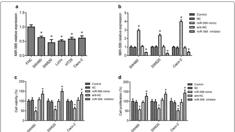
Prior research suggested that miR-566 is decreased in CRC [15]. In the in vitro cell assay, we measured miR-566 levels in the five CRC cell lines SW480, SW620, LoVo, Caco-2 and HT29 and the control colon epithelial cell lines FHC by RT-PCR assay. The results showed that miR-566 was significantly lower in SW480, SW620, LoVo, Caco-2 and HT29 cells, when compared to the control FHC cells (Fig. 1a, p < 0.05). To confirm the function of miR-566 in CRC cells, loss-of-function and gain-of-func-tion experiments were conducted for miR-566 in SW480, SW620 and Caco-2 cells through transfecting cells with miR-566 mimic and inhibitor, respectively. As shown in Fig. 1b, the miR-566 mimic markedly increased the level of miR-566 in SW480, SW620 and Caco-2 cells, whereas the miR-566 inhibitor decreased the levels of miR-566 in of the three cell lines (p < 0.05). Further study indicated
that miR-566 overexpression inhibited cell survival and proliferation in SW480, SW620 and Caco-2 cells com-pared to the control group (p < 0.05), whereas inhibition of miR-566 expression by using miR-566 inhibitor pro-moted survival and proliferation of these cells (Fig. 1c, d, p < 0.05).
MiR‑566 overexpression inhibits CRC cell migration and invasion
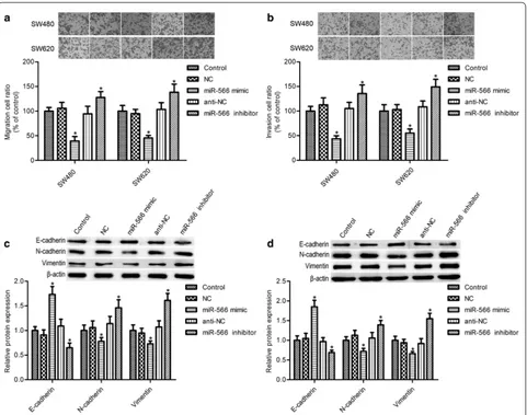
As shown in Fig. 2a, b, miR-566 mimic transfection markedly suppressed SW480 and SW620 cell migration and invasion (p < 0.05), while miR-566 inhibition signifi-cantly increased them (p < 0.05). Epithelial–mesenchymal transition (EMT) plays important roles in cell migration and invasion. We found that miR-566 overexpression
markedly elevated the epithelial marker E-cadherin expression and impeded the mesenchymal markers vimentin and N-cadherin levels, whereas its inhibition had opposite effects on E-cadherin, vimentin, and N-cad-herin expression (Fig. 2c, d, p < 0.05).
PSKH1 is a target of miR‑566
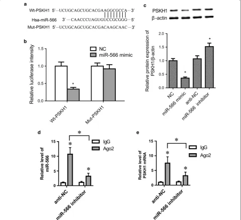
Computational analysis suggested putative binding sites between miR-566 and 3ʹ-UTR of human PSKH1, as shown in Fig. 3a. To confirm whether PSKH1 is a target of miR-566, we used luciferase reporter assays. Compared to the NC group, the relative luciferase activity was significantly decreased in miR-566 mimic and wild-type PSKH1 3ʹ-UTR (Wt-PSKH1) co-trans-fected cells (p < 0.05), while no significant differences
were found between the miR-566 mimic and mutant PSKH1 3ʹ-UTR (Mut-PSKH1) co-transfected group and NC group (Fig. 3b, p > 0.05). Western blot assay further showed that PSKH1 expression was inhib-ited by miR-566 overexpression (Fig. 3c, p < 0.05). RNA immunoprecipitation assay was performed using anti-Ago2 in SW480 cells transfected with anti-NC or
Reintroduction of PSKH1 reversed the effects of miR‑566 on CRC cell migration and invasion
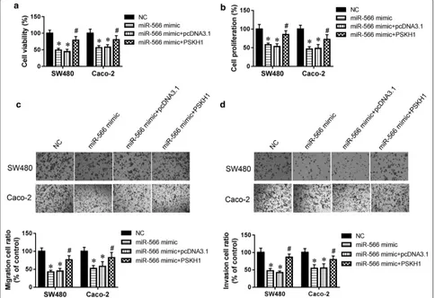
To further validate PSKH1 as a mediator of the effects of miR-566 on CRC growth, migration, and invasion, SW480 cells were co-transfected with miR-566 mimic and pCMV-PSKH1 vector for 48 h. The results sug-gested that the PSKH1 overexpression vector reversed the miR-566-mediated inhibition of SW480 cell survival (Fig. 4a). Furthermore, the PSKH1 overexpression vector increased SW480 cell proliferation, promote cell migra-tion and invasion compared with the miR-566 overex-pression group (Fig. 4b–d). It was reported that both SW480 and SW620 cell lines have TP53 and KRAS muta-tions, but the Caco-2 cells has only one mutation in TP53 [22]. So, Caco-2 cells were also employed in our study. The data from Caco-2 cells were in accordance with SW480 cells (Fig. 4a–d). Above results indicate that the effects of miR-566 in CRC cell growth, migration, and invasion depend on PSKH1 inhibition.
Discussion
Colorectal cancer is ranks the third diagnosed among cancer types worldwide, and it is the main cause of mor-tality from cancer [23]. Cancer metastasis plays roles in the advance of cancer, and in this study, we first con-firmed that miR-566 is involved in colon cancer metasta-sis through targeting PSKH1.
MiRNAs are single-stranded, 20- to 23-nucleotide-long, noncoding RNAs that are involved in the regula-tion of carcinogenic pathways [24, 25]. Compared with mRNAs, miRNAs are less likely to be degraded by Ranse because of their stem-loop and small-size structure [26]; thus, this conserved molecule is easy to detect in tissue and body fluids, including saliva, urine and blood [27]. miRNAs have been well-documented to act as tumor biomarkers, and they have been shown to be associated with the development of cancer, including colorectal cancer [28, 29]. Prior reports indicated that miRNAs are potential tumor suppressor or oncogenes by regulating
Fig. 4 PSKH1 was involved in the effect of miR-566 on CRC cell migration and invasion. a CellTiter 96 AQueous One Solution Cell Proliferation kit was used to determine survival of SW480 and Caco-2 cells. b Proliferation of SW480 and Caco-2 cells was determined by MTT assay. c Representative images and quantitative results for migration assay of SW480 and Caco-2 cells by transwell. d Representative images and quantitative results for invasion assay of SW480 and Caco-2 by transwell. All experiments were performed in triplicate. *p < 0.05 vs. NC, #p < 0.05 vs.
many cell processes including the cell cycle, proliferation, apoptosis, and invasion. Pan et al. [19] reported that the oncogene miR-566 is a potential biomarker of renal cell carcinoma prognosis.
MiR-566 was aberrantly expressed in many tumor types, such as lung adenocarcinoma [30], glioblastoma [31, 32], and colon cancer [15]. Consistent with these previous reports, our study demonstrated that miR-566 had a low expression level in several CRC cell lines (SW480, SW620, LoVo, Caco-2 and HT29) compared to the control colon epithelial cell line (FHC). In addition, Xiao et al. [32] previously found that miR-566 inhibition impeded glioblastoma migratory and invasive abilities through decreased VEGF expression and increased VHL expression. We also observed that miR-566 overexpres-sion suppressed survival, proliferation, migration, and invasion in SW480 and SW620 cells.
Protein kinases play important roles in many cellular processes, such as cell cycle progression, apoptosis, cell movement, metabolism, cytoskeletal rearrangement, transcription, and cell differentiation [33, 34]. PSKH1 is an autophosphorylating human protein serine kinase with 424 amino acids that has been suggested to play structural and regulatory roles in cells and to be involved in the maintenance of the Golgi apparatus [35]. Kim et al. [16] suggested that PSKH1 is highly expressed in patients with colon cancer. To search the specific potential tar-get gene for miR-566, an online computational algo-rithm TargetScan (http://www.targe tscan .org) was used. PSKH1 was a potential target gene for miR-566, and potential binding sites were found in its 3ʹ-UTR region. Our study first demonstrated that PSKH1 is a direct tar-get of miR-566, and its expression was influenced by the level of miR-566.
Conclusion
In conclusion, the current study demonstrated that miR-566 is decreased in colon cancer cells, and overexpression of miR-566 inhibited cancer cell growth and metastasis. In addition, we found that PSKH1 is a target of miR-566, and reintroduction of PSKH1 expression reversed the effects of miR-566 on cancer cell growth and metastasis.
Authors’ contributions
YZ and RX conceived of the study. SZ contributed the materials used in this study. YZ, SZ and JY performed research and analyzed data. YZ and RX wrote the paper. All authors read and approved the final manuscript.
Funding
None.
Availability of data and materials
All data generated or analyzed during this study are included in this published article.
Ethics approval and consent to participate
Not applicable.
Consent for publication
Not applicable.
Competing interests
The authors declare that they have no competing interests.
Author details
1 Endoscopy Center, China-Japan Union Hospital of Jilin University, No. 126
Sendai Street, Changchun 130033, Jilin, China. 2 Department of Nephrology,
China-Japan Union Hospital of Jilin University, Changchun 130033, Jilin, China.
3 Department of Vascular Surgery, China-Japan Union Hospital of Jilin
Univer-sity, Changchun 130033, Jilin, China.
Received: 6 September 2019 Accepted: 30 November 2019
References
1. Fitzmaurice C, Dicker D, Pain A, Hamavid H, Moradi-Lakeh M, MacIntyre M, Allen C, Hansen G, Woodbrook R, Wolfe C, et al. The global burden of cancer 2013. JAMA Oncol. 2015;1(4):505–27.
2. Fitzmaurice C, Allen C, Barber R, Barregard L, Bhutta Z, Brenner H, Dicker D, Chimed-Orchir O, Dandona R, Dandona L, et al. Global, regional, and national cancer incidence, mortality, years of life lost, years lived with disability, and disability-adjusted life-years for 32 cancer groups, 1990 to 2015: a systematic analysis for the global burden of disease study. JAMA Oncol. 2017;3(4):524–48.
3. Li Z, Li N, Wu M, Li X, Luo Z, Wang X. Expression of miR-126 suppresses migration and invasion of colon cancer cells by targeting CXCR4. Mol Cell Biochem. 2013;381(1–2):233–42.
4. Kudo-Saito C, Shirako H, Takeuchi T, Kawakami Y. Cancer metastasis is accelerated through immunosuppression during Snail-induced EMT of cancer cells. Cancer Cell. 2009;15(3):195–206.
5. Soreide K, Berg M, Skudal B, Nedreboe B. Advances in the understanding and treatment of colorectal cancer. Discov Med. 2011;12(66):393–404. 6. Wang J, Wang X, Liu F, Fu Y. microRNA-335 inhibits colorectal cancer
HCT116 cells growth and epithelial–mesenchymal transition (EMT) process by targeting Twist1. Pharmazie. 2017;72(8):475–81.
7. Yao G, Zhang Y, Chen P, Ren X. MicroRNA-544 promotes colorectal cancer progression by targeting forkhead box O1. Oncol Lett. 2018;15(1):991–7. 8. Cellura D, Pickard K, Quaratino S, Parker H, Strefford J, Thomas G, Mitter R,
Mirnezami A, Peake N. miR-19-mediated inhibition of transglutaminase-2 leads to enhanced invasion and metastasis in colorectal cancer. Mol Cancer Res. 2015;13(7):1095–105.
9. Hu W, Coller J. What comes first: translational repression or mRNA degradation? The deepening mystery of microRNA function. Cell Res. 2012;22(9):1322–4.
10. Wu N, Fesler A, Liu H, Ju J. Development of novel miR-129 mimics with enhanced efficacy to eliminate chemoresistant colon cancer stem cells. Oncotarget. 2018;9(10):8887–97.
11. Li J, Mao X, Wang X, Miao G. miR-433 reduces cell viability and promotes cell apoptosis by regulating MACC1 in colorectal cancer. Oncol Lett. 2017;13(1):81–8.
12. Qin Y, Huo Z, Song X, Chen X, Tian X, Wang X. mir-106a regulates cell proliferation and apoptosis of colon cancer cells through targeting the PTEN/PI3K/AKT signaling pathway. Oncol Lett. 2018;15(3):3197–201. 13. Chen Z, Han S, Huang W, Wu J, Liu Y, Cai S, He Y, Wu S, Song W.
Micro-RNA-215 suppresses cell proliferation, migration and invasion of colon cancer by repressing Yin-Yang 1. Biochem Biophys Res Commun. 2016;479(3):482–8.
14. Sun J, Zhou J, Dong M, Sheng W. Dysregulation of MicroRNA-543 expres-sion in colorectal cancer promotes tumor migration and invaexpres-sion. Mol Carcinog. 2017;56(1):250–7.
•fast, convenient online submission •
thorough peer review by experienced researchers in your field • rapid publication on acceptance
• support for research data, including large and complex data types •
gold Open Access which fosters wider collaboration and increased citations maximum visibility for your research: over 100M website views per year •
At BMC, research is always in progress.
Learn more biomedcentral.com/submissions
Ready to submit your research? Choose BMC and benefit from: 16. Kim S, Ahn T, Lee E, Do I, Lee S, Park S, Park J, Park Y, Lim H, Kang W, et al.
Exploratory biomarker analysis for treatment response in KRAS wild type metastatic colorectal cancer patients who received cetuximab plus irinotecan. BMC Cancer. 2015;15:747.
17. Brede G, Solheim J, Tröen G, Prydz H. Characterization of PSKH1, a novel human protein serine kinase with centrosomal, golgi, and nuclear locali-zation. Genomics. 2000;70(1):82–92.
18. Brede G, Solheim J, Prydz H. PSKH1, a novel splice factor compartment-associated serine kinase. Nucleic Acids Res. 2002;30(23):5301–9. 19. Pan X, Quan J, Li Z, Zhao L, Zhou L, Jinling X, Weijie X, Guan X, Li H,
Yang S, et al. miR-566 functions as an oncogene and a potential biomarker for prognosis in renal cell carcinoma. Biomed Pharmacother. 2018;102:718–27.
20. Zhou W, Li X, Liu F, Xiao Z, He M, Shen S, Liu S. MiR-135a promotes growth and invasion of colorectal cancer via metastasis suppressor 1 in vitro. Acta Biochim Biophys Sin. 2012;44(10):838–46.
21. Wang B, Sun F, Dong N, Sun Z, Diao Y, Zheng C, Sun J, Yang Y, Jiang D. MicroRNA-7 directly targets insulin-like growth factor 1 receptor to inhibit cellular growth and glucose metabolism in gliomas. Diagn Pathol. 2014;9:211.
22. Berg KCG, Eide PW, Eilertsen IA, Johannessen B, Bruun J, Danielsen SA, Bjornslett M, Meza-Zepeda LA, Eknaes M, Lind GE, et al. Multi-omics of 34 colorectal cancer cell lines—a resource for biomedical studies. Mol Cancer. 2017;16(1):116.
23. Marikar F, Jin G, Sheng W, Ma D, Hua Z. Metallothionein 2A an interactive protein linking phosphorylated FADD to NF-κB pathway leads to colorec-tal cancer formation. Chin Clin Oncol. 2016;5(6):76.
24. Catto J, Alcaraz A, Bjartell A, De Vere White R, Evans C, Fussel S, Hamdy F, Kallioniemi O, Mengual L, Schlomm T, et al. MicroRNA in prostate, blad-der, and kidney cancer: a systematic review. Eur Urol. 2011;59(5):671–81. 25. Yang J, Ma D, Fesler A, Zhai H, Leamniramit A, Li W, Wu S, Ju J.
Expres-sion analysis of microRNA as prognostic biomarkers in colorectal cancer. Oncotarget. 2017;8(32):52403–12.
26. Manne U, Jadhav T, Putcha B, Samuel T, Soni S, Shanmugam C, Suswam E. Molecular biomarkers of colorectal cancer and cancer disparities: current status and perspective. Curr Colorectal Cancer Rep. 2016;12(6):332–44.
27. Tutar L, Tutar E, Tutar Y. MicroRNAs and cancer; an overview. Curr Pharm Biotechnol. 2014;15(5):430–7.
28. Tan W, Liu B, Qu S, Liang G, Luo W, Gong C. MicroRNAs and cancer: key paradigms in molecular therapy. Oncol Lett. 2018;15(3):2735–42. 29. Bhome R, Goh R, Bullock M, Pillar N, Thirdborough S, Mellone M,
Mirn-ezami R, Galea D, Veselkov K, Gu Q, et al. Exosomal microRNAs derived from colorectal cancer-associated fibroblasts: role in driving cancer progression. Aging. 2017;9(12):2666–94.
30. Rani S, Gately K, Crown J, O’Byrne K, O’Driscoll L. Global analysis of serum microRNAs as potential biomarkers for lung adenocarcinoma. Cancer Biol Ther. 2013;14(12):1104–12.
31. Zhang K, Zhou X, Han L, Chen L, Chen L, Shi Z, Yang M, Ren Y, Yang J, Frank T, et al. MicroRNA-566 activates EGFR signaling and its inhibition sensitizes glioblastoma cells to nimotuzumab. Mol Cancer. 2014;13:63. 32. Xiao B, Zhou X, Ye M, Lv S, Wu M, Liao C, Han L, Kang C, Zhu X.
Micro-RNA-566 modulates vascular endothelial growth factor by targeting Von Hippel-Landau in human glioblastoma in vitro and in vivo. Mol Med Rep. 2016;13(1):379–85.
33. Manning G, Whyte D, Martinez R, Hunter T, Sudarsanam S. The protein kinase complement of the human genome. Science. 2002;298(5600):1912–34.
34. Blume-Jensen P, Hunter T. Oncogenic kinase signalling. Nature. 2001;411(6835):355–65.
35. Brede G, Solheim J, Stang E, Prydz H. Mutants of the protein ser-ine kinase PSKH1 disassemble the Golgi apparatus. Exp Cell Res. 2003;291(2):299–312.
Publisher’s Note