PRIMARY EPITHLELIOID ANGIOSARCOMA OF THE MAXILLA
MASQUERADING AS A POORLY-DIFFERENTIATED CARCINOMA :
CASE REPORT
1 2 2 3 2
Mounesh kumar C D David P. Tauro Rajay Kamat Suresh K V Rajendra Desai
1
Department of Oral & Maxillofacial Surgery,School of Dental Sciences, Krishna Institute of Medical Sciences, 2
Maharashtra, India. Department of Oral & Maxillofacial Surgery, College of Dental Sciences, Davangere, 3
Karnataka, India. Department of Oral Medicine and Radiology, School of Dental Sciences, Krishna Institute of Medical Sciences, Maharashtra, India.
Corresponding author: Suresh K V, Department of Oral Medicine and Radiology, School of Dental Sciences, Krishna Institute of Medical Sciences, Deemed University, Karad, Satara, Maharashtra, India. Email: dr_suri88@yahoo.co.in / dr.suri88@gmail.com Ph- 09890130227
Abstract
Epithlelioid angiosarcoma (EA) is a rare high grade malignant neoplasm arising from vascular endothelium. Amongst epithelioid endothelial cell tumors, EA is rare to occur. It is characterized by atypical, multilayered or solid endothelial proliferation and vasoformative architecture is a major finding in EA. This tumor exhibits a great degree of nuclear pleomorphism and mitotic activity with areas of necrosis. A case report of Primary Epithlelioid Angiosarcoma of the Maxilla in a 13 year old male patient is being reported.
Key words: Angiosarcomas, Sarcoma, Primary Epitheliod Angiosarcomas
Introduction history of difficulty in breathing through the right nostril.
Angiosarcomas are rare malignant
Upon detailed extra oral examination, tumors of the vascular endothelium,
the child had a diffuse mid- facial swelling on characterized by the formation of irregular
the right side. Intraoral examination revealed vascular channels lined by atypical endothelial
an irregular but well-defined, sessile growth, 1
cells. They comprise only 2% of soft tissue The surface was smooth, with areas of sarcomas. These tumors arise within bone hemorrhage and ulceration, presumably
2
with an incidence rate of about 0.5%. In the resultant to incessant occlusal trauma (fig. 1). head and neck area, most of these lesions It measured 6cms x 7cms in dimension and affect the scalp and the facial soft tissues, and was confined to the buccal vestibule and only uncommonly, the oral cavity is the site of alveolar region of the maxilla, extending from
3, 4
primary location. Most of the intraoral the right permanent maxillary lateral incisor to tumors are located in the mandible and the right permanent maxillary first molar, with occurrence in the maxilla and the maxillary extension beyond the mid-palatal raphe.
5
sinus is rare. There was shift in the maxillary dental
mid-line with marked displacement of the
Case Report
st deciduous canine and second molar; The 1 A 13-year-old male patient, with a right premolar of the patient was embedded moderate built, yet poorly nourished, almost completely within the swelling.
reported to the department with a complaint
The inspection findings were of a swelling associated with pain in the right
confirmed by palpation. There was a severe front region of the upper jaw with inability to
tenderness with profuse bleeding on close the mouth since 20 days. He gave a
maxillary sinus was warm and tender. However, we failed to notice any infraorbital nerve paraesthesia or anesthesia.
A differential diagnosis of sarcoma, aggressive odontogenic tumor and sqamous cell carcinoma were made at the conclusion of examination. Conventional and advanced imaging investigations revealed a diffuse increase in attenuation of maxilla with evidence of osteolytic and destructive lesion. Severe expansion of maxilla and displacement of lateral incisor, canine and premolars were noticed. (Fig. 2, 3) Under local anesthesia, an incision biopsy was performed which concluded a poorly-differentiated carcinoma. Under general anesthesia a wide local resection (subtotal maxillectomy) of the tumor was done. (Fig. 4) The oncologic defect, however, was not reconstructed and was allowed to heal by secondary intention. (Fig. 5) Neck dissection was not contemplated at surgery, but instead a wait-and-watch policy was adopted. The patient's recovery was uneventful. Later, an obturator was fabricated to obturate the defect, facilitate speech and restore masticatory function. The resected specimen was subjected to histopathological examination and a final diagnosis of poorly-differentiated carcinoma was made. (Fig. 6)
Immunohistochemical analysis was performed on the tumor to study its immunoreactivity and various tumor markers were elaborated.
(Fig. 7,8) At this stage, a definitive diagnosis of epithelioid angiosarcoma was made. In view of the reported resistance of the tumor to radiotherapy, the patient was commenced on a course of post-operative chemotherapy consisting of three cycles each of Vincristine (2mg x3) and Methotrexate (50mg x3). The postoperative period was uneventful.
After 6 months of follow-up, no recurrence was seen.
The tumor cells show cytoplasmic positivity for vimentin and CD 31 with rare cells showing positivity for CD 34.
The patient was advised to visit regularly for examination.
Fig 1 – Intraoral view showing irregular sessile growth, with areas of hemorrhage and ulceration.
Fig 2 – Maxillary occlusal radiograph showing irregular radilucent area with teeth floating in air appearance
Fig 4 - Intraoperative Photograph
Fig 5 – postoperative photograph
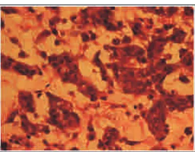
Fig 6 – H & E staining - Epithelioid cells show features like large, pleomorphic vesicular nuclei, many exhibiting nucleoli, and typical and atypical mitosis.
Fig 7 & 8 – IHC - Tumor cell shows cytoplasmic positivity for vimentin and CD 31 with rare cells showing positivity for CD 34
Discussion
Epithelioid angiosarcoma (EA) is a rare malignant tumor of the vascular endothelial cells. The classification of epithelioid endothelial cell tumors includes e p i t h e l i o i d h e m a n g i o m a , e p i t h e l i o i d hemangioendothelioma and epithelioid
6, 7, 8 angiosarcoma .
Most patients with maxillar y angiosarcoma have as short a history of 4 to 8 weeks to being asymptomatic as long as 3 years
4
similar symptoms with short duration of Histopathological diagnosis, particularly with
swelling. poorly differentiated angiosarcomas and the
epithelioid phenotypic variant of the tumor Many theories have been proposed to
may be confused with spindle cell sarcomas, explain the development of angiosarcomas.
carcinomas or a melanoma. Previously, tumor 6
Mc Carthy and Park hypothesized that these markers like factor VIII-related antigen and tumors arise from the capillaries of ulex europeus agglutinin lectin were used to granulation tissue in traumatized areas. The assist in immunohistochemical diagnosis. same authors have reported 3 patients whose However recently, the vascular and lymphatic previously benign angiomas underwent endothelial expression of another antigen, malignant transformation after radiotherapy. CD31, has been proven to be the most
Vinyl chloride has been implicated as a 11,12
sensitive endothelial marker . CD 31 yields possible etiologic factor in 30 patients with
strong labeling of the lesional cells with low angiosarcoma of the liver, as reported by
background immunoreactivity. Difficulties 7
Duck in 1975. Williamson and Ramsden, in with background staining have plagued the 1988, have also reported of a 48-year-old man use of lectin while factor VIII-related antigen who had a history of handling or being although specific, has low sensitivity.
exposed to the chemical for 6 years before an
As far as treatment is concerned, most angiosarcoma of the maxillary sinus
authors believe that surgery, in combination developed.
with radiotherapy and chemotherapy, offers
Clinically, oral lesions usually appear 8
the best chance of survival . Zachariades and as painless, sessile-based masses, soft and
Economopoulou, however, have advocated compressible to moderately fir m in
treatment consisting of wide surgical removal consistency and bleed spontaneously, similar
of the tumor regardless of the combination
to present case. The surface may be ulcerated 8
of irradiation . and an erythematous ring is often present
8, 9
around its periphery. EA, as a whole, have a strong
tendency to recur locally and to metastasize A variety of lesions may be
indicating their highly aggressive nature which considered in the differential diagnosis,
explains the low survival rates. McCarthy and including hemangioma, pyogenic granuloma,
Park reported a 3-year survival rate of 17% p a p i l l a r y e n d o t h e l i a l h y p e r p l a s i a ,
and a 5-year survival rate of 9% with an hemangiopericytoma, angiolymphoid
average survival time of 2½ years. The hyperplasia with eosinophilia, Kaposi's
survival rate for oral and jaw angiosarcomas, sarcoma, malignant melanoma, metastatic
however, is better than that for angiosarcomas disease (renal cell carcinoma) and
in general and, in particular, for those fibrosarcoma and histiocytic lymphoma in
involving the skin. Many investigations in 1, 9,10
some instances recent years have indicated that one-half of
the patients with angiosarcomas die within 15 The radiographic appearance of
months from the time of initial diagnosis, with angiosarcomas involving the facial bones is
approximately 12% surviving 5 years or generally that of a destructive osteolytic lesion
11, 12 with mild periosteal reaction sometimes longer. mimicking benign lesions such as a cyst or
Conclusion
odontogenic tumor. Jaw lesions may show
widening of the periodontal ligament and The frequency of Angiosarcomas in erosion of the alveolar bone producing a the maxillofacial region is exceedingly
4
sporadic. Since Angiosarcomas offer a grave “teeth floating in space” appearance.
genetics of these tumors is paramount to their better management in the future.
Reference
1. Bankaki M, Myers EN, Barnes L, DuBois P.
Angiosarcoma of the maxillary sinus: literature 9. Haustein UF. Angiosarcoma of the face and review and case report. Head Neck Surg 1979; scalp. Int J Dermato 1991; 30:851-6.
1:274-80. 10. Loudon J A, Billy ML, De Young BR, Allen
2. Enzinger FM,Weiss SW; Soft tissue tumors. CM. Angiosarcoma coma of the mandible: a St LouisMosby, 1983 p 430. case report and review of the literature Oral Surg Oral Med Oral Pathol Oral Radiol Endoc 2000;89:471-6.
11. Epitheloid angiosarcoma of maxillary sinus and maxilla; A case report and review of literature; Oral Surg,Oral Med Oral Pathol Oral Radiol Endo2002;94:333-337.
5. Angiosarcoma of maxilla and maxillary sinus; JOMS; 1989; 47; 747-753.
6. McCarthy WD, Pack GT. Malignant blood vessel tumors. A report of 56 cases of angiosarcoma and Kaposi's sarcoma. Surg Gynecol Obstetric 1950; 91:465-82.
7. Mark RJ, Poen JC, Tran LM, Fu YS, Juillard GF. Angiosarcoma. report of 67 patients
and a review of the 1996;
77:2400-6.
8. Zachariades N, Papadakou A, Koundouris J, Constandinidis J, Angelopoulos AP. Primary hemangioendotheliosarcoma of the mandible: review of the literature and report of a case. J Oral Surg 1980; 38:288-96.
3. Zachariades N, Economopoulou P. Maxillary angiosarcoma. Int 1 J Oral Maxillofac Surg 1986; 15:357-60.
4. Aust MR, Olsen KD, Lewis JE, Nascimento AG, Meland NB Foote RL, et al. Angiosarcomas of the head and neck: clinical
and pathologic characteristics. Ann Otol Rhino 12. Kempson RL, Fletcher CD, Evans HL, Laryngol 1997 106:943-51. Hendrickson MR, Sibley RK. Atlas of tumor pathology. Tumors of the soft tissues. Wash-ington, DC: Armed Forses Institute of Pathology; 2001. p. 353.
literature. Cancer A
Source of Support - Nil
Conflict of Interest - None declared
How to cite this article: Mounesh Kumar C. D., David P. Tauro, Rajay Kamat, Suresh K V, Rajendra Desai: Primary epithlelioid