Izabela Łaczmańska
A–G, Małgorzata Szczepaniak
B, C, F, G,
Aleksandra Jakubiak
B, C, F, Agnieszka Stembalska
A–FExonic Deletions in the
NF1
Gene
in Patients with Neurofibromatosis Type I
from the Lower Silesian Region of Poland
Department of Genetics, Wroclaw Medical University, PolandA – research concept and design; B – collection and/or assembly of data; C – data analysis and interpretation;
D – writing the article; E – critical revision of the article; F – final approval of article; G – other
Abstract
Background. Neurofibromatosis type I(NF1, Recklinghausen’s disease) is an autosomal dominant disorder char-acterized by the following clinical features: café au lait spots, neurofibromas, Lisch nodules, freckling of the axillary and inguinal regions, optic nerve gliomas, bone dysplasia and increased risk of certain tumors. NF1 is diagnosed on the basis of clinical criteria, while identifying the genetic background of the disease is important mainly for genetic counseling. NF1 genetic analysis is based on searching for NF1 exon deletions/duplications using Multiplex ligation-dependent probe amplification (MLPA), searching for microdeletions of the critical region using fluorescence in situ hybridization (FISH), searching for point mutations by gene sequencing (in most cases) and analyzing mRNA.
Objectives. The aim of this study was to estimate the frequency of single and multi-exon deletions/duplications in the NF1 gene in Polish patients, and to evaluate the usefulness of MLPA as a cheap and easy method for NF1 molecular diagnosis, despite the fact that such changes may be found in only a small group of NF1 patients.
Material and Methods. The study included 65 patients suspected of NF1 or with recognized NF1 on the basis of clinical criteria. Cytogenetic analysis were carried out for all the patients, and for one patient with a translocation [46,XY,t(17;22)(q11.2;q11.2)], a FISH analysis was performed. All patients were tested for deletions/duplications in the NF1 gene using two MLPA kits for neurofibromatosis I.
Results. The MLPA analysis showed deletions in the NF1 gene in 7.7% of the cases (5/65).
Conclusions. The results indicate that an MLPA analysis may be performed in patients with a clinical diagnosis of NF1 or patients with suspected NF1 as an easy and inexpensive first molecular test, enabling the exclusion of about 7% of NF1 patients from expensive and time-consuming molecular diagnosis by DNA sequencing (Adv Clin Exp Med 2014, 23, 4, 517–521).
Key words: NF1,Recklinghausen’s disease, MLPA.
Adv Clin Exp Med 2014, 23, 4, 517–521 ISSN 1899–5276
ORIGINAL PAPERS
© Copyright by Wroclaw Medical University
Neurofibromatosis type I(NF1), also known as Recklinghausen’s disease, is one of the most com-mon phakomatoses (neurocutaneous syndromes). The term phakomatoses refers to a large group of independent clinical multi-system disorders. In these syndromes, lesions affecting the central ner-vous system and the skin are dominant. Moreover, neurocutaneous disorders are associated with a predisposition to malignancies [1, 2].
The frequency of NF1 is about 1 in 3000– –4000 births worldwide. NF1 is an autosomal dominant disorder, which means there is a 50%
risk of inheriting the disease from an affected parent [2].
cardiovascular diseases [2–4]. Symptoms of NF1 differ from patient to patient even within the same family. Moreover, the features change with the age of the patient. Clinical diagnosis of young children may be difficult, as characteristic NF1 features rare-ly occur in children under 4–5 years of age [2, 4].
A diagnosis of NF1 is based on the presence of two clinical criteria from the following list, for-mulated by Griffiths et al. [2], Ferner [3] and Jett & Friedman [4]:
– six or more café au lait spots > 15 mm in adults; five or more > 5 mm in children,
– two or more neurofibromas or one plexi-form neurofibroma,
– axillary or inguinal freckling, – at least two Lisch nodules, – optic nerve glioma,
– a first-degree relative with NF1,
– a distinctive osseous lesion such as sphenoid wing dysplasia or thinning of the long bone cortex with or without pseudoarthrosis.
As NF1 patients may be at risk for, or pres-ent with, a variety of severe clinical symptoms, a precise schedule of surveillance should be of-fered to them, including ophthalmologic, derma-tologic, neurologic and orthopedic consultations. Moreover, resonance imaging, computer tomog-raphy, ultrasonography and X-rays should be em-ployed to screen for tumors (e.g. in the spinal cord or brain) and skeletal changes [4].
In 90% of cases NF1 is caused by point mu-tations in the NF1 gene, located at 17q11.2; in 5% of cases it results from deletion/duplication (copy number variants or CNVs); in less than 1% of cas-es chromosomal aberrations involving 17q11.2 are observed. Genetic diagnosis of NF1 is difficult be-cause of the size of the NF1 gene and the lack of hotspot point mutations [5, 6]. So far over 1000 dif-ferent NF1 point mutations have been found. About 90% of them can be detected using DNA sequenc-ing. To identify NF1 deletion/duplication (CNVs) the method of choice is multiplex ligation-depen-dent probe amplification (MLPA) [7, 8]. To detect chromosomal aberrations and/or NF1 microdele-tion, chromosomal banding and/or fluorescence in situ hybridization (FISH) are used. Other pathogen-ic changes, like splpathogen-icing defects, can be detected by analysis of mRNA variants using reverse transcrip-tion polymerase chain reactranscrip-tion (RT-PCR), denatur-ing high pressure liquid chromatography (DHPLC) and sequencing of selected fragments. All the listed tests have a total sensitivity of about 95% [4, 6].
The aim of the present study was to estimate the frequency of NF1 gene single and multi-exon deletions and duplications in patients from Low-er Silesia, Poland, and the usefulness of the MLPA test as a convenient first-line screen.
Material and Methods
Patients
The study included 65 Caucasian patients (41 fe-males and 24 fe-males) from the Lower Silesia region of Poland, with suspected or clinically diagnosed NF1, who had been referred to a genetic counselor. The age of the patients ranged from 4 weeks to 60 years (mean age 17.28 years). Most of the cases (46 out of 65) were isolated, unrelated to each other. In 19 other cases, 2 or 3 members of a single family were exam-ined. In all, nine family cases were observed.
All the patients were examined ophthalmo-logically. MRI tests were performed on about two-thirds of the patients.
Out of the study population 40 patients fulfilled the clinical criteria for NF1. Among these patients, only 25 had a family history of NF1. The age of the patients with two clinical criteria ranged from 12 months to 60 years (mean age 22.29 years). The age of the patients with one NF1 criterion ranged from 4 weeks to 49 years (mean age 9.25 years). The clinical data are presented in Table 1.
In 25 patients only one NF1 criterion was ob-served. In most of them café au lait spots were present. Some patients displayed additional features that are not diagnostic criteria but which occur in NF1, such as scoliosis (3 cases), short stature (4 cases), addition-al brain MR abnormaddition-alities such as hamartoma or de-myelinating changes (3 cases), developmental delay/ /mental retardation (11 cases), epilepsy (4 cases) and nonspecific headaches (3 cases). In individu-al cases macrocephindividu-aly, a congenitindividu-al heart defect, hy-dronephrosis, psoriasis and immune disorder were observed.
Methods
Cytogenetic analyses were carried out for all patients according to standard proce-dures [9, 10]. For one patient with translocation 46,XY,t(17;22)(q11.2;q11.2) a FISH analysis was performed using a KBI-40114 MD NF1 (17q11) / MPO (17q22) probe (Kreatech) according to the manufacturer’s instructions.
MLPA
The PCR products were separated using an ABI 310 Genetic Analyzer with GeneScan Analysis software (version 3.1.2) with POP-4 Polymer and LIZ 500 size standard (Applied Biosystems). The analysis of the results was performed using Gene-Marker software (version 1.85, SoftGenetics LLC). The MLPA ratio was used as a validation method. A synthetic control probe as a control.
A change in peak values over ±0.3 was consid-ered a duplication (an increase in value) or a dele-tion (a decrease in value).
Samples with deletions or duplications were analyzed by MLPA in 2 separate experiments us-ing the same MLPA kits (there is no alternative MLPA probe mix for analyzing exonic deletions of the NF1 gene), and only confirmed results were re-garded as reliable.
Results
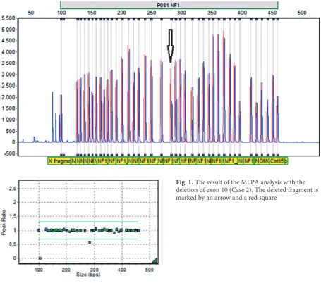
Exonic deletions of the NF1 gene were observed in five cases (7.7%) (Table 2, Fig. 1). There were four single exonic deletions and one multiexonic
deletion covering exons from 1 to 12. Three out of the five patients with deletions (positive ML-PA tests) fulfilled the criteria for a clinical diagno-sis of NF1: In patient number 2 café au lait spots and cutaneous neurofibromas were found; patient number 5 had café au lait spots, cutaneous neuro-fibromas and a plexiform neurofibroma; and pa-tient number 46 had café au lait spots, cutaneous neurofibromas, optic nerve glioma, axillary freck-ling and a first-degree relative with NF1. In the re-maining two cases (patients number 17 and 55, a 10-year-old boy with dysmorphic features and intellectual disability and a 6-month-old girl) only café au lait spots were observed.
In one patient a translocation [46,XY,t(17;22) (q11.2;q11.2)] was found, but FISH and MLPA analyses showed no deletions of the whole NF1
gene or of parts of it. This patient had only café au lait spots.
All in all, genetic changes were found in 7.5% of the patients fulfilling the clinical criteria for NF1 (three cases out of 40 patients) and in 12% of pa-tients with one criterion of NF1 (three cases out of 25 patients).
Table 1. Clinical features (according to clinical criteria) of 65 patients suspected of NF1
Clinical features Total number
of patients Case number
Café au lait spots 63 1, 2, 4–52, 54–65 Cutaneous neurofibromas or plexiform
neuro-fibroma 28 1, 2, 5, 7–10, 16, 21, 31, 34, 35, 38, 39, 41, 46, 47, 48, 50–54, 56, 57, 61, 62, 64 Axillary or inguinal freckling 16 8, 14, 16, 21, 23, 25, 34, 35, 46, 50, 51, 53, 57, 58, 61, 64
Lisch nodules 3 48, 62, 63
Optic nerve glioma 4 13, 46, 58, 60
A first-degree relative with NF1 25 1, 3, 4, 6–10, 20, 21, 30, 31, 34–38, 46–48, 50, 54, 62–64
A distinctive osseous lesion (sphenoid wing dysplasia, thinning of the long bone cortex with or without pseudoarthrosis)
1 42
Table 2. Patients with lesions detected using MLPA
Case Fulfilled clinical criteria for NF1
(at least two criteria required) Family history MLPA result
Case 2 + – deletion of exon 10
Case 5 + – deletion of exon 33
Case 17 – – deletion of exons 1–12
Case 46 + + deletion of exon 28*
Case 55 – – deletion of exon 16
Discussion
In the present study 65 patients meeting at least one criterion for NF1 were tested. In 7.7% of the cases, exonic deletions in the NF1 gene were identified. In four cases (patients number 2, 5, 46 and 55) a single exonic deletion was observed (ex-ons 10, 16, 28, 33), and in one case (patient num-ber 17) a multiexon deletion (exons from 1 to 12) was found. The size of the deletion can affect the additional presence of abnormalities such as facial dysmorphism and mental retardation, both seen in patient number 17. It is believed that by the age of eight years, almost all children with NF1 meet the clinical criteria for diagnosis [11]. In patient 17 it cannot be excluded that other symptoms will oc-cur with age, or that the patient may present tissue mosaicism or incomplete penetrance (diverse ex-pression of NF1).
In one patient with a reciprocal transloca-tion [46, XY, t(17;22)(q11.2;q11.2)] with the break point within the region containing the NF1 gene, no changes in this gene were found in either the
FISH or the MLPA analysis. Presently the patient has only café au lait spots, and further observation as well as NF1 gene sequencing is necessary. The examination using FISH and MLPA techniques does not exclude very small deletions or NF1 gene point mutations.
Neurofibromatosis type I is characterized by intrafamilial and interfamilial clinical variability. A clinical diagnosis of NF1 is unequivocal in most patients, but not in very young children. Some fea-tures of NF1 emerge as the patients age, so further prophylactic examinations are indicated in patient number 55, in whom only café au lait spots were observed [2, 4]. In a study published by de Lu-ca et al., 201 unrelated patients were tested using MLPA. In 63 of them, point mutations were ex-cluded using DHPLC analyses [7]. All of them ful-filled the clinical criteria for NF1, except for three children who presented only café au lait spots. In 36.5% (23 cases, including two children not fully matching the clinical criteria for NF1), deletions were found. When analyzing the whole group of 201 patients, whole or partial gene deletions were
detected in about 7%. This is a higher percent-age than presented by Wimmer et al., who studied a cohort of 1100 patients fulfilling NF1 criteria and found that only 2% of them had single- and/or mul-tiexon deletion/duplication [8]. However, Griffiths et al. found whole or partial gene deletions in 5.9% of the NF1 cases they studied [2]. These results are consistent with those in the present study (7.7%). Moreover, as in other studies, the exonic deletions detected in the present study were unique. In all the cases where alterations in MLPA were found, genetic counseling was performed, including test-ing other members of the family and offertest-ing the family the option of prenatal diagnosis.
The current study did not identify any mosaic deletions in NF1 genes. Mosaicism may affect ge-netic counseling because a change that is present in
only some of cells may cause a milder phenotype and may reduce the risk of transmission to off-spring. The normal range in the MLPA method is from 0.7 to 1.3, which means that a 30% difference between the patient and a control probe is treated as a normal result. Therefore the presence of mo-saicism might be undetected by this test [8,12].
Confirmatory diagnostic testing is indicat-ed both in patients who do not fulfill the diagnos-tic criteria of NF1 and in patients who do fulfill them. Although the MLPA method detects only copy-number variations (CNVs), it is a relatively cheap and easy technique. Therefore, despite the fact that NF1 results from deletion/duplication in fewer than 10% of the patients, in the current au-thors’ opinion, MLPA analysis is worthwhile as a first step in detecting NF1 alterations.
Acknowledgements. The authors are grateful to Prof. Maria M. Sąsiadek for her support in preparing the manuscript and to Prof. Nicolaus Blin for proofreading.
References
Dahan D, Fenichel GM, El-Said R:
[1] Neurocutaneous syndromes. Adolesc Med 2002, 13, 495–509.
Griffiths S, Thompson P, Frayling I, Upadhyaya M:
[2] Molecular diagnosis of neurofibromatosis type 1: 2 years experience. Fam Cancer 2007, 6, 21–34.
Ferner RE:
[3] Neurofibromatosis 1. EJHG 2007, 15, 131–138.
Jett K, Friedman JM:
[4] Clinical and genetic aspects of neurofibromatosis 1. GeneTest Rev 2010, 1, 1–11.
Trovó-Marqui AB, Tajara EH:
[5] Neurofibromin: a general outlook Clin Genet 2006, 70, 1–13.
Valero MC, Martín Y, Hernández-Imaz E, Marina Hernández A, Meleán G, Valero AM, Javier Rodríguez- [6]
-Álvarez F, Tellería D, Hernández-Chico C: A highly sensitive genetic protocol to detect NF1 mutations. J Mol Diagn 2011, 13, 113–122.
De Luca A, Bottillo I, Dasdia MC, Morella A, Lanari V, Bernardini L, Divona L, Giustini S, Sinibaldi L, [7]
Novelli A, Torrente I, Schirinzi A, Dallapiccola B: Deletions of NF1 gene and exons detected by multiplex liga-tion-dependent probe amplification. J Med Genet 2007, 44, 800–808.
Wimmer K, Yao S, Claes K, Kehrer-Sawatzki H, Tinschert S, De Raedt T, Legius E, Callens T, Beiglböck H, [8]
Maertens O, Messiaen L: Spectrum of single- and multiexon NF1 copy number changes in a cohort of 1,100 unse-lected NF1 patients. Genes Chromosomes Cancer 2006, 45, 265–276.
Gustashaw KM:
[9] Chromosome stains In: The AGT Cytogenetics Laboratory Manual. Barch MJ, Knusten T, Spurbeck J. Lippincot-Raven Publishers, Philadelphia 1997, 259–324.
Shaffer LG, Slovak ML, Campbell LJ:
[10] ISCN 2009, An International System for Human Cytogenetic Nomenclature 2009. Karger, Basel 2009.
DeBella K, Szudek J, Friedman JM:
[11] Use of the national institutes of health criteria for diagnosis of neurofibroma-tosis 1 in children. Pediatrics 2000, 105, 608–614.
MRC Holland
[12] www.mrc-holland.com
Address for correspondence:
Izabela Łaczmańska Department of Genetics Wrocław Medical University Marcinkowskiego 1
50-368 Wroclaw Poland
Tel: +48 71 784 1258
E-mail: [email protected]
Conflict of interest: None declared