K
RZYSZTOFS
KIBA1, R
OMANR
UTOWSKI1, 2, K
RZYSZTOFD
UDEK3, P
AWEŁR
EICHERT1,
R
OMANW
IĄCEK1, K
RZYSZTOFB
OGDAN1Late Estimation of Sensibility Loss
after Sural Nerve Autografting
Odległa ocena utraty czucia po pobraniu
nerwu łydkowego
1Department of Traumatology and Hand Surgery, Silesian Piasts University of Medicine in Wrocław, Poland
2 Chair of Sport Medicine of Wrocław University, Poland
3Institute of Machine Design and Operation, Wrocław University of Technology, Poland
Adv Clin Exp Med 2007, 16, 2, 229–237 ISSN 1230−025X
ORIGINAL PAPERS
© Copyright by Silesian Piasts University of Medicine in Wrocław
Abstract
Background. Surgery requiring complementing peripheral nerve injury with a graft of another nerve, usually the
sural nerve, has been performed at the Department of Traumatology and Hand Surgery, Silesian Piasts University of Medicine in Wrocław, since 1981. In the vast majority of cases the use of autografts of the sural nerve involved traumatic injuries of the brachial plexus or nerves of the upper and lower extremity. The area of sensibility loss, which decreases in the course of time thanks to other nerves taking over sensibility functions, is a consequence of sural nerve removal. On the basis of examinations, the authors assessed the degree of sensibility restoration in the sural nerve’s innervation area in the extremity.
Objectives. Learning about the mechanisms of peripheral compensation of sensibility through quantitative and
qualitative assessment of sensibility restoration in the innervation area of the sural nerve lost as a result of its removal in the reconstruction of brachial plexus and peripheral nerves.
Material and Methods. Fifty−two patients were examined in selected age groups operated on at the department
because of traumatic injuries to peripheral nerves. Examination of two−point, soft, and vibration sensibility were performed. Additionally, the patients were tested for subjective assessment of sensibility restoration in the area of innervation by the sural nerve.
Results. On the basis of the performed quantitative examination and patients’ subjective estimation of sensibility
restoration, statistically significant correlations between sensibility restoration in the examined area and the time from surgery to the examination, patient age, or the length of the sural nerve graft were not observed. Much quick−
er sensibility restoration was observed in the areas of the 2ndand 4thinnervation by the sural nerve, i.e. the skin of
the heel from the medial side and of the fifth toe.
Conclusions. In assessing sensibility restoration after taking sural nerve grafts for the reconstruction of nerves
(peripheral and of the brachial plexus), examination of soft sensibility with the help of Semmes−Weinstein fila− ments, two−point sensibility, and vibration sensibility play an essential role. The results of the above examinations showed very good and good results of sensibility restoration in 75% patients, while only 2% had unsatisfactory
results (Adv Clin Exp Med 2007, 16, 2, 229–237).
Key words: sural nerve, microsurgical reconstruction of nerves (peripheral and of brachial plexus), soft sensibili−
ty, vibration sensibility.
Streszczenie
Wprowadzenie. W Klinice Chirurgii Urazowej i Chirurgii Ręki Akademii Medycznej we Wrocławiu od 1981 roku
Reconstructive surgery requiring comple− menting peripheral nerve loss with a graft of an autogenic nerve, usually the sural nerve, has been performed at the Department of Traumatology and Hand Surgery, Silesian Piasts University of Medicine in Wrocław, since 1981. In the place innervated by a nerve there is consequently an area of sensibility loss. This area should decrease in the course of time thanks to surrounding nerves taking over sensibility functions. However, this process is not always favorable and there are sometimes ail− ments in the form of painful neuroma in the stump of the sural nerve or an area of loss of sensibility or, also, an area of hyperaesthesia.
The purpose of this study was to assess qualita− tive and quantitative sensibility restoration in the area innervated by the sural nerve lost as a result of its removal in a group of patients, performed on the basis of a qualitative examination of pressure sensi− bility with the Semmes−Weinstein Monofilaments aestesiometer examination of discrimination of two−point sensibility and vibration sensibility.
In the majority of cases (66%) the use of auto− genic grafts of sural nerve involved the recon− struction of traumatic injuries to the brachial plexus or the upper extremity, and, to a lesser degree (15%), isolated injuries to other peripheral nerves. The sural nerve is the most frequently applied as an autogenic sensory nerve graft in the reconstruction of peripheral nerves and those of the brachial plexus because of its length, thick− ness, easy operational harvesting (Figs. 1, 2), and the slight decrease in sensibility in the area of the foot [1, 9, 11].
The sural nerve is a sensory nerve innervating the lateral ankle and the external edge of the foot. It begins as a ramification of the medial sural nerve in the popliteal fossa and passes down between the heads of gastrocnemius muscle, lying
under deep fascia. After leaving the fascia at the medial level of the calf, it joins the lateral sural nerve, creating the sural nerve. The sural nerve runs along the lateral edge of the Achilles (cal− caneal) tendon to the lateral ankle, winding around it, and runs to the foot as the lateral cutaneous dor−
Cel pracy. Poznanie mechanizmów obwodowej kompensacji czucia przez ilościową i jakościową ocenę powrotu
czucia, w okolicy unerwienia przez nerw łydkowy, utraconego w wyniku jego pobrania, w zabiegach rekonstruk− cyjnych splotu ramiennego oraz nerwów obwodowych.
Materiał i metody. Badaniu poddano 52 pacjentów w wybranych grupach wiekowych operowanych w Klinice
Chirurgii Urazowej i Chirurgii Ręki AM we Wrocławiu z powodu urazowych uszkodzeń nerwów obwodowych. Przeprowadzono badanie czucia dwupunktowego, czucia delikatnego oraz czucia wibracji. Dodatkowo pacjentów poddano ocenie subiektywnej powrotu czucia w obszarze unerwienia przez nerw łydkowy.
Wyniki. Na podstawie przeprowadzonych badań ilościowych i subiektywnej oceny przez chorego powrotu czucia,
nie wykazano statystycznie istotnego związku między czasem powrotu czucia w badanych obszarach a czasem od operacji do badania, wiekiem pacjentów lub długością wszczepu nerwu łydkowego. Zaobserwowano znacznie szybszy powrót czucia w obszarze 2. i 4. unerwienia przez nerw łydkowy, tj. skóry pięty od strony przyśrodkowej i palca V.
Wnioski. W ocenie powrotu czucia po pobraniu wszczepów nerwu łydkowego do zabiegów rekonstrukcyjnych
nerwów (obwodowych oraz splotu ramiennego) istotną rolę odgrywają badania czucia delikatnego za pomocą fil− amentów Semmes−Weinsteina, czucia dwupunktowego oraz czucia wibracji. Zebrane przez autorów wyniki powyższych badań wykazały bardzo dobry i dobry wynik powrotu czucia u 75% pacjentów, podczas gdy wynik
niezadowalający zaledwie u 2% (Adv Clin Exp Med 2007, 16, 2, 229–237).
Słowa kluczowe: nerw łydkowy, mikrochirurgiczna rekonstrukcja nerwów (obwodowych i splotu ramiennego),
czucie dotyku, czucie wibracji.
Figs. 1 and 2. Stages of taking the sural nerve by cut−
ting just behind the lateral ankle
Ryc. 1 i 2.Etapy pobrania nerwu łydkowego z cięcia
sal nerve to the edge of the foot and the little toe. It gives off lateral calcaneal branches to the skin of the heel and medial calcaneal branches which pass through the retinaculum of the flexors to the skin of the heel and the back part of the foot (Fig. 3). It consists, as authors have found, in the majority of cases (40.2–83.7%) of medial and lateral sural nerve and in rarer cases (0–16%) of lateral sural nerve [2, 5, 6, 12].
Material and Methods
Fifty−two patients (including 8 women) from 10 to 56 years old (mean: 29 years) operated on in the years 1982–2005 at the Clinic of Trauma and Hand Surgery, Silesian Piasts University of Medicine, were investigated because of traumatic injury to the brachial plexus and injuries to periph− eral nerves. The basic statistical characteristic of the tested group of patients are gathered in Table 1 (p. XX) and presented in Figs. 4–7.
Pressure sensibility, performed with Semmes− Weinstein monofilaments, discrimination exami−
Fig. 3.Anatomy of the sural nerve: course of the sural
nerve [2]
Ryc. 3.Anatomia nerwu łydkowego – przebieg nerwu
łydkowego
Explanation (objaśnienie): common peroneal n. – nerw strzałkowy wspólny; lateral, median, sural nerve – nerw łydkowy boczny, przyśrodkowy; anastomotic branch – gałązka łącząca; medial, lateral cutaneus branch – przyśrodkowa, boczna gałązka skórna; sural n. – nerw łydkowy; exits deep fascia – wyjście nerwu łydkowego spod powięzi głębokiej)
Shapiro−Wilk's test (Test Shapiro−Wilka):
W= 0,966,p= 0,139 expected standard (oczekiwana normalna)
0 10 20 30 40 50 60 0
2 4 6 8 10 12 14 16 18 20
age of patient – year of life wiek pacjenta – rok życia
number of observations
liczba obserwacji
Fig. 4. Histogram of the patients’ ages at admission to
the clinic and the result of the normality test
Ryc. 4.Histogram wieku pacjentów w chwili przyję−
cia do kliniki oraz wynik testu normalności
21
9
2
5
6
1
3
5
0 5 10 15 20 25
shoulder bark
arm ramię
elbow łokieć
forearm przedramię
wrist nadgarstek
hand dłoń
fingers palce
shank podudzie
number of patients liczba pacjentów localization
umiejscowienie
Fig. 5.Numbers of patients and injury location
Ryc. 5.Liczba pacjentów w zależności od umiejs−
cowienia uszkodzenia
0 2 4 6 8 10 12 14 16 18 20 22 24 26
2 3 4 5 6
number of grafts liczba wszczepów 40%
44%
10%
6% number of patients liczba pacjentów
Fig. 6. Numbers of patients and grafts
Ryc. 6.Liczba pacjentów w zależności od liczby
nation of two−point sensibility, as well as vibration sensibility were assessed in the area innervated by the sural nerve after taking it for reconstruction of the brachial plexus and peripheral nerves. For the purpose of comparative analysis, the lower extremity from which the sural nerve was not taken was also examined.
To assess soft sensibility, a Semmes−Weinstein aesthesiometer (Roylan Semmes−Weinstein Mono− filaments, Smith and Nephew Royal) (Fig. 8) was used. The examination consisted of determining the threshold value pressure that the patient is able to feel in the area innervated by the sural nerve. In Table 2, the literal and numerical (logarithmic) des−
Table 1.Clinical characteristics of the 52 patients
Tabela 1.Charakterystyka kliniczna 52 pacjentów
Females Males Total
(Kobiety) (Mężczyźni) (Razem)
n= 8 (15%) n= 44 (85%) n= 52 (100%)
Age – years (Wiek – lata):
mean (średnia) x– 24.4 30.0 29.1
standard deviation (odchyl. standard.) SD 17.5 10.5 11.8
median (mediana) xMe 14.5 29 28
minimum (wartość minimalna) xmi 10 13 10
maximum (wartość maksymalna xmax 51 56 56
Operated extremity and location of nerve reconstruction (Operowana kończyna i lokalizacja miejsca rekonstrukcji nerwu) n(%):
upper (górna) 4 (50%) 43 (98%) 47 (90%)
shoulder – brachial plexus (bark – splot ramienny) 1 (12%) 20 (45%) 21 (40%)
arm (ramię) 0 (0%) 9 (20%) 9 (2%)
forearm (przedramię) 0 (0%) 5 (11%) 5 (10%)
elbow (łokieć) 0 (0%) 2 (5%) 2 (4%)
wrist (nadgarstek) 2 (25%) 4 (9%) 6 (12%)
hand (dłoń) 1 (12%) 0 (0%) 1 (2%)
fingers (palce) 0 (0%) 3 (7%) 3 (6%)
lower (dolna) 4 (50%) 1 (2%) 5 (10%)
shank (podudzie) 4 (50%) 1 (2%) 5 (10%)
Number of grafts (Liczba wszczepów):
mean (średnia) x– 3.5 2.7 2.8
standard deviation (odch. standard.) SD 1.7 0.7 1.0
median (mediana) xMe 3 3 3
minimum (wartość minimalna) xmi 2 2 2
maximum (wartość maksymalna xmax 6 4 6
Graft length (Długość wszczepów) – cm:
mean (średnia) x– 5.2 3.6 3.9
standard deviation (odch. standard.) SD 1.3 1.8 1.8
median (mediana) xMe 5 3.2 3.8
minimum (wartość minimalna) xmi 4 1 1
maximum (wartość maksymalna xmax 7 8 8
Time from surgery to investigation – years (Czas od operacji do momentu badań – lata):
mean (średnia) x– 9.5 5.9 6.5
standard deviation (odch. standard.) SD 5.4 4.9 5.1
median (mediana) xMe 10.3 5.6 5.8
minimum (wartość minimalna) xmi 0.9 0.02 0.02
maximum (wartość maksymalna xmax 15.1 14.6 15.1
0 2 4 6 8 10 12 14 16
1 2 3 4 5 6 7 8
length of grafts długość wszczepów [cm]
[cm] number of patients liczba pacjentów
Fig. 7.Numbers of patients and graft lengths
Ryc. 7.Liczba pacjentów w zależności od długości
ignations and the corresponding pressure forces of the monofilaments are presented. Sensibility in the field innervated by the sural nerve was estimated according to the rules of Bell−Krostoski [3, 4]. After informing the patient, the test was begun, which consisted of softly and perpendicularly plac− ing the particular monofilaments, from the thinnest to the thickest, and pressing them down onto the foot skin in the chosen fields until the hair bends and slowly sets back. The smallest pressure felt by the patient was noted in the logarithmic form.
The examination of statistical discrimination of two−point sensibility was performed using a sensibility discriminator of Mackinnon−Dellon (Disc−CriminatorTM) [8] (Fig. 8). Putting one− or
two−point impulse to the skin of the foot in the chosen fields of innervation by the sural nerve, the smallest distance between the pins, from 2 to 15 mm, then from 20 and 25 mm, was noted [10].
Assessment of vibration sensibility was defined according to the rules given by Dellon [7] with the help of tuning forks with frequencies of 32 and 256 Hz which were put onto the examined innervation fields of the foot of the extremity from which the sural nerve was taken and the opposite, healthy extremity (Fig. 9). The achieved results were recorded.
Results
The results of the qualitative assessment of soft and two−point sensibility as well as those of
Table 2.Semmes−Weinstein monofilament designations and pressure forces corresponding with them
Tabela 2. Oznaczenia monofilamentów Semmes−Weinsteina i odpowiadające im siły nacisku
Literal designation Numerical designation Pressure force F (in grams) (Oznaczenie literowe) (Oznaczenie cyfrowe) Siła nacisku (w gramach)
= log10(F(mg) × 10)
Correct sensibility A 1.65 0.0045
(Czucie prawidłowe) B 2.36 0.023
C 2.44 0.0275
D 2.83 0.0677
Weakened sensibility E 3.22 0.166
(Czucie osłabione) F 3.61 0.4082
Protective sensibility G 3.84 0.6958
(Czucie ochronne) H 4.08 1.194
I 4.17 1.494
J 4.31 2.052
Lack of protective sensibility K 4.56 3.632
(Brak czucia ochronnego) L 4.74 5.5
M 4.94 8.65
N 5.07 11.7
O 5.18 15
P 5.46 29
Q 5.88 75
R 6.1 127
S 6.45 281.5
T 6.65 447
Fig. 8. Above: Semmes−Weinstein Monofilament,
below: Mackinnon−Dellon sensibility discriminator
Ryc. 8.U góry ryciny Monofilament Semmes−
−Weinstein, poniżej dyskryminator czucia Mackinnon− −Dellon
Fig. 9. Tuning forks with frequencies of 32 and 256 Hz
quantitative examinations (subjective sensibility of patients) are presented in Tables 3–6 and Figs. 10–15.
Discussion
In this study, the degree of sensibility restora− tion was assessed in the innervation area of an
extremity after removal of the sural nerve for reconstructive procedures of the brachial plexus and peripheral nerves. The aim of this study was to learn about the mechanisms of peripheral compen− sation of sensibility in reconstructive procedures and to use the study results in clinical practice with regard to autografts of the sural nerve. Assessment of sural nerve function after its removal to make an autogenic graft was of particular interest.
Table 3. Qualitative examination of sensibility restoration in the area innervated by the sural nerve after its removal
Tabela 3. Ocena ilościowego powrotu czucia w okolicy unerwienia przez nerw łydkowy, po jego pobraniu
Examination place – area Extremity
(Miejsce badania – obszar) (Kończyna)
from which the nerve symmetrical was taken N (%) N (%)
1. Skin at and below the lateral ankle in the direction of the heel, lateral side
(Skóra – na wysokości i poniżej kostki bocznej w kierunku do pięty – strona boczna):
correct sensibility (czucie prawidłowe (A–D)) 10 (20) 38 (74) weakened sensibility (czucie osłabione (E–F)) 12 (23) 5 (9) protective sensibility (czucie ochronne (G–J)) 12 (23) 3 (6) lack of protective sensibility (brak czucia ochronnego (K–T)) 18 (34) 6 (11)
2. Heel skin, medial side
(Skóra pięty – strona przyśrodkowa)
correct sensibility (czucie prawidłowe (A–D)) 24 (46) 37 (71) weakened sensibility (czucie osłabione (E–F)) 9 (17) 5 (9) protective sensibility (czucie ochronne (G–J)) 6 (11) 6 (11) lack of protective sensibility (brak czucia ochronnego (K–T)) 13 (26) 5 (9)
3. Skin of external surface of foot edge (Skóra zewnętrznej powierzchni brzegu stopy)
correct sensibility (czucie prawidłowe (A–D)) 18 (34) 37 (71) weakened sensibility (czucie osłabione (E–F)) 10 (20) 5 (9) protective sensibility (czucie ochronne (G–J)) 10 (20) 5 (9) lack of protective sensibility (brak czucia ochronnego (K–T)) 13 (26) 6 (11)
4. Skin of toe V (Skóra palca V)
correct sensibility (czucie prawidłowe (A–D)) 22 (43) 42 (80) weakened sensibility (czucie osłabione (E–F)) 13 (26) 3 (6) protective sensibility (czucie ochronne (G–J)) 10 (20) 3 (6) lack of protective sensibility (brak czucia ochronnego (K–T)) 6 (11) 4 (9)
Table 4. Results of the vibration sensibility examination at 32 Hz
Tabela 4.Wyniki badania czucia wibracji – 32 Hz
Vibration sensibility Area 1 Area 2 Area 3 Area 4
(Czucie wibracji) (Obszar 1) (Obszar 2) (Obszar 3) (Obszar 4)
o. n.o. o. n.o. o. n.o. o. n.o.
Lack of sensibility 9 (17%) 2 (3%) 7 (14%) 0 (0%) 4 (9%) 3 (6%) 1 (3%) 0 (0%) (Brak czucia)
Weak positive 25 (49%) 44 (85%) 36 (69%) 51 (97%) 36 (69%) 46 (88%) 42 (80%) 46 (88%) (Słabe dodatnie)
Correct 18 (34%) 6 (12%) 9 (17%) 1 (3%) 12 (22%) 3 (6%) 9 (17%) 6 (12%) (Prawidłowe)
Table 5. Results of vibration sensibility examination at 256 Hz
Tabela 5. Wyniki badania czucia wibracji – 256 Hz
Vibration sensibility Area 1 Area 2 Area 3 Area 4
(Czucie wibracji) (Obszar 1) (Obszar 2) (Obszar 3) (Obszar 4)
o. n.o. o. n.o. o. n.o. o. n.o.
Lack of sensibility 10 (20%) 7 (14%) 7 (14%) 4 (9%) 12 (23%) 1 (3%) 7 (14%) 1 (3%) (Brak czucia)
Weak positive 15 (29%) 9 (17%) 10 (20%) 6 (11%) 10 (20%) 10 (20%) 10 (20%) 9 (17%) (Słabe dodatnie)
Correct 27 (51%) 36 (69%) 35 (66%) 42 (80%) 30 (57%) 41 (77%) 35 (66%) 42 (80%) (Prawidłowe)
o. – oper. e. (k. nieoper.); n.o. – non−oper. e. (k. nieoper.).
Table 6.Subjective assessment of the degree of sensibility restoration in the area innervated by the sural nerve
Tabela 6. Ocena subiektywna stopnia powrotu czucia w obszarze unerwienia przez nerw łydkowy Result (Wynik) Examination place – area (Miejsce badania – obszar)
1 – below the lateral 2 – heel skin, medial 3 – skin of external 4 – skin of toe V ankle and on the heel side surface of foot edge (skóra palca V) skin, lateral side (skóra pięty – strona (skóra zewnętrznej N (%) (poniżej kostki bocznej przyśrodkowa) powierzchni brzegu
i na skórze pięty – N (%) stopy)
strona boczna) N (%) N (%)
Unsatisfactory 6 (12) 0 (0) 6 (12) 1 (2)
(Niezadowalający)
Sufficient 21 (40) 3 (6) 10 (19) 6 (12)
(Dostateczny)
Good 22 (42) 14 (27) 22 (42) 19 (37)
(Dobry)
Very good 3 (6) 35 (67) 14 (27) 26 (50)
(Bardzo dobry)
0 1 2 3 4 5 6 7
0 5 10 15 20
time [years] (czas [lata]) operated extremity
kończyna operowana non−operated extremitykończyna nieoperowana
K–T
G–J E, F
A–D
log
(
)*
10 [mg]
10
F
0 1 2 3 4 5 6 7
0 5 10 15 20
log
(
)*
10 [mg]
10
F
time [years] (czas [lata]) operated extremity
kończyna operowana non−operated extremitykończyna nieoperowana
K–T
G–J E, F
A–D
0 1 2 3 4 5 6 7
0 5 10 15 20
log
(
)*
10 [mg]
10
F
time [years] (czas [lata]) operated extremity
kończyna operowana non−operated extremitykończyna nieoperowana
K–T
G–J E, F
A–D
0 1 2 3 4 5 6 7
0 5 10 15 20
log
(
)*
10 [mg]
10
F
time [years] (czas [lata]) operated extremity
kończyna operowana non−operated extremitykończyna nieoperowana
K–T
G–J E, F
A–D
Fig. 10.Correlation diagram of sensibility dependence in areas A, B, C, and D innervated by the sural nerve at the
time of its removal. Examination place – see Table 3
Ryc. 10.Diagram korelacyjny zależności czucia w obszarze A, B, C. i D unerwienia przez nerw łydkowy od czasu
In the cross−sectional examinations of soft sen− sibility, statistically significant correlations (p > 0.05) between sensibility restoration of the tested areas and the time which passed between surgery
and examination as well as dependence on the age of patients, the number of grafts, and their lengths were not observed. Sensibility in the areas 2 and 4, i.e. in the area of the heel skin from the medial side and toe V, was essentially better than in the areas 1 and 3 (skin below the lateral ankle and the external
area obszar ANOVA Friedmana:χ2
(N=52,ν=3)= 16,6; p= 0,00085
mediana 25%–75% 1 2 3 4
1 2 3 4 5 6 7
lo
g10
(
F
)
x
10
[m
g]
min.–max.
Fig. 11. Comparison of perceptible pressure force in
the tested areas
Ryc. 11. Porównanie odczuwalnej siły nacisku w ba−
danych obszarach
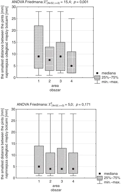
ANOVA Friedmana:χ2
(N=52,ν=3)= 15,4;p= 0,001
mediana 25%–75% 1 2 3 4
0 5 10 15 20 25 30
min.–max.
the smallest distance between the pints [mm] najmniejsza odległosć między bolcami [mm]
area obszar
Fig. 12.Comparison of the smallest distance between
discriminator pins felt in the examined areas of the operated extremity (above) and non−operated (below)
Ryc. 12.Porównanie najmniejszej odległości między
bolcami dyskryminatora odczuwanej w badanych ob− szarach kończyny operowanej (powyżej) i nieopero− wanej (poniżej)
ANOVA Friedmana:χ2
(N=52,ν=3)= 5,0;p= 0,171
mediana 25%–75% 1 2 3 4
0 5 10 15 20 25 30
min.–max.
the smallest distance between the pints [mm] najmniejsza odległosć między bolcami [mm]
area obszar
area obszar
vibration sensibility 32 Hz czucie wibracji 32 Hz brak
słabe prawidłowe
number of patients liczba pacjentów
Fig. 13.Numbers of patients and vibration sensibility
at 32 and 256 Hz in the examined areas of the foot and the results of the chi−squared test
Ryc. 13.Liczba pacjentów w zależności od czucia
wibracji 32 i 256 Hz w badanych obszarach stopy oraz wyniki testu
area obszar
vibration sensibility 256 Hz czucie wibracji 256 Hz
number of patients liczba pacjentów
unsatisfactory (wynik niezadowalający)
2%
sufficient (wynik dostateczny)
25% good
(wynik dobry) 50%
very good (wynik bardzo dobry)
23%
Fig. 14. Global subjective assessment of sensibility
restoration – result
Ryc. 14. Globalna ocena subiektywna powrotu czucia
surface of the foot edge), as illustrated by Fig. 11. Assessment of two−point sensibility showed signif− icantly better (p< 0.01) restoration of that kind of sensibility in area 4 (skin of toe V) than in the remaining examined areas of the operated extremi− ty. Differences were not observed in the non−oper− ated extremity (Fig. 12).
Similar observations were in assessing vibration sensibility with a frequency of 32 Hz. It was observed that area 4 (skin of toe V) is essentially more sensitive to vibration at that frequency than the other tested areas. A significant difference in sensi− bility of the tested areas to vibration at a frequency of 256 Hz was not observed (Fig. 13). Subjective
assessment of the degree of sensibility restoration in the area innervated by the sural nerve showed a very good result of sensibility restoration in areas 2 and 4 (skin of the heel from the medial side and toe V) (Table 6). However, in the global subjective assess− ment of sensibility restoration reported by the tested patients, a very good result was achieved in 23% cases, good in 50%, satisfactory in 25% and unsatis− factory results in 2% (Fig. 14).
On the basis of this study, the authors observed that: 1) Use of the sural nerve is and will remain a routine procedure in the reconstruction of nerves requiring use of grafts fo the proper length. The sural nerve is the most often used sensibility nerve because of the ease of its removal and the slight deficiencies in sensibility in the foot; 2) A large per− centage (75% of cases) of sensibility restoration in the extremity after nerve removal as well as a lack of motor function defects and efficiency of gait were observed; any essential harms resulting to patients were thus not observed, and this is an essential practical aspect; 3) Examination of soft sensibility, two−point sensibility, and vibration sen− sibility plays an essential role in assessing sensibil− ity restoration after iatrogenic injury of the sural nerve in patients treated with autogenic graft of the sural nerve, allowing learning about the mecha− nisms of the peripheral compensation of sensibility.
lack of neuroma (brak nerwiaka)
63% 29%
neuroma – strong Tinell's symptom (nerwiak – silny objaw Tinella)
8%
33
15 4
neuroma – weak Tinell's symptom (nerwiak – słaby objaw Tinella)
Fig. 15.Complications
Ryc. 15.Powikłania
References
[1] Edward D Kim, JU Tae Seo: Minimally invasive technique for sural nerve harvesting: technical description and
follow−up. Urology 2001, 57, 921–924.
[2] Blackshear MB, Gregory EL, O’Brien Stephen J: Sural nerve entrapment after injury to the gastrocnemius:
A case report. Arch Phys Med Rehabil 1999, 80, 604–606.
[3] Bell−Krotoski JA:Sensibility testing: Current concepts. In: Rehabilitation of the Hand: Surgery and Therapy.
Eds.: Hunter JM, Mackin EJ, Callahan AD. St. Louis: Mosby – Year Book Inc., 1995, 1, 109–128.
[4] Bell−Krotoski OTR, FAOT, FAOTA, Judith and Elizabeth Tomancik LOTR:The Repeatability of Testing
with Semmes−Weinstein Monofilaments. J Hand Surg 1987, 12A, 155–161.
[5] De Moura W, Gilbert A:Surgical anatomy of the sural nerve. J Reconstr Microsurg 1984, 1, 31–39.
[6] Coert JH, Dellon AL:Clinical implications of the surgical anatomy of the sural nerve. Plast Reconstr Surg 1994,
94, 850–855.
[7] Dellon AL:Clinical use of vibratory stimuli to evaluate peripheral nerve injury and compression neuropathy. Plast
Reconstr Surg 1980, 65, 4, 466–476.
[8] Dellon AL, Mackinnon SE, Crosby PM:Reliability of two point discrimination measurements. J Hand Surg
1987, 12 A, 693–696.
[9] Kobayashi S, Akizuki T, Sakay Y:Harvest of the sural nerve using the endoscope. Ann Plast Surg 35, 249–253.
[10] Mackinnon SE, Dellon AL:Two−point discriminator test. J Hand Surg 1985, 10 A, 6, 906–907.
[11] Solders G:Discomfort after fascicular sural nerve biopsy. Acta Neurol Scand 1988, 77, 503–504.
[12] Sarrafian SK: Anatomy of the foot and ankle: descriptive, topographic, functional. 2nded. Philadelphia (PA),
Lipinocot 1993.
Address for correspondence:
Krzysztof SkibaDepartment of Traumatology and Hand Surgery Silesian Piasts University of Medicine
ul. Traugutta 57/59 50−417 Wrocław Poland
Tel.: +48 71 7332706 E−mail: [email protected]
Conflict of interest: None declared
![Fig. 3. Anatomy of the sural nerve: course of the suralnerve [2]](https://thumb-us.123doks.com/thumbv2/123dok_us/8772831.1757945/3.595.81.264.181.485/fig-anatomy-sural-nerve-course-suralnerve.webp)