DOI
10.17219/acem/65782
Copyright
© 2017 by Wroclaw Medical University This is an article distributed under the terms of the Creative Commons Attribution Non-Commercial License (http://creativecommons.org/licenses/by-nc-nd/4.0/)
Address for correspondence
Leopold R. Rehan E-mail: [email protected]
Funding sources
None declared
Conflict of interest
None declared
Received on November 21, 2015 Reviewed on September 14, 2016 Accepted on October 12, 2016
Abstract
Pediatric patients suffering from valve bladder syndrome (VBS) are at risk of developing chronic kidney dis-ease (CKD) and renal failure in later life. Therefore, it is of vital importance to determine the risk factors and the best possible strategies for diagnosis and treatment in patients with VBS that would minimize the risk of developing CKD. In this review we have presented the current knowledge of CKD risk factors in patients with posterior urethal value (PUV). We have also discussed possible recommendations for prenatal diag-nostics procedures to be undertaken in patients with PUV, postnatal monitoring and therapeutic strategies that could reduce the risk of developing CKD in this population. Although in most cases there are no clear guidelines for appropriate clinical actions that can be undertaken in patients with PUV to minimize the risk of kidney failure, we have tried to present concise and accurate advice for physicians taking care of patients with PUV.
Key words: chronic kidney disease, valve bladder syndrome, bladder dysfunction, posterior urethral valve
Valve bladder syndrome in children: On the trail of the best
strategies to prevent chronic kidney disease
Dorota Polak-Jonkisz
1, A, B, D–F, Leopold R. Rehan
2, B, D, Konstancja Fornalczyk
1, A, B, Paweł Hackemer
3, B, D, Danuta Zwolińska
1, A, E, F1 Department of Pediatric Nephrology, Wroclaw Medical University, Poland
2 Department of Internal Medicine, Occupational Diseases and Hypertension, Medical University Hospital, Wrocław, Poland 3 Department of Urolody, Medical University Hospital, Wrocław, Poland
A – research concept and design; B – collection and/or assembly of data; C – data analysis and interpretation; D – writing the article; E – critical revision of the article; F – final approval of the article
Introduction
In children, congenital obstructive uropathies are the cause of 30–60% of chronic kidney disease (CKD) cas-es. Among these, posterior urethral valve (PUV) in boys is the most prevalent. Obstructions in the urinary tract lead to its functional and anatomical changes, and then to the development of CKD.1,2
Fetal development of the urinary tract is a very com-plex process, involving a number of genes.3 The process of nephrogenesis begins between the 3rd and the 4th week of gestation, and according to Hennus et al., the onset of histological changes that lead to PUV formation can be observed already in the 4th week of the fetal life.4 Around 95% cases of PUVs result from abnormal insertion of the mesonephric ducts into the cloaca, followed by formation of anomalous ridges or folds.5 PUV increases the residual urine volume after bladder voiding. In 1919, Young dis-tinguished 3 types of PUV: type I (folds which originate from the distal edge of the colliculus) – most often ob-served, type II (folds, originating from the proximal edge of the colliculus), and type III – with the worst progno-sis (a membrane with a central hole, not linked with the colliculus).6 Modern therapeutic procedure involve en-doscopic valve ablation (through urethra) shortly after the birth, but in some cases, especially in prenatal babies with too small urethras, temporary vesicostomy may be necessary.7,8 Even early surgical intervention is usually in-sufficient, and patients with PUV develop valve bladder syndrome (VBS); in 25–45% CKD will develop eventu-ally.7 Obstruction of the urine flow resulting from PUV leads to various, sometimes life-threatening consequenc-es (Fig. 1).
Valve bladder syndrome (VBS) was described by Mitchell (1982) as a complication of PUV, resulting in a combination of persistent upper urinary tract dila-tation, hypertrophy of the bladder walls, vesicoureteral reflux, and hydronephrosis. All these changes may lead to various kidney injuries and compromise urinary blad-der functions.1 Patients with VBS demonstrate con-siderably reduced contractility of detrusors, increased bladder compliance and a higher post-voiding residual (PVR). These parameters are described as the risk factors of dilatation of the upper urinary tract. Phases of valve bladder development can be described as follows: ini-tially, in infants, the bladder is growing in mass, has small capacity and increased contractility. It is usually almost empty, and the voiding pressure is high.9 In the next 3 years from valve ablation, in compensated stage, volume of the bladder increases, and the pressures are maintained above the control level, because of abnormal-ity in smooth muscles, contractilof abnormal-ity gradually decreases leading to a bladder instability.9 In the last phase, decom-pensation of bladder functioning is clinically apparent; the bladder capacity progressively increases, but bladder compliance, contractility and emptying are decreased (so called myogenic bladder insufficiency).10
Another serious complication of PUV exerting adverse effects on fetal development is oligohydramnios, which can cause the death of the fetus.
In almost two-thirds of the children with PUV, CKD will develop, and 11–51% of this population will progress to ESRD around the age of puberty.11
Optimal diagnostic and therapeutic procedures that will reduce the complications of PUV are of vital impor-tance. For example, Bhadoo D. et al. proposed the “step ladder” of PUV treatment:
• valve ablation/vesicostomy,
• USPCN: Ultrasound guided percutaneous nephros-tomy,
• ureterostomy.
In the analyzed cases, the primary endoscopic valve ab-lation was the most common initial procedure. Chronic renal failure was seen in 42.7% of the patients. In their study, the high prognostic significance of initial serum creatinine, PRA levels and GFR for developing CKD in patients with PUV was confirmed.12
CKD risk factors
Many prognostic factors for future renal function in patients with PUV have been proposed.1,2,8 A system-atic monitoring of these indicators is necessary for the appropriate selection of treatment methods and the time of their implementation, optimal for delaying the on-set of CKD or slowing its progression. Most important of those prognostic factors include: the age at the time of diagnosis, the serum creatinine level, considerable
vesicoureteral reflux, renal dysplasia, and the presence or lack of the bladder dysfunction (Table 1).
A separate problem concerns renal prognosis for patients diagnosed in utero with PUV. Some authors suggest the important role of fetal urine analysis. According to Lipitz et al., sodium, calcium, and beta 2-microglobulin in fetal urine were the best predic-tors for fetal survival.13 Muller et al. have assessed that the prediction of postnatal renal function can be based on the fetal urinary concentration of beta 2-mi-croglobulin as it was both specific and sensitive test, whereas other parameters lacked either specificity (so-dium, chloride, and urea levels) or sensitivity (fetal uri-nary glucose, phosphorus, calcium, ammonium, and total proteins).14 Another investigated parameter is fe-tal serum beta 2-microglobulin, whose concentration might be independent from gestational age.15 Neverthe-less, there is still no consensus over the value of fetal urine assessment in CKD prognosis. For example, Mor-ris et al. in their systematic review defined calcium and sodium levels as the most accurate predictors, and at the same time stated that current research is insufficient to determine the optimal, biochemical prognostic markers of postnatal renal function in PUV patients.16
Age at the time of diagnosis
The diagnosis of PUV at an early age is associated with a poor prognosis of further renal function and high in-cidence of CKD development. It is possible that this as-sociation is due to the fact that early diagnosis is often a result of more severe anatomical anomalies.7,9,17,18
Nadir serum creatinine concentration
It is accepted that the nadir serum creatinine concen-tration equal to or higher than 1 mg/dL in the 1st year of life demonstrates a strong association with a higher probability of developing CKD and worse prognosis of its progression to ESRD.7,9,17–19 Some researchers postulate the importance of high serum creatinine concentration on the diagnosis of PUV as a negative prognostic factor of developing CKD or nadir serum creatinine concentra-tion after valve ablaconcentra-tion.20,21
Glomerular filtration rate
Also, the decline in eGFR in the 1st year of life is wide-ly reported as a negative prognostic factor of developing
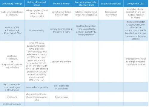
Table 1. The risk factors of developing chronic renal disease (patients before and after PUV ablation)
Laboratory findings Kidney ultrasound image Patient’s history Co-existing anomalies of urinary tract Surgical procedures Urodynamic tests
nadir serum creatinine ≥ 1.0 mg/dL
kidney dysplasia (small renal volume (<3 percentile)
age at presentation before 1 year
bilateral vesicoureteral reflux, hydronephrosis
late surgical intervention
excessive bladder contraction and low
bladder capacity in infants
reduced eGFR at 1 year of age < 90 mL/min/1.73 m2
kidney scarring urinary incontinence at the age > 5 years
bladder dysfunction – low susceptibility, detrusor overactivity,
urinary retention
increase in bladder capacity, resolution
of excessive contraction, unstable bladder function over 3 years from the valve
ablation
creatinine > 1.0 mg/dL
at diagnosis of posterior
urethral valves
small RPA (renal parenchymal area) –
RPA’s growth of 1 cm2 correlated with
a decrease in the risk of ESRD), the cut-off point in the study assumed at the size of 12.4 cm2 (boys with
RPA < 12.4 cm2 showed
progression to ESRD 10 times more likely
than those with RPA ≥ 12.4 cm2)
growth impairment progression with age to a large myogenic
insufficient bladder
high concentration of urea nitrogen
in blood serum increased echogenicity
over 3 episodes of febrile UTI
proteinuria
abnormal distribution of core / kidney cortex
ratio hypertension
diagnostic indicators of PUV in prenatal US imaging are: the increased thickness of the bladder wall and the dila-tation of the bladder. Another US measurement is longi-tudinal bladder dimension (associated with a poor renal prognosis when it exceeds 15 mm).11,26
Assessment of fetal renal function
Fetal urine analysis is being currently investigated as an excellent diagnostic tool for predicting the future renal function in patients with PUV. One of the laboratory values used as a predictor is the urinary sodium concentration, which helps to determine the function of renal tubules. The normal value for fetal urinary sodium concentration is below 90 mmol/L between the 20th and the 30th week of gestation. Sodium levels in urine above 100 mmol/L have been found to indicate a condition threatening fetal life (usually either renal or pulmonary failure).13,27
Beta 2-microglobulin is freely filtered by the normal fe-tal glomeruli and nearly in 100% reabsorbed in the proxi-mal tubules, thus it can be useful as an indicator of fetal glomerular filtration rate (GFR). It also has a predictive value as to postnatal GFR. Beta 2-microglobulin detect-ed in the urine suggests an impairment in renal tubules function.13,20,27 Other prognostic factors in fetal urine analysis, such as its specific gravity, osmolality, and the excreted volume have been defined by Glick et al.28
Tests assessing the beta 2-microglobulin in fetal urine and N-acetyl-beta-D-glucosaminidase (NAG) concentra-tion in serum have been recently used to evaluate the de-gree of damage of renal parenchyma (their concentration is proportional to the extent of damage). The concentra-tion of beta 2-microglobulin above 4 mg/L and cystatin C levels above 1 mg/L, and the NAG levels above 6 mg/dL are thought to be poor prognostic factors for postnatal renal function.13,20,27,29,30
Postnatal monitoring
Voiding cystourethrography
In male newborns with prenatally diagnosed hydrone-phrosis and a “keyhole” image in US examination, asso-ciated with a high PUV probability, a prompt, extended diagnostics of the urinary system is recommended. Cys-tography has been established as a diagnostic “golden standard” (Fig. 2). For example, performing voiding cystourethrography (VCUG) in these cases (prenatally diagnosed hydronephrosis with high probability of vesi-coureteral reflux or lower urinary tract pathologies) has been recommended by the Society for Fetal Urology and Canadian Urological association.31 VCUG can be per-formed without anesthesia. It reveals residual urine and vesicoureteral reflux, and is also useful in the assessment of detrusor and bladder neck activity.7,32
CKD.22,23 In 2003, Osama demonstrated that when the
clearance of creatinine was lower than 60 mL/min/1.73 m2, the prognosis for renal failure in the future would be at approximately 50%, while for creatinine clearance above 90 mL/min/1.73 m2, the incidence of renal failure would decrease to 27%.17
Some other factors are also considered in assessing the risk of developing CKD in the future or its progression to ESRD. The most often mentioned amongst them are: the existence of renal dysplasia, vesicoureteral reflux, various bladder dysfunctions (reduced contractility of de-trusors, decreased bladder compliance, etc.), proteinuria and hypertension, more than 3 episodes of urinary tract infections (UTI) and late surgical intervention.1,7,9,17–20
Prenatal diagnostic strategies
Choosing an appropriate diagnostic path is of extreme importance in patients with PUV. Early diagnosis is nec-essary for implementing therapeutic procedures that will minimize CKD development in those patients, and re-duce the rate of progression to ESRD.
Ultrasound
Ultrasound (US) imaging is used to visualize anatomi-cal anomalies of fetal bladder in the 1st trimester of preg-nancy and dilatation in the urinary tract from 18th–20th week of gestation. Such findings usually provide an in-sight into potential pathologies within the urinary tract, and can also be a marker of karyotype abnormalities.24
tion), that PVR and maximum bladder capacity (MBC) increase with age after the procedure. Multiple bladder dysfunctions can be diagnosed accurately using urody-namics.19
Management strategies
An early diagnosis of PUV with its complications, in-cluding VBS, and the application of the right treatment at the right time is of key importance for the prognosis, and may prevent or delay CKD in the future.
Unfortunately, even after successful valve ablation some degree of bladder dysfunction will develop in most of the patients. This is true even for PUV patients who have been surgically treated at a very early stage of the disease.7,19 Therefore, patients with PUV require constant monitoring, also after surgical procedure, in order to de-tect symptoms of VBS early enough to undertake possible preventive methods.
Multi-center observations estimate the incidence of CKD in the population of boys with PUV at 10–30.4% (in the age group from the 1st month of life up to 18 years).18,35 Early diagnosis due to prenatal ex-aminations (diagnostics imaging, laboratory assays, etc.) together with timely surgical interventions in utero have led to a significant decrease of mortality, and improved overall health in this group of patients.20,29 Nevertheless, we still lack the clear guidelines for the best type of surgi-cal procedure, and each case needs individual assessment of possible risks and gains.
Strategies of limiting the risk of developing CKD in pa-tients with VBS include diagnostic imaging, laboratory tests and therapeutic procedures. The latter are not only surgical interventions (including in utero), but also phar-macotherapy and other procedures, which mainly aim at reducing the residual urine in the bladder.
Treatment possibilities
Modern surgical techniques allow for PUV ablation performed in utero. In that case the procedure car-ries a significant risk of complications, such as fetal injury or even fetal or maternal death. For example, Sananes N. et al. reported 2-year outcomes after thera- peutic fetal cystoscopy with ablation of PUV, where only 57% of infants survived.36 This is why intrauterine surgeries are reserved for exceptional cases (Fig. 4, 5). There is still a lack of consensus as to the advantages of intrauterine intervention over surgery after birth.29 Intrauterine treatment currently is limited to cases of fetuses with oligohydramnios and diagnosed before the 2nd trimester.10,19 There are 3 possible treatments: amnio-bladder valve, vesicostomy and fetal endoscopic valve ablation, and none of them has definite advantage supported by evidence.8
Urodynamic studies
Pathophysiological changes leading to urinary blad-der dysfunction and deterioration in upper urinary tract functions in patients with PUV seem to occur even de-spite an early diagnosis and valve ablation.
Urodynamic tests in children are employed as an effec-tive way to establish the urodynamic pattern of the blad-der, and implement appropriate treatment. In their study involving patients with PUV, Capitanucci et al. compared the usefulness of noninvasive urodynamic evaluation vs invasive urodynamics in detecting lower urinary tract dysfunction and preventing CKD development. They have found similar effectiveness and safety of both approaches. Some patients experience strong discomfort during invasive urodynamic studies, and many decline further procedures. This gives the noninvasive approach an advantage. There are no clear guidelines regarding the frequency of performing urodynamic studies. How-ever, it seems to be reasonable to perform them with each change in employed therapeutic method.33
Late diagnosis of PUV
Most of the children with PUV are diagnosed before birth, during a prenatal ultrasound. Sometimes, however, there are no symptoms at this stage, so the disease can go undiagnosed into childhood. Zornoza et al. described 8 cases of late diagnosed PUV in children between 1 and 14 years of age. Five patients had symptoms related to voiding dysfunction, and the other 3 were diagnosed incidentally during cystoscopy performed for another reason. All patients were operated, and after a 20-month follow-up no stenosis were diagnosed. Voiding cystoure-throgram (VCUG) is a much more specific method for diagnosing PUV than an ultrasound. Diagnosis can also be made by cystoscopy, where a small camera is inserted into the urethra for direct visualization of the posteriorly positioned valve, and this method is recommended by Zornoza et al. in any case of urinary retention.34
An urodynamic investigation of valve bladder syn-drome is very important. Wen et al. reported in an urody-namic study, performed in 2 groups of patients (less than 1 year and more than 1 year after urethral valve
• clean, intermittent catheterization (CIC) that allows for effective emptying of the bladder, also preventing urinary blockage and reducing risk of recurrent uri-nary tract infections;9,10,18,22,39,40
• as recurring urinary tract infections (UTI) are one of risk factors for CKD development in VBS patients, some authors concentrated on assessing various pre-ventative methods. The clear rules of antibiotic preven-tive therapy has not been established, some recommend trimethoprim 1–2 mg nightly dose until vesicoureteral reflux recedes or age of 4–5 years.10,37 Interestingly, many authors quote arguments of the positive effects of circumcision in UTI prevention in children with PUV;11,37
• some other methods for reducing urinary retention in the bladder include pelvic floor muscles exercise and Valsalva maneuver.10,18
When all of the methods mentioned above are insuf-ficient in reducing urine retention and improving bladder voiding, surgical procedures are performed:
• bladder augmentation, traditionally enterocystoplasty, but recently more popular ureterocystoplasty, which retains histological structure of the urinary tract;9,10 • high ureter diversion (various versions: vesicostomy,
percutaneous ureterostomy, pyelostomy, nephros-tomy, etc.) is controversial due to the lack of strong evidence that it can prevent CKD;9
• Mitrofanoff appendicovesicostomy is one of the treatments of the valve bladder syndrome that allows patients clean intermittent catheterization and over-night drainage when catheterization is impossible. King et al. reported that this way of treatment can achieve significant improvements in decreasing hy-dronephrosis and urodynamic parameters of bladder dysfunction. However, it does not prevent renal dete-rioration and progression to CKD.1,10,21
All treatment possibilities listed above are illustrated in Fig. 3.
Performing the valve ablation alone, without urody-namic follow-up, is inappropriate. Urinary diversion is still a very popular procedure, but recent studies sug-gest that in most cases in which diversion is considered,
Fig. 3. Management of urine incontinence in VBS
Fig. 4. Steps to take before choosing the intrauterine intervention
Fig. 5. Biochemical markers of good prognosis
A proper understanding of the molecular pathways, which are activated in damaged cells of the bladder wall, allows for appropriate pharmacological intervention in order to improve its function.4,22 Modern pharmaco-therapy in VBS uses drugs like:
• cholinolytics (e.g., oxybutynin – 0.2 mg/kg/24 h), which decrease hyper-contractility of the bladder; therapy should be started right after valve ablation, even when urine incontinence is not presented; their use in the long term leads to a myogenic bladder fail-ure – then it is recommended to use the CIC (clean, intermittent catheterization), but sometimes, after discontinuation of cholinolytic, the bladder function normalizes on its own;9,18,19,37,38
• alpha-blockers (e.g., terazosin – 0.25–2 mg), which help in bladder voiding; their use is particularly ad-visable when obstruction in bladder neck is suspect-ed cause of ineffective voiding.10
Other drugs that are being tested in patients with VBS are aimed at reversing histological changes in the bladder (mainly ACEI and various growth factors inhibitors).39
Non-pharmacological procedures used in VBS man-agement include:
those same patients can be treated with proactive uro-dynamics and anticholinergic therapy achieving simi-lar results. It is still unclear if growth factor inhibitors or angiotensin converting enzyme inhibitors have a role in preventing or reversing the histological changes of the valve bladder.39
Summary
PUV is a congenital, obstructive uropathy leading to the valve bladder syndrome, which in turn results in the development of CKD. Active urodynamic monitor-ing of the PUV patients plays a significant role in the pre-vention of bladder dysfunction, CKD development and its progression to ESRD.
Early diagnosis is currently possible even before birth (thanks to ultrasound, fetal urine analysis and some bio-chemical tests of fetal blood). At later stages cystography and urodynamic tests are essential to monitor the PUV patients, and determine the risk of developing CKD.
The therapeutic methods for preventing or minimizing VBS vary from surgical approach (which can be imple-mented in utero) through some mechanical procedures (such as timed bladder voiding and CIC) to pharmaco-logical management.
Due to the fact that cytological changes in the urinary tract, observed in children with PUV, are irreversible, medical intervention ought to be carried out with due haste after the diagnosis, but prenatal procedures, carry-ing a high risk of complications, are still reserved for the minority of cases.
References
1. Penna FJ, Elder JS. CKD and bladder problems in children. Adv Chronic
Kidney Dis. 2011;18(5):362–369. doi:10.1053/j.ackd.2011.08.001.
2. Ansari MS, Gulia A, Srivastava A, Kapoor R. Risk factors for progression to end-stage renal disease in children with posterior urethral valves.
J Pediatr Urol. 2010;6(3):261–264. doi:10.1016/j.jpurol.2009.09.001.
3. Zwolińska D, Polak-Jonkisz D, Makulska I. Podłoże genetyczne
wrodzonych wad nerek i układu moczowego. Postepy Hig Med
Dosw (Online). 2011;65:829–837. http://www.ncbi.nlm.nih.gov/
pubmed/22173447. Accessed June 24, 2016.
4. Hennus PML, van der Heijden GJMG, Bosch JLHR, de Jong TPVM, de Kort LMO. A systematic review on renal and bladder dysfunc-tion after endoscopic treatment of infravesical obstrucdysfunc-tion in boys.
PLoS One. 2012;7(9):e44663. doi:10.1371/journal.pone.0044663.
5. Krishnan A, De Souza A, Konijeti R, Baskin LS. The anatomy and embryology of posterior urethral valves. J Urol. 2006;175(4):1214– 1220. doi:10.1016/S0022-5347(05)00642-7.
6. Young HH, Frontz WA, Baldwin JC. Congenital obstruction of the posterior urethra. J Urol, 3: 289-365, 1919. J Urol. 2002;167(1):265– 268. http://www.ncbi.nlm.nih.gov/pubmed/11743334. Accessed June 24, 2016.
7. Yohannes P, Hanna M. Current trends in the management of poste-rior urethral valves in the pediatric population. Urology. 2002;60(6): 947–953. http://www.ncbi.nlm.nih.gov/pubmed/12475647. Accessed June 24, 2016.
8. Nasir AA, Ameh EA, Abdur-Rahman LO, Adeniran JO, Abra-ham MK. Posterior urethral valve. World J Pediatr. 2011;7(3):205–216. doi:10.1007/s12519-011-0289-1.
9. DeFoor W, Clark C, Jackson E, Reddy P, Minevich E, Sheldon C. Risk factors for end stage renal disease in children with posterior ure-thral valves. J Urol. 2008;180(Suppl 4):1705–1708; discussion 1708. doi:10.1016/j.juro.2008.03.090.
10. Hutton KAR. Management of posterior urethral valves. Curr
Paedi-atr. 2004;14(7):568–575. doi:10.1016/j.cupe.2004.07.013.
11. Pohl M, Mentzel HJ, Vogt S, Walther M, Rönnefarth G, John U. Risk factors for renal insufficiency in children with urethral valves.
Pedi-atr Nephrol. 2012;27(3):443–450. doi:10.1007/s00467-011-1999-2.
12. Bhadoo D, Bajpai M, Panda SS. Posterior urethral valve:
Prog-nostic factors and renal outcome. J Indian Assoc Pediatr Surg.
2014;19(3):133–137. doi:10.4103/0971-9261.136459.
13. Lipitz S, Ryan G, Samuell C, et al. Fetal urine analysis for the
assess-ment of renal function in obstructive uropathy. Am J Obstet
Gynecol. 1993;168(1 Pt 1):174–179. http://www.ncbi.nlm.nih.gov/
pubmed/8420322. Accessed June 24, 2016.
14. Muller F, Dommergues M, Mandelbrot L, Aubry MC, Nihoul-Fekete C, Dumez Y. Fetal urinary biochemistry predicts postna-tal renal function in children with bilateral obstructive uropathies.
Obstet Gynecol. 1993;82(5):813–820. http://www.ncbi.nlm.nih.gov/
pubmed/8414330. Accessed June 24, 2016.
15. Ciardelli V, Rizzo N, Farina A, Vitarelli M, Boni P, Bovicelli L. Prenatal evaluation of fetal renal function based on serum beta(2)-microglob-ulin assessment. Prenat Diagn. 2001;21(7):586–588. doi:10.1002/pd.90. 16. Morris RK, Quinlan-Jones E, Kilby MD, Khan KS. Systematic review
of accuracy of fetal urine analysis to predict poor postnatal renal function in cases of congenital urinary tract obstruction. Prenat
Diagn. 2007;27(10):900–911. doi:10.1002/pd.1810.
17. Sarhan OM, El-Ghoneimi AA, Helmy TE, Dawaba MS, Ghali AM, Ibra-hiem E-HI. Posterior urethral valves: Multivariate analysis of factors affecting the final renal outcome. J Urol. 2011;185(Suppl 6):2491– 2495. doi:10.1016/j.juro.2011.01.023.
18. Ansari MS, Singh P, Mandhani A, et al. Delayed presentation
in pos-terior urethral valve: Long-term implications and outcome.
Urolo-gy. 2008;71(2):230–234. doi:10.1016/j.urology.2007.09.037.
19. Wen JG, Li Y, Wang QW. Urodynamic investigation of valve bladder syndrome in children. J Pediatr Urol. 2007;3(2):118–121. doi:10.1016/j. jpurol.2006.06.008.
20. Salam MA. Posterior urethral valve: Outcome of antenatal
inter-vention. Int J Urol. 2006;13(10):1317–1322.
doi:10.1111/j.1442-2042.2006.01555.x.
21. King T, Coleman R, Parashar K. Mitrofanoff for valve bladder
syndrome: Effect on urinary tract and renal function. J Urol.
2014;191(Suppl 5):1517–1522. doi:10.1016/j.juro.2013.09.008. 22. Scott JE. Management of congenital posterior urethral valves. Br J
Urol. 1985;57(1):71–77. http://www.ncbi.nlm.nih.gov/pubmed/3971107. Accessed June 24, 2016.
23. Lopez Pereira P, Espinosa L, Martinez Urrutina MJ, Lobato R, Navar-ro M, Jaureguizar E. Posterior urethral valves: PNavar-rognostic
fac-tors. BJU Int. 2003;91(7):687–690. http://www.ncbi.nlm.nih.gov/
pubmed/12699486. Accessed June 24, 2016.
24. Yiee JH, Tasian GE, Copp HL. Management trends in prenatal-ly detected hydronephrosis: National survey of pediatrician
prac-tice patterns and antibiotic use. Urology. 2011;78(4):895–901.
doi:10.1016/j.urology.2011.04.027.
25. Bernardes LS, Aksnes G, Saada J, et al. Keyhole sign: How specific is it for the diagnosis of posterior urethral valves? Ultrasound Obstet
Gynecol. 2009;34(4):419–423. doi:10.1002/uog.6413.
26. Dias T, Sairam S, Kumarasiri S. Ultrasound diagnosis of fetal renal abnormalities. Best Pract Res Clin Obstet Gynaecol. 2014;28(3):403– 415. doi:10.1016/j.bpobgyn.2014.01.009.
27. Abdennadher W, Chalouhi G, Dreux S, et al. Fetal urine biochem-istry at 13-23 weeks of gestation in lower urinary tract obstruc-tion: Criteria for in-utero treatment. Ultrasound Obstet Gynecol. 2015;46(3):306–311. doi:10.1002/uog.14734.
28. Glick PL, Harrison MR, Golbus MS, et al. Management of the fetus with congenital hydronephrosis II: Prognostic criteria and selection for treatment. J Pediatr Surg. 1985;20(4):376–387. http://www.ncbi. nlm.nih.gov/pubmed/3900327. Accessed June 24, 2016.
30. Thomas DFM. Prenatally diagnosed urinary tract abnormalities: Long-term outcome. Semin Fetal Neonatal Med. 2008;13(3):189–195. doi:10.1016/j.siny.2007.10.003.
31. Davenport MT, Merguerian PA, Koyle M. Antenatally diagnosed
hydronephrosis: Current postnatal management. Pediatr Surg Int.
2013;29(3):207–214. doi:10.1007/s00383-012-3258-4.
32. Cozzi DA, Morgante D, Frediani S, et al. Posterior urethral valves: Relationship between vesicoureteral reflux and renal function.
Urology. 2011;77(5):1209–1212. doi:10.1016/j.urology.2010.08.014.
33. Capitanucci ML, Marciano A, Zaccara A, et al. Long-term bladder function followup in boys with posterior urethral valves:
Com-parison of noninvasive vs invasive urodynamic studies. J Urol.
2012;188(3):953–957. doi:10.1016/j.juro.2012.04.121.
34. Zornoza M, Angulo JM, Parente A, Simal S, Burgos L, Ortiz R. Late diagnosis of posterior urethral valves. Actas Urol españolas. 2015;39(10):646–650. doi:10.1016/j.acuro.2015.05.005.
35. Kousidis G, Thomas DFM, Morgan H, Haider N, Subramaniam R, Feather S. The long-term outcome of prenatally detected pos-terior urethral valves: A 10 to 23-year follow-up study. BJU Int. 2008;102(8):1020–1024. doi:10.1111/j.1464-410X.2008.07745.x. 36. Sananes N, Cruz-Martinez R, Favre R, et al. Two-year outcomes after
diagnostic and therapeutic fetal cystoscopy for lower urinary tract obstruction. Prenat Diagn. 2016;36(4):297–303. doi:10.1002/pd.4771. 37. Narasimhan KL, Chowdhary SK, Kaur B, Mittal BR, Bhattacharya A.
Factors affecting renal scarring in posterior urethral valves. J
Pedi-atr Urol. 2006;2(6):569–574. doi:10.1016/j.jpurol.2005.12.003.
38. Koff SA, Mutabagani KH, Jayanthi VR. The valve bladder syndrome: Pathophysiology and treatment with nocturnal bladder
emp-tying. J Urol. 2002;167(1):291–297. http://www.ncbi.nlm.nih.gov/
pubmed/11743343. Accessed June 24, 2016.
39. Glassberg KI. The valve bladder syndrome: 20 years later.
J Urol. 2001;166(4):1406–1414. http://www.ncbi.nlm.nih.gov/
pubmed/11547099. Accessed June 24, 2016.